




ALLICIN IN HEPATOCELLULAR CARCINOMA: MECHANISTIC INSIGHTS INTO ROS-MEDIATED APOPTOSIS AND POTENTIAL P53-INDEPENDENT ANTICANCER EFFECTS
HTML Full TextALLICIN IN HEPATOCELLULAR CARCINOMA: MECHANISTIC INSIGHTS INTO ROS-MEDIATED APOPTOSIS AND POTENTIAL P53-INDEPENDENT ANTICANCER EFFECTS
Suryavardhan Singh *, Rashyap Saraswat, Krishnapal Singh Rathore and Md Mujahedul Islam
Chitkara College of Pharmacy, Chitkara University, Chandigarh, Punjab, India.
ABSTRACT: Hepatocellular carcinoma (HCC) is a significant health problem and a leading cause of cancer-related deaths worldwide, mainly because of delayed diagnosis and the presence of multiple clones and a lack of effective treatment. Targeted therapies and immunotherapy offer limited survival gains, and often result in drug resistance, toxicity and high expense. One of the major factors that contributes to therapeutic failure is the inactivity of the tumor suppressor p53 which leads to resistance and apoptosis. Natural compounds have proven to be multi-targeted agents of interest in this context. The bioactive compound of Allium sativum, allicin (diallyl thiosulfinate), has been noted for its p53-independent anticancer properties. This review compiles the current evidence of allicin's anticancer activities related to its ability to generate ROS, to disrupt mitochondrial functions, to activate caspases, and to regulate key signaling pathways in HCC. It underscores studies suggesting that some of these mechanisms can act without p53 signaling, but there is a lack of direct evidence from the HCC models that have been validated in p53 deficient cells, and the redox modulating, mitochondrial dysfunction, caspase activation and endoplasmic reticulum (ER) stress effects of allicin in HCC are highlighted. Furthermore, allicin can modulate key pathways, such as PI3K/Akt, MAPK, and NF-κB, which are involved in the crosstalk between apoptosis and autophagy, thus further stimulating the killing of tumour cells even in resistant phenotypes. “Importantly, this review provides a ROS-metabolic cell-centric mechanism that elucidates how allicin may be selectively pro-apoptotic to HCC cells, bypassing p53-resistant cells, thereby providing support for the hypothesis that resistance in HCC is correlated with p53 dysfunction. Further research is still required to confirm and provide direct evidence of the selective targeting of p53-resistant HCC.” Although there is promising preclinical evidence potential bioavailability, pharmacokinetics, and clinical validation issues are large hurdles. In conclusion, allicin is a promising therapeutic agent for HCC, and further research is needed using advanced drug delivery systems and clinical trials to establish its efficacy.
Keywords: Allicin, Hepatocellular carcinoma, Apoptosis, p53 mutation, Reactive oxygen species (ROS), Mitochrial dysfunction, Caspase activation, PI3K/Akt pathway, NF-κB signaling, Natural compounds, Drug resistance, Precision oncology
INTRODUCTION: Hepatocellular carcinoma (HCC) is the most common primary liver cancer & a major cause of cancer-related morbidity & mortality 1.
Although diagnostic & therapeutic approaches have improved, HCC remains a clinical challenge because of its late presentation, high recurrence & low response to conventional treatments 2.
A major feature of HCC is its complex molecular heterogeneity, which includes alterations in multiple signaling pathways involved in regulating cell growth, death, blood vessel formation & spread of the tumor. In particular, alterations in the tumor suppressor p53 play an important role in the progression of hepatocarcinoma 3.
Defects in p53 cause dysfunction in the regulation of the apoptotic pathway, allowing cancer cells to evade cell death & become resistant to chemotherapy. This has prompted a recent focus on the development of therapeutic compounds that can induce apoptosis independent of p53 4. Bioactive natural products have emerged as potential candidates in this respect because they can interact with several pathways, are less toxic and can modulate key pathways. In particular, the sulfide compound allicin, found in garlic, has demonstrated potent anti-cancer activity in several cancers, including HCC 5.
Recent studies have demonstrated allicin's ability to induce apoptosis in cancer cells with p53 mutations or inactivation, suggesting its potential as a new therapeutic compound. This review aims to comprehensively explore the molecular responses of allicin-induced apoptosis in HCC, with a special focus on its potential to overcome resistance through p53 mutations 6. This article is intended to be a narrative review that summarizes the current knowledge of molecular mechanisms, therapeutic potential and translational challenges of allicin in HCC, including the differences between Wild-Type p53, p53 Loss-of-Function Mutations, p53-Null Status & Gain-of-Function Mutant p53 conditions; the focus would be on allicin's role in apoptosis & p53 independent signaling pathways 7.
Global Burden, Therapeutic Limitations, and Emerging Role of Natural Compounds in Hepatocellular Carcinomas: The Hepatocellular carcinoma (HCC) is one of the leading causes of cancer-related death worldwide, especially in areas with high prevalence of hepatitis B and C infections, like East Asia and Sub-Saharan Africa 8. Long-term inflammation, fibrosis and cirrhosis of the liver are caused by these infections, which lead to liver cancer. Moreover, the global epidemiology of HCC is changing, as the incidence of metabolic disorders such as NAFLD, alcoholic liver disease and metabolic syndrome continues to increase. Although there have been improvements in diagnosis and therapy, prognosis is poor because the condition is often identified too late and there are few effective treatments 9.
TABLE 1: DIRECT EVIDENCE OF ALLICIN ACTIVITY IN HEPATOCELLULAR CARCINOMA MODELS
| Experimental Model | TP53 Status | Major Findings | Evidence Type | Reference |
| HepG2 | Wild-type p53 | ROS generation and apoptosis | Direct | 7 |
| Hep3B | p53-null | Apoptotic response observed | Direct | 7 |
| Huh7 | TP53-mutant | Reduced viability and apoptosis | Direct | 7 |
Current treatment options (such as surgery, liver transplantation, locoregional and systemic therapy) offer little benefit. Modest improvement in survival with targeted drugs (sorafenib and lenvatinib) that can be resistant, toxic, and costly; Immunotherapy has variable efficacy and immune related side effects. Failure of treatment is associated with p53 dysfunction, tumor heterogeneity and cancer stem cells, all of which can lead to resistance and recurrence 10.
TABLE 2: INDIRECT EVIDENCE SUPPORTING POTENTIAL P53-INDEPENDENT MECHANISMS OF ALLICIN
| Disease Model | Compound | Mechanism Identified | Limitation | Reference |
| Breast cancer | Allicin | ROS-mediated apoptosis | Non-HCC | 11 |
| Colon cancer | Allicin | Caspase activation | Non-HCC | 12 |
| Glioma | Allicin | Mitochondrial dysfunction | Non-HCC | 13 |
Natural compounds are showing potential to become promising multi-target anticancer drugs in HCC. Phytochemicals like curcumin, resveratrol and epigallocatechin gallate have anti-cancer properties at relatively low toxicity levels, through apoptosis, proliferation inhibition and inhibition of angiogenesis 14. In particular, many of them can induce apoptosis p53 independently, which may give a therapeutic advantage in drug-resistant HCC. This underscores the potential of bioactive agents such as allicin as new approaches to overcome resistance and enhance clinical outcomes 6.
TABLE 3: STUDIES EXCLUDED FROM DIRECT INTERPRETATION DUE TO MODEL OR COMPOUND DIFFERENCES
| Original Model | Compound Investigated | Reason for Exclusion | Reference |
| Cardiac hypertrophy | Allicin | Non-cancer model | 15 |
| Cholangiocarcinoma | Allicin | Different malignancy | 16 |
| Colon cancer | DADS | Different compound | 17 |
| Various cancers | Coptisine | Unrelated compound | 18 |
Rationale for Investigating Allicin in HCC: Allicin (diallyl thiosulfinate), a bioactive component of Allium sativum, is a compound with powerful antioxidant, anti-inflammatory and pro-apoptotic anticancer activity 19. Its pleiotropic nature enables the regulation of key signaling pathways that play a role in tumour growth and survival. In hepatocellular carcinoma (HCC), allicin has a selective cytotoxicity in cancer cells, largely due to the induction of oxidative stress through the generation of reactive oxygen species (ROS) resulting in mitochondrial dysfunction and apoptosis 20.
Interestingly, allicin can induce apoptosis independent of p53, thus overcoming a major source of chemoresistance in HCC. Mechanistically, allicin induces both intrinsic & extrinsic apoptosis, including caspase activation, & regulates key pathways such as PI3K/Akt, MAPK & NF-κB 21. Additionally, it has an impact on autophagy & modulates the cell cycle, addressing multiple hallmarks of cancer. While stability and bioavailability pose challenges, the ability of allicin to overcome p53-resistant pathways & to exert multi-pathway anti-cancer effects makes it a promising candidatdie for further research as an agent against HCC 22.
REVIEW METHODOLOGY:
Review Design: This manuscript is a narrative review that critically summarizes the current evidence for the anti-cancer effects of allicin in hepatocellular carcinoma (HCC). It highlights particular mechanisms of p53-dependent and p53-independent apoptosis, signaling pathways, preclinical studies, drug disposition issues and translational barriers.
Literature Search Strategy: A thorough literature search was performed utilizing electronic databases such as PubMed, Scopus, Web of Science, and Google Scholar. The articles were chosen from a period of 20 years spanning from January 2000 to June 2026. The following single and combined terms were used in the search: Allicin, Allium sativum, Garlic-derived organosulfur compounds, Hepatocellular carcinoma, HCC, Liver cancer, p53, TP53 mutation, p53-deficient, p53-null, Apoptosis, Autophagy, Reactive oxygen species, PI3K/Akt/mTOR, MAPK, NF-κB.
Example Search String: Allicin or Allium sativum & Hepatocellular carcinoma or HCC AND p53 or TP53 mutation or p53 deficiency & Apoptosis or Cell death or Anticancer activity.
Inclusion Criteria:
Studies were included if they:
- Takes one or more of the purified forms of allicin.
- Assessed anticancer effects in models of hepatocellular carcinoma.
- Studied molecular mechanisms related to apoptosis, oxidative stress, autophagy, regulation of cell cycle, or signaling pathways.
- Published in an English language, peer-reviewed journal in an in vitro, in vivo or relevant preclinical model.
Exclusion Criteria:
Studies were excluded if they:
- Focused exclusively on the compounds from garlic, not specifically on allicin.
- Did not have any relatives with cancer or HCC.
- Consists entirely of conference abstracts, editorials, letters, patents or unpublished materials.
- Did not present sufficient methodological information or relevance to the topic of the review.
- Made unnecessary duplications of information that had already been published elsewhere.
Study Selection and Data Extraction: The title and abstracts retrieved from the database search were screened for relevance. The next step was to review articles that met the inclusion criteria following the extraction of full text articles.
Data on study design, experimental model, p53 status, concentration/dosage of allicin, treatment time, molecular targets, signaling pathways and key outcomes were extracted and summarized.
Evidence Synthesis:
The studies selected were analyzed qualitatively and categorized into key thematic areas, which include:
- Changes in TP53 and biology of hepatocellular carcinoma.
- The chemical composition and pharmacological actions of allicin.
- The mechanisms involved in the induction of apoptosis.
- Independent and dependent pathways that involve p53.
- Modulation of oncogenic signaling pathways.
- The evidence provided by preclinical studies in HCC models.
- Methods of drug delivery.
- Safety, toxicity and translation considerations.
Limitations of the Review: Preclinical data predominates in this review and the experimental models used are heterogeneous, the formulation and dosage of allicin are variable, and there are few studies focusing on allicin in p53 deficient HCC models.
Biology of Hepatocellular Carcinoma: HCC is a complex &multifaceted cancer, characterised by complex molecular alterations, genetic instability & activation of multiple pathways 23. HCC development is typically a multi-stage process that evolves from chronic liver injury to fibrosis & cirrhosis, and ultimately to cancer. This transformation is driven by ongoing inflammatory stimuli, oxidative stress & the accumulation of genetic & epigenetic changes 24. A hallmark of HCC is the dysfunction of critical regulatory pathways that control cell proliferation, death & differentiation. In particular, alterations in tumor suppressor genes, such as p53, and activation of oncogenic pathways result in sustained cell growth & evasion of apoptosis 25. In addition, the tumor microenvironment plays a pivotal role in disease progression through the processes of angiogenesis, immune evasion and potential metastasis. Understanding the biology of HCC is essential to identify new treatment targets, especially those that may overcome the resistance associated with p53 dysfunction 26.
Molecular Pathogenesis of Hepatocellular Carcinoma: The molecular mechanisms underlying HCC involve a dynamic interplay between genetic & epigenetic alterations & environmental influences 27. The development of chronic liver diseases, including viral hepatitis, alcohol-induced liver injury & metabolic diseases, leads to a cycle of liver damage & repair. This process promotes DNA damage, chromosomal instability & the development of oncogenic mutations 28. Key genetic changes in HCC include mutations in tumor suppressor genes (such as p53) & alterations in pathways regulating cell cycle, apoptosis & DNA repair. The inactivation of p53 is especially important in allowing the cell to evade apoptosis when exposed to stress, allowing the tumor to survive & grow 29. Epigenetic changes, including DNA methylation, histone modifications, & non-coding RNA dysregulation also contribute to altered gene expression in HCC. These changes can lead to the activation of oncogenes or inactivation of tumor suppressors, respectively, without altering the underlying DNA code 30. Oxidative stress is also important in hepatic carcinogenesis. Elevated production of reactive oxygen species (ROS) leads to the peroxidation of lipids, damage of proteins & mutations in DNA, further promoting this transformation. These molecular mechanisms contribute to the development and progression of HCC 31.
Key Oncogenic Signaling Pathways (MAPK, PI3K/Akt, NF-κB): HCC is characterized by aberrations in intracellular signaling pathways, which contribute to enhanced growth, survival, neovascularization, & metastatic potential. In particular, the MAPK, PI3K/Akt & NF-κB pathways play important roles in tumor development 32. The MAPK pathway is involved in regulating cell proliferation, differentiation & survival. Its dysregulation, often through upstream activation by growth factor receptors, leads to increased cell growth & survival. The PI3K/Akt pathway is another significant oncogenic pathway commonly activated in HCC 33. PI3K activation results in the phosphorylation of Akt, promoting cell survival by blocking apoptosis & promoting protein synthesis through downstream effectors such as mTOR. This pathway is also involved in the metabolic reprogramming & angiogenesis of tumors 34. The NF-kappa B pathway is crucial in regulating inflammation and immunity. In HCC, sustained activation of NF-κB promotes the expression of anti-apoptotic proteins, cytokines, and growth factors, thereby promoting tumour cell survival, chronic inflammation & therapeutic resistance 35. These pathways are not isolated but rather interact with each other to form an intricate network that drives hepatocarcinogenesis. This network of pathways is important to consider when designing therapeutic interventions 36. One of the limitations of the current literature is that many pathways that are generally associated with allicin's effects like PI3K/Akt/mTOR, MAPK/ERK, NF-κB, VEGF, EMT, MMP regulation, ER stress, and autophagy pathways have not been comprehensively evaluated in HCC models. Therefore, it is important to interpret the findings in these pathways with caution unless it was validated by direct evidence from an allicin treated HCC system 37.
Critical Considerations of ROS-Mediated Anticancer Activity of Allicin: A common theory of how allicin works against cancer is by producing Reactive Oxygen Species (ROS). However, the relationship between the allicin and cellular redox is complicated and requires careful study. Allicin is a very reactive thiol-modifying agent, which is easily able to interact with any protein containing low molecular thiol (e.g., GSH), which has cysteine residues. Thus, its biological effects may vary depending on concentration, duration of exposure, antioxidant capacity of the cell, and the natural redox state of the cell to which it is applied 38.
Allicin could have antioxidant or cytoprotective effects when used at low concentrations, which is hypothesized to be through the modulation of cellular redox signaling pathways. However, at high concentrations, it can exceed antioxidant capacities which can result in excessive ROS production, oxidative stress, mitochondrial dysfunction and activation of cell-death pathways. Therefore, allicin should not be considered just a pro-oxidant compound, its effect may be context dependent 39.
A proposed explanation for the specificity of anticancer activities of ROS-producing agents is that tumor cells tend to have higher basal ROS levels than normal cells, which is attributed to increased metabolic activity, mitochondrial dysfunction and oncogenic signaling. This means that cancer cells can function nearer to the limit of their tolerance for oxidative stress, making them more sensitive to additional oxidative stress induced by allicin. But the direct evidence on the selective ROS-mediated cytotoxicity of allicin on HCC cells and its protection on normal hepatocytes remains limited. So, the safety of tumor selectivity should be considered a working hypothesis and not confirmed 40.
One of the intracellular antioxidant systems and a main target for the thiol-modification effect of allicin is glutathione. Lowering the cell's glutathione concentrations could increase oxidative stress and help kill cancer cells. However, over-activation of the GSH pathway may make normal tissues more susceptible to oxidative stress. Thus, it is important to understand how glutathione plays a role in both tumour and non-tumour hepatic cells to determine the therapeutic potential and safety profile of allicin 41.
Because hepatocytes have a very high metabolic activity and are involved in a large number of xenobiotic metabolisms, ROS production might result in excessive amount of oxidative damage to healthy liver tissue. Adverse effects include lipid peroxidation, mitochondrial dysfunction, DNA damage and impaired hepatic function. Although many studies are done on the anticancer activity, relatively fewer studies focus on the oxidative damage of normal hepatocytes. Therefore, additional toxicological investigations are needed to ensure the safety margin of allicin based therapy 42.
One of the major difficulties in the use of ROS in anticancer therapy is the narrow window of therapeutic action. The insufficient induction of ROS cannot adequately induce the death of tumor cells, and over production of ROS can cause toxicity in healthy tissues. Thus, the therapeutic efficacy of allicin is likely to depend on balancing between inducing selective oxidative stress in tumor cells and the normal function of normal cells. Optimal therapeutic window for allicin in HCC should be identified by establishing dose response relationship, getting redox biomarkers and safety thresholds 43.
Autophagy: Cytoprotective Versus Cytotoxic Effects in HCC: Autophagy is a cellular process that is still preserved and maintains the balance of the cell, by breaking down and recycling damaged organelles and proteins. Autophagy has a dual role in the context of cancer, either helping to preserve the viability of tumor cells or to kill them, depending on the context 44.
Under metabolic stress, oxidative stress, or anti-cancer therapies, Autophagy acts as a protective mechanism to supply nutrients, remove damaged cellular components, and to help the cells adapt to adverse microenvironments. On the other hand, overactive apoptotic responses can result in an antitumoral effect 45.
Thus, measuring levels of autophagy markers such as accumulation of LC3-II, up-regulation of Beclin-1 or down-regulation of p62 is not sufficient to determine whether autophagy is beneficial or disadvantageous. It is essential to make functional experiments to elucidate the biological role of autophagy activation 46.
Many studies have suggested that allicin can impact pathways related to autophagy, but the current evidence is not conclusive on whether these pathways of autophagy are a survival pathway or cell death pathway in the context of HCC that is treated with allicin. The majority of the research focuses on changes in autophagy-related markers without directly evaluating the consequences of blocking or promoting autophagy. Based on current evidence, it is therefore confirmed that autophagy-related responses exist, but there is no clear distinction between protective and harmful autophagy in HCC 47.
Future studies should employ inhibitors of autophagy, such as chloroquine, bafilomycin A1, 3-methyladenine and knock-down of key autophagy regulators (ATG5, ATG7 and Beclin-1), to determine whether autophagy induced by allicin is beneficial or detrimental. Increased apoptosis and decreased cell survival when autophagy is inhibited would indicate a cytoprotective nature of autophagy. Conversely, if autophagy inhibition is associated with increased cell death and decreased cell survival, it would suggest that autophagy is a cytotoxic/pro-death process 48.
Tumor Microenvironment in HCC Progression: The HCC tumor microenvironment (TME) is a dynamic environment that consists of cancer cells, stromal cells, immune cells, extracellular matrix (ECM) and soluble factors 49. It plays a vital role in tumor initiation, progression and therapeutic resistance. Inflammation is a prominent feature of the HCC microenvironment, often resulting from persistent viral infections or metabolic insults. Inflammatory cells release cytokines and chemokines that promote tumor proliferation, new blood vessel formation and immune escape 50. Moreover, cancer-associated fibroblasts (CAFs) contribute to extracellular matrix remodelling and provide a scaffold for cancer growth. Immune dysregulation is another important aspect of the HCC microenvironment. Tumor cells evade the immune system by activating anti-inflammatory pathways, such as regulatory T cells and suppressing cytotoxic T cells 51. This establishes an immunosuppressive milieu, facilitating tumor progression. Hypoxia in the TME also contributes to the aggressiveness of the disease by promoting the stability of hypoxia-induced factors (HIFs), which triggers new blood vessel formation, metabolic reprogramming and metastasis 52. Increased oxidative stress in the TME also causes DNA damage and genomic instability of cancer cells. Overall, the dynamic relationship between cancer cells and the TME is important in disease development and response to therapy. A dual approach to target cancer cells and the TME is a potential avenue to improve therapy in HCC 53.
The P53 Tumor Suppressor in Cancer: The protein p53 is a tumor suppressor and is often referred to as the "guardian of the genome". It is crucial for maintaining genomic integrity by regulating the cell's response to various stress stimuli, such as DNA damage, oxidative stress and oncogene activation 54. Upon activation, p53 has a range of downstream consequences, including cell cycle arrest, DNA repair, senescence, and apoptosis. In cancer, p53 is one of the most commonly mutated tumor suppressors, particularly in the liver cancer (hepatocellular carcinoma or HCC) 55. This is because mutations in the p53 gene result not only in the loss of its anti-oncogenic activities but can also confer gain-of-function properties, which actively contribute to tumor growth, spread, and drug resistance. Disruption of p53-mediated apoptosis is of particular importance as it enables cancer cells to evade cell death, a hallmark of cancer 56.
Understanding the structural and functional features of p53 protein and how it becomes mutated and deregulated is essential for the development of therapeutic strategies to tackle the issue of resistance to apoptosis in HCC 57.
Distinguishing Wild-Type p53, p53 Loss-of-Function Mutations, p53-Null Status, and Gain-of-Function Mutant p53: While the term "p53 deficiency" is frequently used in cancer studies, it actually encompasses multiple distinct, biologically unique molecular states and their impact on tumor cell behavior, response to therapy and prognosis. Therefore, it is crucial to differentiate these conditions when discussing hepatocellular carcinoma (HCC) 58.
Wild-Type p53: The normal p53 protein acts as a tumour suppressor and plays a pivotal role in the maintenance of genomic stability. The wild-type form of p53, in response to DNA damage or oxidative stress or oncogenic signals, can induce cell cycle arrest, DNA repair, senescence, and/or apoptosis through the activation of genes like p21, Bax, PUMA, and NOXA. Tumors with intact wild-type p53 tend to be more sensitive to therapies that are reliant on p53 apoptotic pathways 59.
Loss-of-Function TP53 Mutations: In TP53, loss-of-function mutations result in a decrease in DNA-binding ability and loss of tumor-suppressive functions. Consequently, cancer cells are less likely to undergo apoptosis when exposed to cellular stress, like anti-cancer therapy. These mutations help tumors to grow, to make cells unstable and to become resistant to treatment 60.
p53-Null Status: p53-null tumors express no p53 protein as a result of deletion of the p53 gene or of nonsense mutations or other factors that prevent the production of p53 protein. In contrast with loss-of-function mutant p53 cells, p53-null cells do not generate any dysfunctional p53 protein.
They are entirely reliant on alternative stress-response and apoptotic pathways and are a distinct biological entity 61.
Gain-of-Function Mutant p53: Some mutations in TP53 result in the formation of long-lived mutant proteins which lose their normal tumor suppressor functions and acquire new oncogenic ones. These gain of function mutants can increase proliferation, invasion, metastasis, metabolic reprogramming, stemness, and resistance to chemotherapy. The occurrence of gain-of-function mutant p53 is often associated with more aggressive disease and poor prognosis 62.
Relevance to Allicin-Based Therapy: These molecular states may have different responses to allicin. While existing evidence suggests that allicin induces apoptosis through multiple mechanisms including ROS production, mitochondrial dysfunction, caspase activation, etc., apart from those that do not involve p53, there is a dearth of direct comparison studies examining the effects of allicin in wild-type p53, loss of function p53 mutant, gain of function p53 mutant, and p53 null HCC. Thus, there are some indications that allicin could be useful in overcoming some of the p53-mediated resistance, but there is no evidence of it being effective in all TP53 molecular variants 21.
Structure and Function of p53: p53 is a transcription factor and comprises of a number of different domains that are involved in its regulation. These include an N-terminal transactivation domain, a middle DNA-binding domain, and a C-terminal oligomerization domain 63. The DNA-binding domain is particularly important, enabling p53 to regulate the expression of its target genes involved in cell cycle arrest and cell death. Under normal conditions, p53 concentrations are kept low through constant degradation by the E3 ubiquitin ligase, MDM2 64. In response to stress, p53 is stabilized and activated by post-translational modifications, such as phosphorylation and acetylation, allowing it to accumulate in the nucleus and function as a transcription factor. Activated p53 induces the transcription of genes that are important for cell cycle arrest (e.g. p21), DNA repair and apoptosis (e.g. Bax, PUMA, NOXA) 65. By doing so, p53 blocks the propagation of genetic errors and blocks tumor growth. In addition, p53 can also regulate mitochondrial apoptosis directly by interacting with both anti- and pro-apoptotic members of the Bcl-2 family of proteins 58.
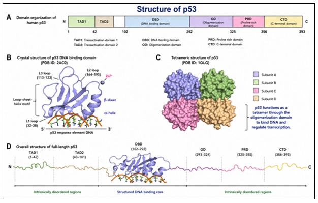
FIG. 1: STRUCTURAL ORGANIZATION OF THE P53 PROTEIN SHOWING MAJOR FUNCTIONAL DOMAINS
Mechanisms of p53 Mutation in Hepatocellular Carcinoma: In hepatocellular carcinoma (HCC), the p53 gene is commonly mutated and this is the result of a combination of environmental, viral and intrinsic factors 29.
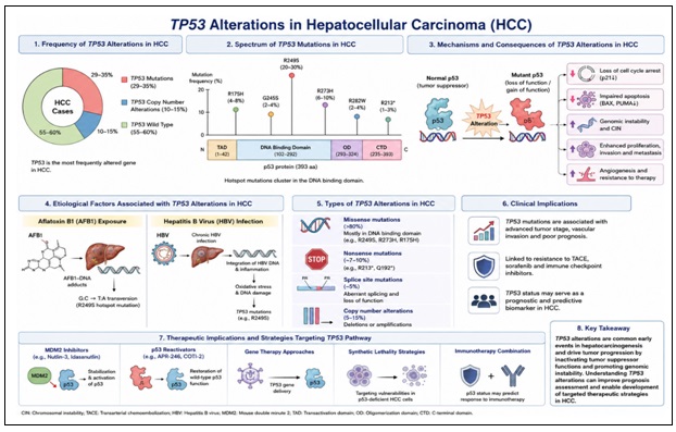
FIG. 2: TP53 ALTERATIONS IN HEPATOCELLULAR CARCINOMA
One common cause is the exposure to aflatoxin B1, a potent hepatocarcinogen, and results in a particular mutation at codon 249 of the p53 gene. p53 dysfunction is also due to chronic hepatitis virus infections. The hepatitis B virus (HBV) encodes the HBx protein, which may bind to p53 and decrease its transcriptional activity, impairing its tumor suppressor function 66. Similarly, the hepatitis C virus (HCV) is associated with indirect mechanisms of p53 inactivation via oxidative stress and inflammation. Apart from mutations, p53 can also be inactivated by elevated levels of negative regulators such as MDM2 that lead to p53 degradation 67. Moreover, epigenetic modifications and alterations in upstream pathways also result in p53 inactivation in HCC. These events lead to either the complete loss of p53 function or production of mutant p53 proteins with altered or oncogenic functions 68.
Impact of p53 Loss on Apoptosis Resistance: p53 alteration & mutation has profound effects on the apoptotic pathway in cancer cells 69. Under normal conditions, p53 is essential in inducing apoptosis in response to stress by inducing the expression of pro-apoptotic genes & suppressing anti-apoptotic proteins 70. But a mutation or deactivation of p53 upsets this balance 71. This results in cancer cells becoming resistant to apoptosis, allowing them to survive despite DNA damage & other forms of stress. This is one of the main reasons why conventional treatment approaches fail, as many of them rely on p53-mediated apoptosis to kill cancer cells 72. Furthermore, mutant p53 proteins can exert dominant-negative effects on any wild-type p53 that remains & may acquire new functions that promote cell survival, invasion & metastasis. This enhances the aggressiveness of the tumour & adds to the complexities of therapy 73. Inactivation of p53 also leads to dysfunction of mitochondrial apoptosis, reduced caspase activation, & increased expression of anti-apoptotic proteins such as Bcl-2. Thus, exploring p53-independent pathways is an important aspect of cancer therapy, particularly in hepatocellular carcinoma (HCC) 74.
Clinical Significance of p53 Mutations: p53 gene mutations play a significant role in hepatocellular carcinoma (HCC). These mutations are often associated with higher disease stage, lower tumor differentiation, increased metastatic potential and reduced survival. As a result, p53 mutations are considered a critical prognostic factor in HCC 75. p53 mutations not only impact prognosis but also therapeutic response. In general, cancers with mutant p53 are less responsive to chemotherapy and radiotherapy, largely because of diminished p53-dependent apoptosis. This highlights the need for new therapies independent of p53 61. Up to now, research has focused on developing targeted treatments that seek to restore p53 activity or exploit deficiencies associated with p53 loss. However, these approaches remain challenging due to the diversity of p53 mutations 76. In this sense, naturally occurring compounds such as allicin might be an alternative to induce apoptosis in a p53-independent manner. This makes them particularly effective in the treatment of HCC patients with p53-mutant tumors that are resistant to conventional treatment 77.
Allicin: Chemistry and Pharmacological Profile: Allicin (diallyl thiosulfinate) is a highly reactive, organosulfur compound that is the primary active ingredient in garlic responsible for its therapeutic effects 78. This compound has attracted significant attention for its diverse biological properties, including its antimicrobial, antioxidant, anti-inflammatory & antitumor effects. With respect to hepatocellular carcinoma (HCC), allicin is particularly important for its ability to regulate the cellular redox state, induce apoptosis & disrupt cancerous pathways 77. Chemically, allicin is a thiosulfinate and has a reactive sulfur atom that binds to hydroxyl groups of proteins and low-molecular-weight antioxidants, such as glutathione. This feature is key to many of its biological activities, including its ability to alter cellular signalling pathways and trigger oxidative stress in cancer cells 78. While allicin has potential therapeutic applications, its potential use in cancer treatment is limited by factors such as chemical instability, rapid metabolism and poor bioavailability. As such, it's imperative to understand its chemistry and pharmacology to optimise its use in cancer therapy 79.
Source and Biosynthesis from Garlic: Allicin is derived from Allium sativum and is not present in intact garlic bulbs. Rather, it is generated enzymatically when garlic tissue is damaged (for example, crushed or chopped) to expose the enzyme alliinase to a substrate (alliin, S-allyl-L-cysteine sulfoxide) 80. Damage to the tissue enables alliinase to rapidly convert alliin to allicin by a series of reactions. This reaction takes place very rapidly and results in the formation of allicin, an unstable and reactive compound 81. Allicin synthesis is part of the plant's natural defence mechanism against pathogens and damage 82. However, because of the instability of allicin, it can also transform into other secondary sulfur compounds, such as diallyl disulfide, diallyl trisulfide and ajoene, which may also contribute to the health effects of garlic extracts 83.
Distinguishing Allicin from Other Garlic-Derived Organosulfur Compounds: Garlic is loaded with a variety of biologically active organosulfur compounds (OSCs), including allicin, ajoene, diallyl sulfide (DAS), diallyl disulfide (DADS) and diallyl trisulfide (DATS).
These compounds have some similarities in biological properties, but differ greatly in chemical structure, stability, metabolism, cellular targets and pharmacological effects. For example, allicin is a highly reactive thiosulfinate that is formed from alliin by the action of enzymes during tissue damage. However, ajoene, DAS, DADS and DATS are secondary sulfur compounds, which are formed by the breakdown or processing of allicin 19. All of these compounds possess distinct pharmacokinetic and molecular characteristics, and can activate different signaling pathways. So, outcomes from the studies performed with ajoene, DAS, DADS, or DATS cannot be equated with activity of allicin. This review provides brief descriptions of the studies on these compounds individually and does not provide the conclusive evidence that the effect of allicin in HCC can be attributed to those of organosulfur compounds of garlic 84.
TABLE 4: COMPARISON OF MAJOR GARLIC-DERIVED ORGANOSULFUR COMPOUNDS
| Compound | Chemical Class | Relationship to Allicin | Direct Evidence for HCC | Interpretation |
| Allicin | Thiosulfinate | Primary active compound | Yes | Direct evidence |
| Ajoene | Sulfur-containing derivative | Allicin degradation product | Limited | Indirect evidence |
| DAS | Monosulfide | Distinct OSC | Limited | Cannot be attributed to allicin |
| DADS | Disulfide | Distinct OSC | Available | Separate pharmacological entity |
| DATS | Trisulfide | Distinct OSC | Available | Separate pharmacological entity |
Chemical Structure and Stability: The chemical name of allicin is diallyl thiosulfinate and it is characterised by the presence of a sulfinyl functional group (–S(O)–S–) bridging two allyl groups 85. This structure confers high reactivity, particularly towards thiol-containing compounds such as cysteine residues in proteins and the tripeptide, glutathione 86.
The reactivity of allicin is important for its biological activity, since it can change the structure and function of proteins and enzymes via thiol-disulfide exchange reactions. Such reactions can disrupt various key processes in cancer cells, culminating in oxidative stress, mitochondrial damage and cell death 87. Despite its potent biological effects, allicin is highly unstable under physiological conditions. It is highly sensitive to temperature, pH and enzymes, and rapidly degrades into a range of organosulfur compounds. This poses significant challenges for its formulation, storage & therapeutic applications 88.
Pharmacokinetics and Bioavailability: Allicin's pharmacokinetics are complex because of its rapid reactivity & instability. After oral administration, allicin is typically not present in its original form in the bloodstream 89. Rather, it is rapidly converted into metabolites, including allyl methyl sulfide, diallyl disulfide & other sulfur compounds. The uptake of allicin-derived compounds mainly occurs in the gut, where they can reach systemic circulation & exert their effects 90. But active allicin is generally thought to be poorly bioavailable due to its metabolism and excretion. The distribution of allicin metabolites in different tissues, such as the liver, is of particular interest for the treatment of HCC 91. These metabolites can retain activity & contribute to the observed in vivo pharmacological effects. To overcome issues related to low bioavailability, recent research has focused on novel drug delivery strategies, including nano-formulations, liposomal formulations and prodrugs, to enhance stability, delivery and bioavailability 88.
Safety and Toxicological Profile: Allicin is generally considered safe for dietary consumption (via garlic, Allium sativum); but its safety at higher doses must be evaluated 92.
Animal studies have indicated that allicin displays selective toxicity towards cancer cells, largely attributable to the heightened susceptibility of cancer cells to oxidative damage compared to healthy cells. This property highlights its potential as a therapeutic agent against cancer, particularly hepatocellular carcinoma (HCC) 93.
On the other hand, allicin can be cytotoxic towards non-malignant cells in a dose-dependent manner. The high thiol group reactivity of allicin can deplete antioxidant stores (such as glutathione) in cells, promoting excessive levels of reactive oxygen species, membrane damage and mitochondrial dysfunction. These effects may play a role in cytotoxicity in normal tissues, especially in the liver and the gastrointestinal system 78.
Animal studies have suggested that high doses of allicin or garlic extracts can cause gastrointestinal irritation, liver toxicity and altered liver enzyme activity, but these are typically associated with supra-physiological concentrations. Additionally, allicin might exhibit mild hemolytic effects at elevated levels due to interactions with red blood cell membranes 94.
Another key consideration is herb–drug interactions. Allicin may influence cytochrome P450 enzymes, potentially altering drug metabolism. It also has an anticoagulant effect due to inhibition of platelet aggregation, which may lead to bleeding when used with antiplatelet or anticoagulant drugs 95.
Toxicological evaluations also highlight the importance of dose & formulation considerations 96. Sustained release formulations may help in minimising systemic toxicity and improving efficacy. Also, allicin content in different garlic products may vary, & hence could lead to variation in pharmacological activity, which requires standardisation 97. Overall, while allicin has a good safety profile at moderate dosages, its dose-dependent toxicity, instability & drug interactions need to be carefully investigated. Targeted drug delivery, pharmacokinetics and clinical safety studies are warranted to fully ascertain its therapeutic potential in HCC 98.
Anticancer Properties of Allicin: Allicin (diallyl thiosulfinate) from Allium sativum has demonstrated a wide range of anticancer effects in various cancers including hepatocellular carcinoma (HCC) 77. Allicin's therapeutic potential stems from its multi-functional mode of action, which facilitates the simultaneous regulation of key cellular processes, such as cell growth, death, invasion and new blood vessel formation. Another distinctive feature of the anticancer effects of allicin is its ability to selectively induce oxidative stress-induced death in cancer cells 99.
Through the elevation of intracellular reactive oxygen species (ROS), allicin alters the cell's redox balance, leading to mitochondrial dysfunction and induction of apoptosis. These processes can occur in the absence of p53, making allicin promising for the treatment of p53-mutant tumors 100. In addition to its ability to induce apoptosis, allicin also targets multiple oncogenic pathways such as PI3K/Akt, MAPK and NF-κB, thereby suppressing tumor growth and enhancing the efficiency of cancer treatment 77.
Overview of Antitumor Activity: Allicin has considerable antitumor activities, which are mediated by a combination of cytotoxic, anti-proliferative and pro-apoptotic effects 38. In a time- and concentration-dependent manner, allicin has been shown to decrease the viability of different cancer cells, including HCC, in vitro. The cytotoxic responses are primarily mediated by oxidative stress, leading to the damage of macromolecules, including DNA, proteins and lipids 101. A key advantage of allicin is its cancer selectivity, which can be attributed to the elevated basal level of oxidative stress and metabolic state of the cancer cells. As a result, allicin-induced reactive oxygen species (ROS) further enhance the vulnerability of cancer cells to oxidative stress 102. In-vivo studies also confirm the antitumor activity of allicin by showing its ability to decrease tumor growth, reduce tumor size, and increase survival in animals. And, allicin has been shown to enhance the efficacy of conventional chemotherapeutic drugs, suggesting its potential use in combination with chemotherapies 103.
Effects on Cell Proliferation and Cell Cycle Arrest: Crucially, the ability of allicin to induce cell cycle arrest may occur independently of p53, which is particularly important in HCC where p53 mutations are prevalent 104. Allicin can engage other pathways in p53-deficient or p53-mutated cells, such as the induction of cyclin-dependent kinase inhibitors, p21 and p27, in a p53-independent manner. Mechanistically, allicin down-regulates key proteins involved in cell cycle progression, such as cyclin D1, cyclin B1 and CDKs, resulting in cell cycle arrest 105. This, in turn, blocks DNA replication and inhibits tumor growth. In addition, allicin-induced oxidative stress results in DNA damage and reinforces the cell cycle arrest 106. Allicin also interferes with signaling pathways that promote cell proliferation, such as PI3K/Akt and MAPK, which can be overactive in HCC. This reduces survival responses and sensitises cancer cells to apoptosis. As a result, allicin is a potent anti-proliferative agent, even in the absence of functional p53 107.
Anti-metastatic and Anti-angiogenic Effects: Metastasis and angiogenesis are critical steps in the development of cancer and play an important role in the poor prognosis of hepatocellular carcinoma. Allicin has demonstrated significant potential in suppressing both processes via multiple mechanisms 108. Allicin's anti-metastatic effect is primarily achieved by inhibiting the migration and invasion of cancer cells. It downregulates matrix metalloproteinases (MMPs), particularly MMP-2 & MMP-9, which are involved in the destruction of the extracellular matrix & metastatic spread of cancer cells 109. Simultaneously, allicin enhances the levels of tissue inhibitors of metalloproteinases (TIMPs), which helps to maintain the integrity of the extracellular matrix and prevents metastasis 110. In addition, allicin affects the epithelial–mesenchymal transition (EMT), which is crucial to metastasis. Allicin downregulates the expression of mesenchymal markers such as N-cadherin & vimentin, and upregulates the expression of epithelial markers such as E-cadherin, thus maintaining the epithelial phenotype and blocking the acquisition of invasive traits 111.
Furthermore, allicin has potent anti-angiogenic effects, crucial for tumor growth and metastasis. It decreases the production of vascular endothelial growth factor (VEGF) and other angiogenic markers, preventing the formation of new blood vessels required for the survival and growth of a tumour 112. Further to this, allicin interferes with hypoxia-inducible signalling pathways, reducing angiogenic responses under hypoxic conditions in the tumor 113. Importantly, allicin's anti-metastatic and anti-angiogenic effects are closely linked to the regulation of major signaling pathways such as PI3K/Akt, MAPK and NF-κB. This disruption of key pathways undermines the molecular underpinnings of tumor growth and metastasis 114. In conclusion, allicin's ability to limit growth, metastasis, and angiogenesis highlights its potential as a multifunctional anti-cancer drug, especially in the case of hepatocellular carcinoma, a highly metastatic and resistant cancer 115.
Mechanisms of Allicin-Induced Apoptosis in Hepatocellular Carcinoma: Allicin (diallyl thiosulfinate) of Allium sativum triggers apoptosis in HCC via p53 independent pathways. It is thiol-reactive and causes cellular redox imbalance, mitochondrial damage and activation of important death pathways such as the caspases, Bcl-2 family proteins and ER stress. These effects allow the chemoresistance induced by p53 to be overcome in HCC 116.
TABLE 5: MOLECULAR MECHANISMS OF ALLICIN-INDUCED APOPTOSIS IN HCC
| Mechanism | Molecular Targets | p53 Dependence | Cellular Outcome | Supporting Evidence | Ref. |
| ROS generation | ROS, GSH depletion | Independent | Oxidative stress, apoptosis | In-vitro studies | [117] |
| Mitochondrial pathway | Bax↑, Bcl-2↓ | Partial | Cytochrome c release | Cell line studies | [118] |
| Caspase activation | Caspase-3, -9 | Independent | Apoptosis execution | Experimental models | [119] |
| ER stress | CHOP, GRP78 | Independent | Apoptosis induction | In-vitro evidence | [120] |
| Cell cycle arrest | Cyclins, CDKs | Dependent/Independent | Growth inhibition | Multiple studies | [121] |
Reactive Oxygen Species (ROS)-Mediated Cytotoxicity: The rapid accumulation of reactive oxygen species (ROS) by allicin is one of the major causes of apoptosis 122. Allicin causes a reduction in antioxidant levels (such as glutathione) and modifies redox-sensitive proteins via thiol-disulfide exchange reactions, thereby placing cancer cells under excessive oxidative stress 87.
Elevated ROS levels result in:
- Oxidative modifications of DNA, proteins and lipids.
- Activation of stress kinases (e.g. JNK and p38 MAPK).
- Loss of mitochondrial membrane potential 123.
HCC cells are generally in a higher basal level of oxidation and are therefore more sensitive to increased ROS. This redox selectivity enables allicin to selectively kill tumor hepatocytes and not normal cells 124.
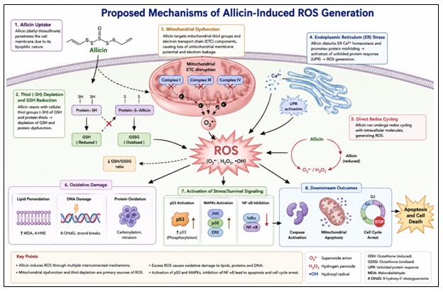
FIG. 3: PROPOSED MECHANISMS OF ALLICIN-INDUCED ROS GENERATION
Mitochondrial Dysfunction and Intrinsic Apoptosis Pathway: Allicin also affects the integrity of mitochondria by thiol modification and ROS generation, playing a key role in intrinsic apoptosis 125. This results in membrane depolarization, permeability changes and the release of cytochrome c that leads to the formation of apoptosomes and hence downstream apoptosis. This is a mitochondrial pathway that is largely independent of p53 which allows for apoptosis in p53-deficient HCC cells 126.
Activation of Caspase Cascade (Caspase-3, -8, -9): The caspases are the key effectors of apoptosis and allicin strongly activates the intrinsic and extrinsic caspase pathways 127. The release of cytochrome c from mitochondria activates the initiator caspase-9 via the formation of the apoptosome, which in turn activates the executioner caspase-3 128.
At the same time, allicin can also activate the extrinsic pathway via activation of death receptors that leads to activation of caspase-8. The extrinsic and intrinsic pathways work together to accelerate apoptosis 21.
Once activated, caspase-3 induces apoptosis by:
- Inducing structural and regulatory protein cleavage.
- Inducing DNA fragmentation.
- Inducing membrane blebbing and apoptotic bodies formation 129.
Allicin's capacity to activate caspase-3, -8 and -9 in a p53-independent manner suggests that it may have therapeutic potential in p53-mutant hepatocellular carcinoma (HCC) 130.
Modulation of Bcl-2 Family Proteins: The Bcl-2 protein family controls the integrity of the mitochondrial membrane, and is involved in the regulation of apoptosis. Allicin shifts the balance of the pro-apoptotic and anti-apoptotic members of this family 131.
In particular, allicin:
- Up-regulates pro-apoptotic proteins such as Bax and Bak.
- Reduces the expression of anti-apoptotic proteins like Bcl-2 and Bcl-xL 132.
This shift in the Bax/Bcl-2 ratio promotes mitochondrial outer membrane permeabilization (MOMP), facilitating the release of cytochrome c and activation of caspases 128.
Significantly, these regulatory effects occur in the absence of p53, implying that allicin can bypass the conventional p53-mediated transcriptional control of Bcl-2 family genes 7.
Endoplasmic Reticulum Stress-Induced Apoptosis: Allicin also induces apoptosis through the induction of endoplasmic reticulum (ER) stress. Protein misfolding and redox dysregulation in the ER activates the unfolded protein response (UPR) 133.
Prolonged or severe ER stress activates the apoptotic pathway via:
- Upregulation of CHOP (C/EBP homologous protein).
- ER-associated caspase activation (e.g. caspase-12 in models).
- ER calcium leakage and further contribution to mitochondrial dysfunction 134.
The allicin-induced ER stress is closely linked to oxidative stress and vice versa, amplifying the caspase-dependent apoptotic process. This mechanism provides a mechanism by which allicin can selectively kill cancer cells, in a p53-independent manner 119.
TABLE 6: MOLECULAR MECHANISMS OF ALLICIN-INDUCED APOPTOSIS IN HCC
| Mechanism | Key Molecular Events | Major Biomarkers/
Proteins |
p53 Dependency | Supporting Evidence (Standardized Findings) | Ref. |
| ROS-Mediated Oxidative Stress | Increased intracellular ROS, depletion of glutathione, oxidative damage | ROS ↑, GSH ↓, lipid peroxidation ↑ | Independent | Observed in HepG2, Hep3B cells; ROS-triggered apoptosis widely reported | [135] |
| Mitochondrial Dysfunction | Loss of mitochondrial membrane potential, cytochrome c release | ΔΨm ↓, Cytochrome c ↑ | Independent | Mitochondrial pathway activation in HCC xenograft models | [136] |
| Caspase Activation | Activation of intrinsic and extrinsic pathways | Caspase-3, -8, -9 ↑, PARP cleavage | Independent | Caspase cascade activation confirmed in multiple in vitro studies | [137] |
| Bcl-2 Family Modulation | Increased pro-apoptotic proteins, decreased anti-apoptotic proteins | Bax ↑, Bak ↑, Bcl-2 ↓, Bcl-xL ↓ | Independent | Bax/Bcl-2 ratio shift reported in HCC models | [138] |
| ER Stress-Induced Apoptosis | Activation of unfolded protein response (UPR) | CHOP ↑, Caspase-12 ↑, Ca²⁺ release ↑ | Independent | ER stress-mediated apoptosis documented in oxidative stress models | [139] |
| Death Receptor Pathway | Activation of extrinsic apoptosis signaling | Fas, TRAIL, Caspase-8 ↑ | Independent | Death receptor signaling reported in natural compound studies | [140] |
P53-Independent Mechanisms of Allicin Action: One of the major problems with treatment of HCC is the inactivation of p53, which decreases the induction of apoptosis and increases the resistance to drugs 141. A promising solution would be the induction of p53 independent cell death by allicin (diallyl thiosulfinate) contained in Allium sativum. It exerts its effect through oxidative stress modulation, activation of alternative tumor suppression mechanisms and induction of apoptosis and autophagy, overcoming resistance in HCC cells 6.
Apoptosis in p53-Mutant and p53-Null Cells: Allicin has been reported to induce apoptosis in cancer cells, regardless of p53 status, including p53-mutant and p53-null models 142. This is particularly relevant in hepatic cancer (HCC) as impairment of the p53 function is often observed and associated with a poor outcome and drug resistance. In terms of mechanism, allicin triggers apoptosis via the generation of reactive oxygen species (ROS) and subsequent mitochondrial dysfunction, leading to the release of cytochrome c, and activation of the caspase-9 and caspase-3 7. These events occur in the absence of p53-mediated transcriptional responses, implying that allicin's interaction with intrinsic apoptotic components can occur independently of p53 signaling. In addition, allicin can also induce extrinsic apoptotic pathways, which involves death receptor stimulation and activation of caspase-8 143. The activation of intrinsic and extrinsic pathways ensures the successful execution of apoptosis, even in the absence of p53. This ability to bypass p53-mediated apoptosis highlights the potential of allicin in treating chemoresistant HCC 144.
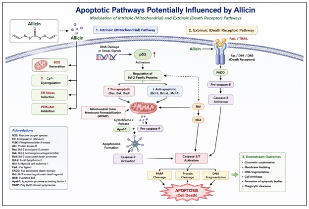
FIG. 4: APOPTOTIC PATHWAYS POTENTIALLY INFLUENCED BY ALLICIN
Alternative Tumor Suppressor Pathways (p73, p21): When p53 is absent or inactivated, other pathways of tumor suppression are able to compensate. One such pathway is mediated by p73, a structural and functional homolog of p53 that is critical for the regulation of apoptosis and cell cycle arrest 145. Allicin has been shown to switch on p73-dependent gene expression, leading to the activation of pro-apoptotic genes, including Bax, PUMA, and NOXA. Unlike p53, p73 is less frequently mutated in cancer, making it an attractive therapeutic target. Furthermore, allicin can activation the expression of the cyclin-dependent kinase inhibitor p21 in a p53-independent manner, resulting in cell cycle arrest and decreased proliferation of tumor cells 146. This could be through transcription factors such as Sp1 or stress-induced signaling pathways in response to oxidative stress. In this way, allicin restores essential regulatory processes that are usually lost in p53-deficient HCC cells, via the alternative tumor suppressor pathways 147.
Role of Autophagy in Cell Death: Autophagy is a highly regulated process that enables the degradation and reutilisation of cellular components. Autophagy in cancer can play a double role, acting as a survival mechanism or an alternative form of cell death, depending on the context 148. In HCC cells, allicin has been found to regulate autophagy, primarily through the induction of oxidative stress and the inhibition of cell survival pathways, including the phosphatidylinositol 3-kinase (PI3K) pathway. Activation of autophagy proteins, such as LC3-II and Beclin-1, has been observed after allicin treatment 149. Autophagy can be used as a backup mechanism to eliminate damaged cells in the absence of p53. Allicin-induced autophagy can lead to excessive cell self-destruct and cell death, especially when it is accompanied by mitochondrial damage and reactive oxygen species (ROS) accumulation. Thus, autophagy is important to the overall antitumor effects of allicin, especially in tumors with defective apoptosis 150.
Crosstalk between Apoptosis and Autophagy: Apoptosis and autophagy are tightly coupled processes of cell survival. Allicin regulates their crosstalk via PI3K/Akt, MAPK and NF-κB pathways. Allicin induces apoptosis and autophagy by inhibiting PI3K/Akt/mTOR and down-regulating Bcl-2 through the activation of Beclin-1 151. Autophagy is protective in the early stages of HCC, but promotes cell death in the later stages. In this way, allicin is able to kill tumor cells even when p53 is not present 152. A comparative overview of p53-dependent and p53-independent apoptotic pathways relevant to hepatocellular carcinoma is shown in Table 7.
TABLE 7: COMPARISON OF P53-DEPENDENT AND P53-INDEPENDENT APOPTOTIC PATHWAYS IN HCC
| Feature | p53-Dependent Pathway | p53-Independent Pathway | Relevance in HCC | Role of Allicin | References |
| Trigger | DNA damage | Oxidative stress, ER stress | p53 often mutated | Activates both | [153] |
| Key Proteins | p53, Bax, p21 | Caspases, ROS, CHOP | Resistance common | Bypasses p53 | [152] |
| Mechanism | Transcription regulation | Direct apoptosis signaling | Impaired in HCC | Alternative pathway | [152] |
| Therapeutic Value | Limited in mutated tumors | Highly valuable | Overcomes resistance | Strong potential | [152] |
TABLE 8: EVIDENCE SUPPORTING THE RELATIONSHIP BETWEEN ALLICIN ACTIVITY AND TP53 STATUS IN HEPATOCELLULAR CARCINOMA
| Experimental Model | TP53 Status | Allicin Purity | Allicin Dose | Treatment Duration | Measured Endpoints | Principal Findings | Evidence Classification | Ref. |
| HepG2 cells | Wild-type p53 | Specify from source | Specify | Specify | Cell viability, apoptosis markers, ROS generation | Apoptosis observed in p53-competent HCC cells | Direct evidence for allicin; not evidence of p53-independence | [154] |
| Hep3B cells | p53-null | Specify from source | Specify | Specify | Cell viability, caspase activation, apoptosis | Activity observed despite absence of p53 | Direct evidence for p53-null HCC | [154] |
| Huh7 cells | TP53-mutant | Specify from source | Specify | Specify | Apoptosis, ROS, mitochondrial dysfunction | Response observed in TP53-mutant cells | Direct evidence for mutant TP53 HCC | [154] |
| HCC animal model | TP53 status reported/not reported | Specify | Specify | Specify | Tumor volume, apoptosis markers | Antitumor effect observed | Direct evidence if allicin used in HCC | [38] |
| Breast cancer model | TP53-mutant or reported status | Specify | Specify | Specify | Apoptosis pathways | Mechanistic support only | Indirect evidence (non-HCC) | [11] |
| Colon cancer model | TP53 status reported | Specify | Specify | Specify | ROS, apoptosis | Suggests p53-independent pathways | Indirect evidence (non-HCC) | [155] |
| DADS/DATS studies | Variable | Not allicin | Specify | Specify | Signaling pathways | Mechanistic insight only | Indirect evidence (different compound) | [156] |
Interpretation of Evidence Categories:
Direct Evidence:
- Allicin was used as the compound tested.
- Hepatocellular carcinoma models were used in the experiments.
- TP53 status was either reported or characterized using experimental methods.
- Apoptosis or other endpoints were measured directly 154.
Indirect Evidence
- Research was carried out in cancer models other than HCC.
- Other garlic compounds in addition to allicin (DADS, DATS, ajoene) were also studied.
- No specific information about TP53 status was given.
- Research was able to give mechanistic information and was not directly used to test the central hypothesis in HCC 103.
Available data suggest that allicin can induce apoptosis and/or oxidative stress in multiple models of cancer. However, there has been little direct evidence of selective p53-independent function in HCC. While some studies using p53-null and TP53-mutant HCC models support this proposed mechanism, the majority of the literature is indirect, examining other forms of cancer, p53-competent systems or examining other garlic-derived compounds other than allicin.
Based on this, the available evidence suggests that allicin can trigger apoptotic pathways independent of p53, although there is no clear evidence of being selectively effective on p53-deficient HCC 154.
Modulation of Cell Signaling Pathways by Allicin: Allicin (diallyl thiosulfinate) from Allium sativum exhibits its chemopreventive effects through efficient modulation of several of these intercellular pathways of signaling that regulate survival, proliferation, inflammation and stress responses 157. For HCC, these pathways are frequently dysregulated, contributing to the growth, spread and resistance of the tumors. A key feature of allicin is its ability to modulate multiple oncogenic signaling pathways, thus disrupting the complex network of signaling pathways that drive malignant phenotypes 158. Many of these functions are independent of p53, thus overcoming a significant barrier to the treatment of HCC. These actions involve the regulation of PI3K/Akt/mTOR, MAPK, NF-κB and oxidative stress response pathways 159.
PI3K/Akt/mTOR Pathway Inhibition: PI3K/Akt/mTOR pathway is a critical mediator of cell survival, growth, metabolism and angiogenesis and is frequently dysregulated in HCC 160. When this pathway is activated, it allows tumors to grow by inhibiting cell death and increasing the synthesis of proteins. Allicin has been shown to block this pathway at multiple points. It decreases PI3K activation, which causes a reduction in the phosphorylation of Akt and therefore decreases the downstream target mTOR 161. This leads to a decrease in protein synthesis, impairs cell proliferation and increases apoptosis. In addition, allicin's inhibition of the PI3K/Akt/mTOR pathway facilitates the induction of autophagy, particularly under stress. Interestingly, this suppression occurs even in p53-deficient cells, suggesting allicin's ability to bypass p53-mediated survival mechanisms 160.
MAPK Pathway Regulation: The mitogen-activated protein kinase (MAPK) pathway plays an essential role in regulating a range of cellular processes, including growth, differentiation and stress responses 162. This pathway includes sub-pathways such as ERK, JNK and p38 MAPK, which have distinct functions. Allicin has context-specific effects on MAPKs. It blocks the ERK pathway, which is typically associated with cell growth and survival, thereby impeding tumor development 163. At the same time, allicin activates stress-response kinases such as JNK and p38 MAPK that are involved in the induction of apoptosis. JNK and p38 pathway activation leads to an up-regulation of pro-apoptotic genes and enhanced mitochondrial-mediated apoptosis 164. These effects are strongly linked to the reactive oxygen species (ROS) and oxidative stress that allicin induces. Allicin, through its differential modulation of the MAPK sub-pathways, shifts the survival-apoptosis equilibrium in cells, enhancing its anti-cancer activities in hepatocellular carcinoma (HCC) 165.
NF-κB Suppression: The NF-kappa B pathway is a key transcriptional regulator for inflammation, immunity and survival. In HCC, sustained activation of the NF-κB pathway contributes to the growth of tumors by upregulating the expression of anti-apoptotic proteins, cytokines, and growth factors 166. Allicin has been shown to suppress NF-κB pathway by preventing the phosphorylation and degradation of NF-kappa B inhibitor (IκBα).
This blocks NF-κB from entering the nucleus, reducing the expression of NF-κB target genes that promote survival and inflammation. As a result, allicin downregulates the pro-survival proteins Bcl-2 and survivin, as well as pro-inflammatory cytokines involved in tumor growth 167. This facilitates the induction of cancer cell death and may reduce inflammation of the tumor. Importantly, allicin's suppression of NF-κBsignaling is p53-independent, further highlighting its therapeutic potential for overcoming apoptosis resistance in HCC 7.
Regulation of Oxidative Stress Signaling: Signaling by oxidative stress plays a dual role in cancer, both promoting tumorigenesis and driving cell death when the levels of reactive oxygen species (ROS) become excessive. Allicin plays a major role in modulating this balance through its effects on redox signaling 168. Alicin uses its reactive sulfur atom to interact with thiol groups, reducing the concentration of intracellular antioxidants such as glutathione. This, in turn, increases ROS levels, triggering stress-sensing pathways and apoptosis. Allicin also modulates redox-sensitive transcription factors and pathways, including Nrf2, JNK and p38 MAPK. While there may be an initial induction of antioxidant responses, sustained oxidative stress leads to mitochondrial dysfunction and induction of apoptosis 169. Hepatocellular carcinoma (HCC) cells already exhibit elevated basal oxidative stress, and the production of ROS by allicin exceeds the tolerance of the cells, leading to selective cytotoxicity. This approach is particularly effective in cells lacking p53, where other mechanisms of stress response play an important role in cell survival 170.
TABLE 9: MODULATION OF KEY ONCOGENIC SIGNALING PATHWAYS BY ALLICIN
| Pathway | Normal Role in HCC | Effect of Allicin | Downstream Impact | Therapeutic Relevance | Ref. |
| PI3K/Akt/mTOR | Promotes survival, growth, angiogenesis | Inhibition of PI3K and Akt phosphorylation | Reduced protein synthesis, increased apoptosis | Overcomes drug resistance | [171] |
| MAPK (ERK, JNK, p38) | Regulates proliferation and stress response | ERK ↓, JNK/p38 ↑ | Suppression of proliferation, induction of apoptosis | Enhances stress-mediated cell death | [172] |
| NF-κB | Promotes inflammation and anti-apoptotic signaling | Inhibits NF-κB activation | Reduced cytokines, Bcl-2 ↓ | Reduces tumor survival and inflammation | [173] |
| Oxidative Stress Pathway | Maintains redox balance | ROS overproduction | DNA damage, apoptosis induction | Selective cytotoxicity in cancer cells | [152] |
| Nrf2 Pathway | Antioxidant defense | Dysregulation by allicin | Redox imbalance | Enhances ROS-mediated killing | [174] |
| Autophagy (PI3K/mTOR axis) | Survival or death mechanism | Induces autophagy | Autophagic cell death | Alternative pathway in p53-deficient cells | [153] |
Direct Versus Indirect Evidence for p53-Independent Activity of Allicin In HCC: Although allicin has been well documented to induce apoptosis in many cancer models, there have been very few reports of allicin-induced p53-independent apoptosis in hepatocellular carcinoma (HCC). To distinguish between the direct evidence from validated p53-deficient HCC models and indirect evidence from other experimental systems 154.
Direct evidence includes studies showing that allicin-induced apoptosis in HCC cell lines with well-known TP53 alteration, including TP53-mutant (e.g., Huh7) and p53-null (e.g., Hep3B), and mechanistic validation that apoptosis can be induced by allicin without p53 signaling. The studies provide the strongest support for a p53-independent mechanism 154.
On the other hand, much of the current literature is indirect evidence and comprises:
- The research was carried out in non-HCC cancer cell lines.
- Studies of other organosulfur compounds from garlic besides allicin.
- Experiments showing general ROS induced apoptosis but not assessing p53 status.
- Reports of apoptotic signaling pathways as the mechanism linking to p53 independence without direct evidence 175.
Conclusive evidence of the selective efficacy of allicin against HCC cells that lack p53 is still lacking, but current research suggests that allicin can act through alternative apoptotic pathways, which are p53 independent. Additional studies using genetically characterized HCC models and direct comparisons of wild-type and p53 deficient models are required 154.
TABLE 10: DIRECT AND INDIRECT EVIDENCE SUPPORTING P53-INDEPENDENT ANTICANCER ACTIVITY OF ALLICIN IN HEPATOCELLULAR CARCINOMA
| Evidence Category | Evidence Strength | Example Models | Interpretation |
| HCC with wild-type p53 | Direct | HepG2 | Demonstrates apoptosis but not p53 independence |
| HCC with p53-null status | Direct | Hep3B | Supports possible p53-independent activity |
| HCC with TP53 mutation | Direct but limited | Huh7 | Suggests activity despite altered p53 |
| Non-HCC cancer models | Indirect | Colon, breast, lung cancer cells | Cannot be directly extrapolated to HCC |
| Garlic compounds other than allicin | Indirect | DADS, DATS, ajoene studies | Mechanistically informative but not proof for allicin |
| General ROS/apoptosis studies | Indirect | Various cell systems | Support mechanism but not HCC-specific p53-independent activity |
Preclinical Evidence: Preclinical data provide important information about the anti-cancer potential of allicin (diallyl thiosulfinate) from Allium sativum 176. Allicin has been shown to be anti-proliferative, pro-apoptotic, anti-metastatic and anti-angiogenic in both in-vitro and in-vivo models of hepatocellular carcinoma (HCC). Crucially, this is also observed in p53 wild-type and mutant disease, highlighting its potential role in resistant disease 116.
Overall, preclinical studies indicate that allicin exerts its anti-tumor effects by generating reactive oxygen species (ROS), mitochondrial dysfunction, caspase activation, and regulation of survival pathways (PI3K/Akt, MAPK and NF-κB). This evidence is highly supportive for translational studies 132.
In-vitro Studies in HCC Cell Lines: Considerable in-vitro studies show that allicin decreases the survival and proliferation of HCC cell lines in a dose- and time-dependent manner. Commonly used models include HepG2 (wild-type p53), Hep3B (no p53) and Huh7 (mutant p53) to directly test p53-independent effects 7.
Notable findings from in-vitro studies include:
- Apoptosis is observed with condensation of nuclei, the fragmentation of DNA, and increased Annexin V staining 177.
- Caspase activation (caspase-3, -8, -9) and PARP cleavage 178.
- Depolarization of the mitochondrial membrane and release of cytochrome c 179.
- Generation of reactive oxygen species (ROS) and induction of oxidative stress 180.
Importantly, allicin induces similar proapoptotic effects in p53-wild and p53-null cell lines, thus validating its p53-independent mechanism of action.
Additionally, allicin curbs cell growth by arresting the cell cycle (at G0/G1 or G2/M phases) and suppressing cyclins and CDKs 142.
Research also shows the suppression of PI3K/Akt pathways, activation of JNK/p38 MAPK pathways and inhibition of NF-κB, which effectively switch the survival pathway to the apoptotic pathway 181.
In-vivo Animal Models: In-vivo studies conducted using xenograft and chemically induced HCC models also confirm the anti-cancer activity of allicin 182. In mice, treatment with allicin or garlic extracts is associated with:
- Reduced tumor size and weight.
- Slowed tumor growth and development.
- An increased apoptotic index in tumor tissues.
- Suppression of angiogenesis, with decreased microvessel density and VEGF levels 182.
Microscopic studies show increased apoptosis in tumor tissues, with condensed chromatin and high caspase activity. Additionally, allicin-treated animals often show changes in oxidative stress indices, with elevated lipid peroxidation levels in tumors, and enhanced antioxidant capacity in non-tumoral tissues 183.
More importantly, allicin shows a favourable toxicity profile when administered in-vivo with low systemic toxicity at therapeutic concentrations. This specificity enhances its prospects as a lead compound for further preclinical and clinical development 184.
Comparative Efficacy with Standard Chemotherapeutics: Comparative research on allicin combined with conventional chemotherapies highlight the potential of allicin as an independent or adjunct therapeutic agent 103.
Conventional therapies for advanced HCC, like Sorafenib, primarily target angiogenesis and cell proliferation but often encounter resistance and adverse effects 185.
Allicin has multiple comparative advantages:
- Broad range of targets, reducing the risk of drug resistance.
- Activity against p53-mutant or deficient cells, in contrast to many drugs.
- Lower toxicity towards healthy cells, improving selectivity 186.
In combination with other treatments, allicin has been shown to:
- Enhance the efficacy of chemotherapeutic drugs by increasing their ability to induce cancer cell death.
- Reduce the required drug dosages, thereby minimising side effects.
- Reverse resistance by regulating survival signalling and oxidative stress 132.
These findings suggest that allicin may act as a chemosensitizer, improving the effectiveness of conventional therapies. However, issues of formulation, dose and bioavailability remain to be resolved in future studies 132.
TABLE 11: SUMMARY OF PRECLINICAL STUDIES ON ALLICIN IN HCC
| Study Type | Model/System | Dose/Concentration | Key Findings | Outcome | References |
| In-vitro | HepG2 (p53 WT) | 10–100 µM | ROS generation, caspase activation | Apoptosis induction | [152] |
| In-vitro | Hep3B (p53 null) | 20–80 µM | p53-independent apoptosis | Confirms resistance bypass | [152] |
| In-vitro | Huh7 (p53 mutant) | 25–100 µM | Cell cycle arrest (G2/M), mitochondrial dysfunction | Reduced proliferation | [187] |
| In-vivo | Xenograft mouse model | 5–50 mg/kg | Tumor volume ↓, apoptosis ↑ | Tumor suppression | [20] |
| In-vivo | Chemically induced HCC | Variable | Reduced angiogenesis, VEGF ↓ | Anti-angiogenic effect | [188] |
| Combination Study | Allicin + Sorafenib | Variable | Enhanced apoptosis, reduced resistance | Synergistic effect | [189] |
| Mechanistic Study | Multiple HCC lines | Dose-dependent | PI3K/Akt inhibition, NF-κB suppression | Multi-target action | [190] |
Clinical Potential and Therapeutic Implications: The growing body of preclinical evidence suggests that allicin (diallyl thiosulfinate) from Allium sativum has the potential to treat hepatocellular carcinoma (HCC). Its ability to induce apoptosis through multi-targeted and, mainly, p53-independent pathways distinguish it from the majority of conventional agents that rely on p53 functionality 107. This feature is particularly important in HCC, as p53 gene mutations are common and confer resistance. Allicin has the potential to be used in clinical practice, not only as an effective anticancer agent, but also as a modulator of response to existing therapies, thereby enhancing their efficacy. Given its favourable safety profile seen in preclinical investigations, and its capacity to target several hallmarks of cancer, future research into allicin's clinical potential is needed 29.
Allicin as a Standalone Therapeutic Agent: The characteristics of allicin support its potential use as a standalone treatment for HCC. Allicin's multifunctional properties enable it to interfere with multiple processes that are vital to tumor progression, such as cell proliferation, apoptosis, angiogenesis & metastasis. Unlike conventional chemotherapies that typically have one target, allicin disrupts complex signaling pathways, including PI3K/Akt, MAPK, NF-κB, and oxidative stress 107.
Importantly, allicin induces apoptosis in both p53-wild-type and p53-mutant cancer cells, making it particularly relevant for p53-mutant HCC patients, with limited therapeutic options. Its selective toxic effect on cancer cells, depending on the different redox status, also adds to its therapeutic appeal 7.
Clinically, allicin could be explored as:
- A first-line treatment for early- or low-burden disease.
- A maintenance therapy to prevent recurrence.
- A prophylactic treatment for at risk populations (e.g., those with chronic liver disease) 103.
However, its development as a single agent will depend on resolving pharmacokinetic issues and determining dosing guidelines 191.
Combination Therapy with Chemotherapy/ Targeted Drugs: Perhaps the most exciting application for allicin is as an adjunct or combination therapy. Current therapies for advanced HCC, such as Sorafenib and Lenvatinib, are often plagued with issues such as resistance, side effects and short survival 192.
Allicin can enhance treatment outcomes by:
- Enhancing the sensitivity of cancer cells to chemotherapy through increased oxidative stress and reduced threshold for apoptosis.
- Suppressing survival pathways (such as PI3K/Akt and NF-κB), often associated with resistance.
- Lowering the effective doses of drugs, potentially reducing side effects.
- Inhibiting resistant cancer cells, such as p53-deficient cells 103.
In addition, allicin may synergise with the novel immunotherapies by regulating the tumor microenvironment and suppressing inflammation, but this remains to be investigated 193. Therefore, using allicin as part of a combination therapy may provide a multimodal approach, which could increase the effectiveness and decrease the resistance 77.
Challenges in Clinical Translation: Despite allicin's potential therapeutic effects in cancer, there are a number of issues that must be resolved before its effective delivery can be applied in the clinic 103. A major challenge is its inherent instability and instability, which results in poor bioavailability of the active molecule following administration. This is exacerbated by unfavourable pharmacokinetic characteristics such as rapid metabolism and erratic absorption 194. A further key consideration is standardization. The amount of allicin can vary depending on the source, preparation and processing of garlic products, thereby producing varied effects. Therefore, precise formulation and quality control are crucial for its clinical advancement 97.
Additional challenges include:
- Insufficient clinical trial data on efficacy and safety.
- Herb–drug interactions, particularly with warfarin and drugs metabolised by liver enzymes.
- Therapeutic dose and toxicity studies 195.
To overcome these challenges, future studies should focus on:
- Generating new drug delivery systems (nanoparticles, liposomes) for enhanced stability and targeting.
- Well-designed clinical trials to validate efficacy and safety.
- The discovery of biomarkers to predict response and enable personalised treatment 196.
Allicin-Specific Drug Delivery Systems: Current Evidence and Limitations: Allicin has been hindered from clinical development because of its chemical instability, degradation and low bioavailability. For this reason, a number of delivery strategies have been investigated to increase the stability, circulation time, tumor accumulation, and prevent off-target effects of allicin. It is important, however, to distinguish between experimental systems for delivering allicin and between general nanocarrier systems that have not been specifically tested with allicin 197.
At present, the evidence for the delivery of allicin to the HCC is limited. Thus, conclusions for the delivery of allicin deduced from the nanocarriers developed for other drugs should not be considered as conclusive for hepatocellular carcinoma. Instead, these systems should be considered as platforms that need to be further validated with allicin 198.
TABLE 12: ALLICIN-SPECIFIC DRUG DELIVERY SYSTEMS AND EXPERIMENTAL EVIDENCE
| Formulation Type | Composition | Allicin Loading/
Encapsulation |
Stability Findings | Release Characteristics | Experimental Model | Therapeutic Outcome | Safety Findings |
| Liposomal allicin | Specify original study | Report actual value | Report actual value | Report actual value | Cell line/animal model | Observed outcome | Report findings |
| Polymeric nanoparticles | Specify original study | Report actual value | Report actual value | Report actual value | Experimental model | Observed outcome | Report findings |
| Nanoemulsion formulation | Specify original study | Report actual value | Report actual value | Report actual value | Experimental model | Observed outcome | Report findings |
| Other validated allicin systems | Specify original study | Report actual value | Report actual value | Report actual value | Experimental model | Observed outcome | Report findings |
Liposomal Delivery Systems: Encapsulation of allicin in liposomes is proposed to improve the stability of allicin and protect it from premature degradation. But the therapeutic potential of liposomal allicin depends on the experimental validation of enhanced encapsulation efficiency, chemical stability, controlled release, pharmacokinetics and anti-tumor activity.
Thus, claims about the effect of liposomal allicin should be supported by actual studies of the formulation, and not be extrapolated from liposomal delivery systems used for other anticancer drugs 199.
Polymeric and Ligand-Targeted Systems: Other interesting nanosystems, such as polymeric nanoparticles and ligand-targeted ones, have been pursued as possible ways to improve the tumor-selective delivery. Much of the available data, however, is from research on therapeutic ingredients other than allicin.
These delivery systems should be accepted as potential options for future treatment of HCC, but are not specific to allicin. The formulation composition, bioavailability of allicin, its loading efficiency, release rate, biodistribution, therapeutic effects, and toxicity are important factors to consider when evaluating these technologies 200.
Limitations: In the existing literature, there are no studies on allicin delivery in the context of hepatocellular carcinoma. A considerable amount of the conversation on delivery using nanotechnology has come from other drugs, unrelated disease models, or other compounds found in garlic.
Thus, the studies cannot be considered as unequivocal proof of the delivery of allicin to HCC. Stabilized allicin formulations should be further evaluated in clinically-relevant animal models of HCC using standardized characterization methods, pharmacokinetic analysis, efficacy studies, and safety evaluations 201.
Safety, Toxicity, and Pharmacological Considerations: Allicin, a bio-active organosulfur compound derived from garlic (Allium sativum), has demonstrated promising anti-cancer activity in hepatocellular carcinoma (HCC) by induction of apoptosis and regulation of oxidative stress 116.
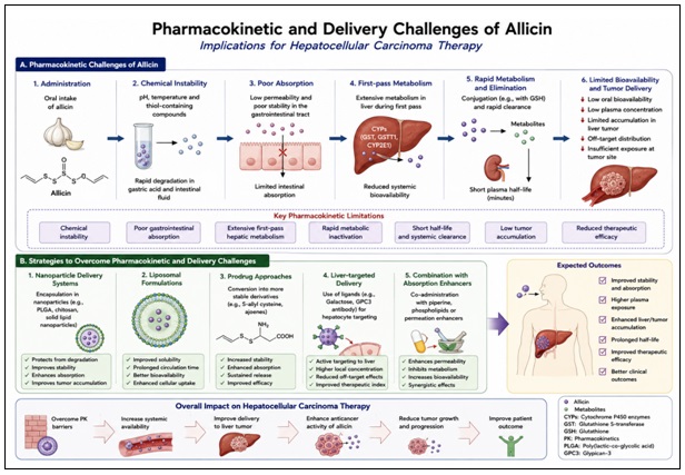
FIG. 5: PHARMACOKINETIC AND DELIVERY CHALLENGES
However, its therapeutic use is constrained by problems like pharmacokinetic instability, dose-dependent toxicity and potential interactions with conventional chemotherapeutic agents. Allicin is highly unstable and readily metabolises into secondary sulfur metabolites, such as diallyl sulfide and diallyl disulfide, which can contribute to its therapeutic and toxic effects 202. Furthermore, its high level of reactivity with thiol-containing proteins can cause non-specific cell damage at high concentrations. Therefore, it is important to properly evaluate the safety, toxicity and pharmacology of allicin, especially in chronic liver disease and impaired liver function observed in HCC 203.
Dose-Dependent Effects: Allicin shows a biphasic dose-dependent effect on its biological activity. Low to moderate doses of allicin offer cytoprotective and anti-cancer effects primarily via:
- Generation of reactive oxygen species (ROS)-induced apoptosis.
- Mitochondrial activation (release of cytochrome c, caspase-9).
- Manipulation of tumor suppressor pathways, including the partial re-establishment of p53-independent apoptosis 103.
In hepatocellular carcinoma models, low concentrations of allicin have been shown to induce apoptosis in cancer cells without affecting normal hepatocyte cells, which may be explained by the differences in the redox status of cancer cells and normal cells 7.
In contrast, at higher concentrations, allicin can:
- Generate excessive oxidative stress culminating in necrosis.
- Damage mitochondria of healthy cells.
- Alter the redox balance and damage proteins 204.
This limited window of efficacy highlights the need for dose optimization strategies. Animal studies suggest that delivery via controlled-release systems (such as nanoparticles and liposomes) may enhance therapeutic effectiveness and minimise toxicity 205.
Toxicological Concerns: Allicin is naturally derived, but it's not without its toxic side. There are a number of toxicological concerns 206.
Hepatotoxicity and Cytotoxicity: Allicin is toxic to cancer cells, but can also exacerbate liver damage, particularly in people with cirrhosis or hepatitis. High doses have been linked to:
- Increased liver enzymes (ALT, AST)
- Lipid peroxidation and damage to membranes
- Mitochondrial swelling and dysfunction 132
Gastrointestinal and Systemic Effects: Oral administration of allicin or garlic extracts can lead to irritation of the gastrointestinal tract, including:
- Nausea, vomiting, and stomach discomfort
- Changes in gut microbiota at elevated doses 92
Genotoxicity and Oxidative Damage: While allicin produces reactive oxygen species (ROS) for its anti-cancer effect, excess ROS can cause:
- DNA strand breaks
- Oxidation of lipids and proteins in healthy tissues
However, recent studies suggest that genotoxicity is mainly dose-dependent and reversible 207.
Pharmacokinetic Limitations:
Allicin demonstrates:
- Low bioavailability
- Quick metabolism and clearance from the system
- Instability under physiological conditions
This makes it less therapeutic and suggests formulation approaches to increase stability and delivery 98.
Herb–Drug Interactions: The concurrent use of allicin and conventional anti-cancer drugs or other medications could pose considerable risks of herb–drug interactions, particularly in multi-medicated HCC patients 208.
Cytochrome P450 Modulation: Allicin and its metabolites may affect liver enzymes that metabolise drugs, including:
- Activation or deactivation of CYP450 isoenzymes (e.g. CYP3A4, CYP2E1).
- Altered drug elimination and concentrations.
This could result in:
- Higher toxicity of chemotherapy agents.
- Reduced efficacy as a result of accelerated metabolism 209.
Interaction with Chemotherapeutic Agents: Allicin may also synergise or antagonise with drugs like:
- Sorafenib (the current treatment for HCC).
- Doxorubicin and cisplatin.
Possible outcomes may include:
- Increased apoptosis via ROS enhancement and mitochondrial sensitisation.
- Increased risk of hepatotoxicity and oxidative stress 20.
Anticoagulant and Antiplatelet Effects: Garlic-derived compounds (such as allicin) have antiplatelet activity, which may:
- Amplify the effects of anticoagulants (such as warfarin).
- Increase the risk of bleeding (especially in patients with liver disease and coagulopathy) 210.
Clinical Implications: Due to these interactions, it is necessary to closely monitor when allicin is used as an adjunct. Suggestions include dose reduction, therapeutic drug monitoring and avoiding self-supplied supplements 98.
TABLE 13: PHARMACOLOGICAL, TOXICOLOGICAL, AND DRUG DELIVERY CONSIDERATIONS OF ALLICIN
| Parameter | Description | Challenges | Proposed Solutions | Clinical Implication | Ref. |
| Bioavailability | Rapid metabolism, low systemic availability | Poor absorption | Nanoformulations, liposomes | Improved therapeutic levels | [211] |
| Chemical Stability | Highly unstable (thiol-reactive) | Rapid degradation | Encapsulation strategies | Sustained drug release | [212] |
| Dose-Dependent Effects | Low dose = therapeutic; high dose = toxic | Narrow therapeutic window | Controlled dosing systems | Safer clinical use | [132] |
| Toxicity | Hepatotoxicity, oxidative damage at high doses | Off-target effects | Targeted delivery | Reduced systemic toxicity | [132] |
| Herb–Drug Interactions | CYP450 modulation, anticoagulant effects | Drug interactions | Monitoring & dose adjustment | Safer combination therapy | [213] |
| Nanotechnology Delivery | Enhanced targeting and stability | Scale-up challenges | Advanced nanocarriers | Precision oncology application | [214] |
| Targeted Therapy | Tumor-specific delivery (e.g., GPC3 receptors) | Limited clinical validation | Ligand-based targeting | Improved selectivity | [215] |
Critical Analysis and Research Gaps: There are hardly any studies in the literature to explain the different redox responses of HCC cells versus normal hepatocytes to allicin exposure. Future studies should investigate glutathione depletion, markers of oxidative damage, antioxidant responses, and cell viability in both cancerous and healthy liver models to quantify these. It is important to conduct these studies to determine if there is a clinically relevant therapeutic window for allicin administered through ROS 216.
Critical Appraisal of Current Evidence: Although a number of studies have demonstrated the therapeutic efficacy of allicin in hepatocellular carcinoma (HCC), the available data are mostly preclinical and are of variable nature, especially under p53 deficient conditions 152. The majority of studies are based on in-vitro cell lines of cancer cells (such as HepG2, Huh7) that fail to fully reflect the tumor microenvironment, tumor genetic heterogeneity and metabolic complexity of human HCC. Therefore, the implications of these results for translation are somewhat restricted. In addition, a major drawback in all of the aforementioned studies is the use of different allicin concentrations, different exposure times and different assay procedures 217. In many reports, the effective concentrations of allicin are above the physiologically achievable plasma levels (usually in the low micromolar range, 10-100 µM), and there are concerns about the clinical feasibility and dose extrapolation 89.
Limitations in Mechanistic Interpretations: The anticancer activity of allicin, which has been attributed to a variety of pathways including ROS production, mitochondrial dysfunction, activation of caspases and NF-κB and modulation of signaling pathways such as PI3K/Akt and MAPK, has been studied separately, without a holistic biological context. Importantly, the causal relationship of these pathways has yet to be adequately validated, with the majority of studies using indirect markers, not pathway-specific inhibition and/or genetic validation 132. This suggests that some mechanisms reported might be secondary or non-specific stress mechanisms that are not the primary therapeutic targets. Further, the focus on p53-independent apoptosis, while conceptually intriguing, is not well-validated in various HCC models. The evidence that allicin is specific in this regard is less conclusive because of the limited evidence that it targets p53 deficient tumours over p53 normal counterparts 218.
Limitations regarding p53-Specific Targeting: Although there are several reports that allicin can inhibit the proliferation of cancer cells via apoptosis mechanisms that do not necessarily require the p53 activity, there is no direct evidence supporting the selective activity of allicin in p53-null and p53-mutant HCC models. The majority of the literature focuses on the general apoptotic and oxidative stress pathways but does not directly compare the response of p53-defective and p53-silent HCC models. However, the current evidence suggests that allicin could overcome p53-mediated resistance pathways, but does not rule out the use of allicin as a targeted drug for p53 non-functional HCC 219. This theory needs to be proven by future studies in genetically modified p53-null and p53 mutant HCC models. One important piece of the puzzle is the missing mechanism of action of autophagy in HCC under the effect of allicin. The majority of existing studies are based on autophagic marker measurements, but lack autophagic flux and loss-of-function studies.
Thus, the role of autophagy as a protective response to the stress response caused by allicin or as a mechanism for killing tumour cells is not yet certain. The application of both pharmacological and genetic approaches to autophagy modulation should be included in future studies to resolve this issue 220.
Gaps in Preclinical and In-vivo Evidence: The move from in-vitro results to in-vivo confirmation has not been sufficiently covered. Some animal studies indicate a tumor-suppressive effect of allicin, but these studies have a number of limitations:
- Small sample sizes.
- Lack of proper controls.
- Lack of pharmacokinetic and toxicity studies 16.
Furthermore, the immunological and stromal interactions seen in human HCC are not recapitulated by commonly used tumor xenograft models in these studies. There has been a lack of studies based on orthotopic or genetically engineered mouse models which further restricts the strength of current preclinical evidence 221.
Pharmacokinetic and Bioavailability Challenges:
Allicin Instability and Chemical Reactivity: Allicin's chemical instability is a major hurdle to its therapeutic use. When the garlic tissue is injured, allicin, a highly reactive thiosulfinate, is rapidly synthesized from alliin by the enzyme alliinase. Allicin, as soon as it is produced, rapidly reacts with thiolcontaining molecules and automatically breaks down into several other secondary sulfur compounds. Consequently, the biological exposure of allicin exhibits poor stability in storage, formulation, and biological exposure. This instability makes it difficult to interpret the experimental results, and the biological activity noted after allicin treatment could be due to metabolites produced during the conversion of allicin. Thus, the pharmacological activities attributed to allicin must be interpreted realizing that the exposure can be a complex combination of sulphur-containing compounds instead of a single molecule 78.
Formation of Secondary Sulfur Metabolites: Allicin can rapidly convert to a number of secondary organosulfur compounds, including ajoene, diallyl sulfide (DAS), diallyl disulfide (DADS), diallyl trisulfide (DATS) and other sulfur containing compounds. Because of these metabolites' distinct pharmacological properties and biological targets, it can be difficult to determine the importance of whether the therapeutic effects are due to allicin or its downstream metabolites. This challenge represents a major drawback in the evaluation of the mechanism and comparison of experimental data from different studies 84.
Challenges in Measuring Systemic Exposure: Due to the quick chemical changes of intact allicin, accurate evaluation of systemic allicin exposure is challenging. Therefore, it is difficult to accurately measure the circulating levels of allicin, and the pharmacokinetic data have been reported on an indirect basis that is based on measurements of sulfur-containing metabolites rather than allicin itself. The absence of standardized analytical methods further adds to the difficulties of comparison between different studies and makes the concept of dose-exposure relationships, indispensable for clinical applications, difficult to understand 222.
Formulation-Dependent Variability: There may be major differences between garlic preparations, methods of extraction, formulation and the way in which it is administered. The different forms of garlic extracts, purified allicin products, aged garlic formulations, encapsulated products and synthetic allicins may differ significantly in allicin content, stability, and bioavailability. Thus, the results from one formulation cannot be reliably applied to another. It is therefore critical to standardize formulation methods and employ quality-control measures to improve clinical relevance and reproducibility 223.
Experimental Concentrations versus Clinically Achievable Exposure: There is another translational challenge in experimental studies, which is the difference between the concentration used in experimental studies and the concentration that could be achieved in-vivo. Many in-vitro studies employ micromolar levels of allicin that are able to promote apoptosis, oxidative stress and growth inhibition in cancer cells. But, it remains uncertain whether such levels can be attainably achieved and maintained in tumor tissues after oral or systemic administration in humans. Therefore, conclusions about the mechanisms of action from experiments should not be used to infer the mechanisms of action in humans unless supported by experimental pharmacokinetic and pharmacodynamic data 193.
Translational and Clinical Limitations of Allicin Therapy:
Limited Clinical Evidence: Although promising preclinical data, there are yet significant clinical findings to back the use of allicin as a treatment for hepatocellular carcinoma (HCC). The vast majority of the information available is limited to in vitro studies, animal research or mechanistic studies. There is currently a lack of clinical evidence supporting the efficacy, optimal dosing protocols, safety and survival benefits of allicin for HCC patients. Therefore, the existing theories about its therapeutic effects need to be taken with a grain of salt and should be considered to be hypotheses at this point 103.
Uncertainty Regarding Effective Dose: One major obstacle to translation is the lack of well-defined therapeutic dosing guidelines. Many experimental trials have been done at concentrations that are often not achievable in human tissues because of the instability of allicin, its rapid metabolism and low bioavailability. This means that it is uncertain whether the concentrations used in experiments are concentrations that can be used clinically. Further pharmacokinetic and dose escalation studies are required to determine safe and biologically effective dose regimens 98.
Hepatotoxicity Considerations: Since the liver is already compromised in HCC patients due to cirrhosis, chronic hepatitis or a diminished hepatic reserve, it is important to evaluate hepatotoxicity. Although allicin is generally considered to be safe as a natural compound, there remains a lack of comprehensive studies on liver-specific toxicity, biochemical abnormalities, histopathological changes and chronic liver effects. At present, there is no evidence that it is not hepatotoxic 224.
Bleeding Risk: The chemicals found in garlic have been associated with both antiplatelet and anticoagulant actions. Therefore, high doses of allicin may theoretically increase the risk of bleeding, especially for people using blood-thinning medications, who have had invasive procedures, and have very low blood platelet counts (thrombocytopenia) as a result of more advanced cancer treatments. This is especially relevant with patients who have hepatocellular carcinoma (HCC), who frequently have additional coagulation disorders due to chronic liver disease 225.
Gastrointestinal Adverse Effects: In addition, potential gastrointestinal side effects like abdominal pain, nausea, dyspepsia, bloating, and gastrointestinal tract irritation are to be considered. These effects are generally thought to be manageable, but the incidence and intensity of these reactions in those taking concentrated allicin preparations remains poorly characterized 226.
Herb–Drug Interactions: Another important potential translation challenge is the potential for herb-drug interactions. Garlic products might impact drug metabolizing enzymes, transport proteins and platelet activity, and alter the pharmacokinetics or pharmacodynamics of drugs used concurrently. When considering the complexity of treatment regimens often used in oncology, a comprehensive evaluation of clinically relevant interactions is warranted prior to broad clinical application 227.
Interactions with Anticancer Therapies: The possible interactions between allicin and the standard chemotherapy drugs are not fully understood. Experimental studies have indicated some synergistic effects, but there is not clinical evidence available to confirm that allicin increases the effectiveness of treatment, changes toxicity profiles, affects treatment resistance or affects the pharmacologic effects of the approved systemic therapies. Future studies should examine the interactions of the chemotherapy, targeted therapy and immunotherapy, and locoregional treatment options that are frequently used to manage HCC 103.
Product Standardization and Quality Control: One of the challenges in standardization is the wide variation in the amount of allicin that can be found in garlic depending on the garlic source, processing methods, storage conditions, extraction methods and formulation technologies. Reproducibility and therapeutic outcome can be significantly affected by differences in purity, stability and active compound concentration. Therefore, developing standardized manufacturing processes and rigorous quality control will be important to successful clinical use of allicin 228.
Risk of Overgeneralization and Publication Bias: The vast majority of published studies have reported favorable results, suggesting that publication bias may have occurred. Few negative or inconclusive results are reported, which may lead to an overestimation of the therapeutic effects of allicin. In addition, overgeneralization of the findings across different types of cancer and experimental systems occurs without adequate evidence to support the extrapolations. It compromises the scientific integrity & can mislead the future course of research 229.
Future Research Directions to Address Current Gaps: Further research works are needed to develop stabilized allicin formulations, targeted delivery system, controlled release system & comprehensive pharmacokinetic studies. Improved knowledge of absorption, distribution, metabolism and elimination, as well as tumor specific exposure will play an important role when determining if pharmacologically effective exposure levels can be safely achieved in patients with HCC. These studies are crucial to bridge the gap between the encouraging preclinical results and clinical implementation. Additional studies are needed to reinforce the scientific & practical value of allicin therapy, including:
- Experiments with standard and repeatable designs.
- The use of clinically relevant in vivo models.
- Conducting in-depth pharmacokinetic and toxicity analysis.
- Designing new drug delivery systems to enhance product stability and bioavailability.
- Start structured and safe clinical trials for safety and effectiveness.
- Combination of multi-omics approaches for mechanistic validation.
- Comparative studies with existing anticancer drugs 230.
Summary of Critical Insights: To conclude, allicin has demonstrated a wide range of molecular targets & mechanisms that show promise for anticancer activity, but the existing evidence is biased against its preclinical use, lacks proper validation of mechanisms, suffers from pharmacokinetic limitations, and lacks clinical evidence. These challenges must be overcome to make allicin a therapeutic prospect of HCC.
TABLE 14: RESEARCH GAPS AND FUTURE OPPORTUNITIES IN ALLICIN-BASED HCC THERAPY
| Identified Gap | Evidence Limitation | Impact on Therapy | Proposed Solution |
| Lack of clinical trials | Mostly preclinical data | No clinical validation | Conduct RCTs |
| Poor bioavailability | Rapid degradation | Low efficacy | Nanocarriers |
| Mechanistic ambiguity | Incomplete pathways | Weak targeting | Multi-omics studies |
| Toxicity data lacking | Limited safety studies | Clinical risk | Long-term evaluation |
| Standardization issues | Variable dosing | Inconsistent results | Protocol harmonization |
Future Perspectives and Research Directions: While preclinical studies strongly suggest that allicin has pro-apoptotic effects in hepatocellular carcinoma (HCC), particularly in overcoming resistance associated with p53 mutations, its clinical use remains limited. Recent research highlights the need for a translation from mechanistic insights to clinical relevance. The next steps should focus on integrating allicin with modern approaches in cancer treatment, including targeted therapy, biomarker-driven patient stratification, & precision medicine. An interdisciplinary approach that combines molecular biology, pharmacology and clinical oncology is essential to exploit the therapeutic potential of allicin. Furthermore, advances in drug delivery & systems biology may help to better & safer use allicin for the treatment of HCC.
Need for Clinical Trials in HCC: To date, the majority of evidence regarding the anti-cancer effects of allicin is based on in vitro and animal studies, with limited human data. The need for well-designed clinical trials to address its safety, tolerability, pharmacokinetics and efficacy in HCC patients is pressing.
Clinical studies should examine:
- Phase I studies to determine the maximum tolerated dose (MTD) & dose-limiting toxicities.
- Phase II/III studies to evaluate efficacy measures, such as overall survival (OS) & progression-free survival (PFS).
- Assessment of combination treatments, including with current therapies, such as sorafenib or immunotherapies.
- Recruitment of patients with p53-mutated cancers, as a result of the mechanistic importance derived from allicin's apoptotic pathways.
In addition, it is critical to establish formulations of allicin with improved stability & bioavailability to ensure uniform conditions for clinical use.
Identification of Novel Molecular Targets: Although allicin is known to induce apoptosis through oxidative stress and mitochondrial pathways, the full spectrum of allicin targets is unknown. Future research should investigate and validate other targets that are involved in the survival, proliferation and resistance of the tumor, particularly those in p53-negative HCC.
Potential targets include:
- Oxidation-sensitive pathways (e.g., Nrf2/Keap1, MAPK, NF-κB).
- Epigenetic enzymes, like histone deacetylases (HDACs) and DNA methyltransferases (DNMTs).
- Autophagy proteins and their role in survival or death.
- Cancer stem cell (CSC) markers, which are associated with relapse and resistance.
High-throughput technologies such as proteomics, transcriptomics, and CRISPR screening may help with the identification of new targets and understanding the multi-targeted actions of allicin.
Biomarker-Based Patient Stratification: HCC is a highly heterogeneous disease, with a wide range of responses to treatment. It's essential to use biomarker-based stratification to identify the patients most likely to benefit from allicin treatments.
Future efforts should include:
- Discovering predictive biomarkers such as p53 mutation status, markers of oxidative stress & gene expression patterns indicative of apoptosis.
- Developing companion diagnostics to guide treatment.
- Utilising liquid biopsy technologies (circulating tumour DNA, exosomes) for monitoring treatment response in real time.
Through molecular stratification of patients, we can enhance treatment outcomes & avoid unnecessary toxic effects.
Advances in Precision Oncology: The era of precision oncology offers a unique opportunity to incorporate allicin in personalized treatment strategies for HCC. Precision medicine can be used to align treatment strategies with genetic & molecular traits to improve efficacy and reduce toxicity.
Future prospects include:
- Multimodal approaches that combine allicin with targeted therapies, immunotherapy (such as immune checkpoint inhibitors) & epigenetic drugs.
- Nanotechnology-based drug delivery systems (such as nanoparticles and liposomes) to enhance targeted delivery to tumors and pharmacokinetics.
- Use of artificial intelligence (AI) & bioinformatics approaches to predict patient outcomes & optimize treatment strategies.
- Exploration of multi-omics (genomics, proteomics, and metabolomics) to understand complex tumor mechanisms & guide treatment.
Importantly, precision oncology approaches may exploit allicin's ability to bypass impaired p53 pathways and initiate alternative apoptotic pathways to overcome a major challenge in anti-HCC therapy.
CONCLUSION: To conclude, HCC remains a therapeutic challenge because of the late detection of tumors, tumor heterogeneity and the high incidence of loss of p53 function, resulting in resistance to treatment. Traditional treatments do not prove very effective, making it necessary to look for other, more effective, methods. Allicin is an organosulfur compound found naturally, with great potential to be a multi-targeted, p53-independent anticancer agent. The versatility of its mechanisms is reflected in its capacity to disrupt redox homeostasis, to cause mitochondrial dysfunction, to activate caspase-dependent apoptosis, and to modulate endoplasmic reticulum stress. In addition, the modulation of apoptosis/autophagy balance by allicin increases its therapeutic effects especially in HCC resistant phenotypes. Therapeutic treatment based on ROS, proposed in this study, is a novel concept that can be used to explain allicin's anticancer effects and its ability to overcome chemoresistance. Some of these drawbacks to translation into clinical application include instability, limited bioavailability and lack of clinical evidence. Further studies will be needed to validate the therapeutic potential of allicin through standardized experimental studies, development of more sophisticated drug delivery systems, and properly designed clinical trials. These developments suggest that allicin could be a new promising approach for HCC management or a complement of the current therapeutic strategies.
ACKNOWLEDGEMENTS: Nil
CONFLICTS OF INTEREST: Nil
REFERENCES:
- Asafo-Agyei KO and Samant H: “Hepatocellular Carcinoma,” Jun. 2023, Accessed: Apr. 28, 2026. [Online]. Available: https://www.ncbi.nlm.nih.gov/books/NBK559177/
- Shukla PK, Dallio M and Chemello L: “Editorial: Hepatocellular carcinoma: from diagnostic approaches to surgical and systemic therapies,” Front. Med., vol. 12, p. 1633587, 2025, doi: 10.3389/FMED.2025.1633587.
- Z. Ding, L. Wang, J. Sun, L. Zheng, Y. Tang, and H. Tang, “Hepatocellular carcinoma: pathogenesis, molecular mechanisms, and treatment advances,” Front. Oncol., vol. 15, p. 1526206, 2025, doi: 10.3389/FONC.2025.1526206.
- T. Ozaki and A. Nakagawara, “Role of p53 in Cell Death and Human Cancers,” Cancers (Basel)., vol. 3, no. 1, p. 994, Mar. 2011, doi: 10.3390/CANCERS3010994.
- M. T. El-Saadony et al., “Garlic bioactive substances and their therapeutic applications for improving human health: a comprehensive review,” Front. Immunol., vol. 15, p. 1277074, 2024, doi: 10.3389/FIMMU.2024.1277074.
- Y. L. Chu, C. T. Ho, J. G. Chung, R. Raghu, Y. C. Lo, and L. Y. Sheen, “Allicin induces anti-human liver cancer cells through the p53 gene modulating apoptosis and autophagy,” J. Agric. Food Chem., vol. 61, no. 41, pp. 9839–9848, Oct. 2013, doi: 10.1021/JF403241S.
- Y. L. Chu, C. T. Ho, J. G. Chung, R. Raghu, Y. C. Lo, and L. Y. Sheen, “Allicin Induces Anti-human Liver Cancer Cells through the p53 Gene Modulating Apoptosis and Autophagy,” J. Agric. Food Chem., vol. 61, no. 41, pp. 9839–9848, Oct. 2013, doi: 10.1021/JF403241S.
- E. Amponsah-Dacosta, “Hepatitis B virus infection and hepatocellular carcinoma in sub-Saharan Africa: Implications for elimination of viral hepatitis by 2030?,” World J. Gastroenterol., vol. 27, no. 36, p. 6025, Sep. 2021, doi: 10.3748/WJG.V27.I36.6025.
- Z. M. Younossi and L. Henry, “Epidemiology of non-alcoholic fatty liver disease and hepatocellular carcinoma,” JHEP Reports, vol. 3, no. 4, p. 100305, Aug. 2021, doi: 10.1016/J.JHEPR.2021.100305.
- S. Sadrinasab, S. Saket, N. Pourmohammadi, F. Khosravi, and M. S. Fakhr, “Modern therapeutic approaches for hepatic tumors: progress, limitations, and future directions,” Discov. Oncol., vol. 16, no. 1, p. 959, Dec. 2025, doi: 10.1007/S12672-025-02773-Z.
- K. H. Kim, S. J. Cho, B. O. Kim, and S. Pyo, “Differential pro-apoptotic effect of allicin in oestrogen receptor-positive or -negative human breast cancer cells,” J. Funct. Foods, vol. 25, pp. 341–353, Aug. 2016, doi: 10.1016/J.JFF.2016.06.019.
- S. Oommen, R. J. Anto, G. Srinivas, and D. Karunagaran, “Allicin (from garlic) induces caspase-mediated apoptosis in cancer cells,” Eur. J. Pharmacol., vol. 485, no. 1–3, pp. 97–103, Feb. 2004, doi: 10.1016/J.EJPHAR.2003.11.059.
- J. H. Cha, Y. J. Choi, S. H. Cha, C. H. Choi, and W. H. Cho, “Allicin inhibits cell growth and induces apoptosis in U87MG human glioblastoma cells through an ERK-dependent pathway,” Oncol. Rep., vol. 28, no. 1, pp. 41–48, Jul. 2012, doi: 10.3892/OR.2012.1772.
- A. Chimento, M. D’Amico, A. De Luca, F. L. Conforti, V. Pezzi, and F. De Amicis, “Resveratrol, Epigallocatechin Gallate and Curcumin for Cancer Therapy: Challenges from Their Pro-Apoptotic Properties,” Life, vol. 13, no. 2, p. 261, Feb. 2023, doi: 10.3390/LIFE13020261.
- C. Liu et al., “Allicin protects against cardiac hypertrophy and fibrosis via attenuating reactive oxygen species-dependent signaling pathways,” J. Nutr. Biochem., vol. 21, no. 12, pp. 1238–1250, Dec. 2010, doi: 10.1016/J.JNUTBIO.2009.11.001.
- H. Chen et al., “Allicin Inhibits Proliferation and Invasion in Vitro and in Vivo via SHP-1-Mediated STAT3 Signaling in Cholangiocarcinoma,” Cell. Physiol. Biochem., vol. 47, no. 2, pp. 641–653, Jun. 2018, doi: 10.1159/000490019.
- J. S. Yang et al., “Diallyl disulfide induces apoptosis in human colon cancer cell line (COLO 205) through the induction of reactive oxygen species, endoplasmic reticulum stress, caspases casade and mitochondrial-dependent pathways,” Food Chem. Toxicol., vol. 47, no. 1, pp. 171–179, Jan. 2009, doi: 10.1016/j.fct.2008.10.032.
- J. Wu et al., “Coptisine from Coptis chinensis exerts diverse beneficial properties: A concise review,” J. Cell. Mol. Med., vol. 23, no. 12, pp. 7946–7960, Dec. 2019, doi: 10.1111/JCMM.14725.
- A. Shang et al., “Bioactive Compounds and Biological Functions of Garlic (Allium sativum L.),” Foods, vol. 8, no. 7, p. 246, Jul. 2019, doi: 10.3390/FOODS8070246.
- X. Zou et al., “Allicin sensitizes hepatocellular cancer cells to anti-tumor activity of 5-fluorouracil through ROS-mediated mitochondrial pathway,” J. Pharmacol. Sci., vol. 131, no. 4, pp. 233–240, Aug. 2016, doi: 10.1016/j.jphs.2016.04.017.
- C. Li, H. Jing, G. Ma, and P. Liang, “Allicin induces apoptosis through activation of both intrinsic and extrinsic pathways in glioma cells,” Mol. Med. Rep., vol. 17, no. 4, pp. 5976–5981, Apr. 2018, doi: 10.3892/MMR.2018.8552.
- M. A. Rahman et al., “p53 Modulation of Autophagy Signaling in Cancer Therapies: Perspectives Mechanism and Therapeutic Targets,” Front. Cell Dev. Biol., vol. 10, p. 761080, Jan. 2022, doi: 10.3389/FCELL.2022.761080.
- J. H. Hung et al., “Genomic instabilities in hepatocellular carcinoma: biomarkers and application in immunotherapies,” Ann. Hepatol., vol. 29, no. 6, p. 101546, Nov. 2024, doi: 10.1016/J.AOHEP.2024.101546.
- M. G. Refolo, C. Messa, V. Guerra, B. I. Carr, and R. D’alessandro, “Inflammatory Mechanisms of HCC Development,” Cancers (Basel)., vol. 12, no. 3, p. 641, Mar. 2020, doi: 10.3390/CANCERS12030641.
- J. Zheng et al., “Hepatocellular carcinoma: signaling pathways and therapeutic advances,” Signal Transduct. Target. Ther. 2025 101, vol. 10, no. 1, pp. 35-, Feb. 2025, doi: 10.1038/s41392-024-02075-w.
- V. Hernandez-Gea, S. Toffanin, S. L. Friedman, and J. M. Llovet, “Role of the Microenvironment in the Pathogenesis and Treatment of Hepatocellular Carcinoma,” Gastroenterology, vol. 144, no. 3, p. 512, 2013, doi: 10.1053/J.GASTRO.2013.01.002.
- P. Merle and C. Trepo, “Molecular Mechanisms Underlying Hepatocellular Carcinoma,” Viruses, vol. 1, no. 3, p. 852, Nov. 2009, doi: 10.3390/V1030852.
- H. K. Lim, G. P. Jeffrey, G. A. Ramm, and C. Soekmadji, “Pathogenesis of Viral Hepatitis-Induced Chronic Liver Disease: Role of Extracellular Vesicles,” Front. Cell. Infect. Microbiol., vol. 10, p. 587628, Nov. 2020, doi: 10.3389/FCIMB.2020.587628.
- H. B. Choudhary, S. K. Mandlik, and D. S. Mandlik, “Role of p53 suppression in the pathogenesis of hepatocellular carcinoma,” World J. Gastrointest. Pathophysiol., vol. 14, no. 3, p. 46, Jun. 2023, doi: 10.4291/WJGP.V14.I3.46.
- M. G. Fernández-Barrena, M. Arechederra, L. Colyn, C. Berasain, and M. A. Avila, “Epigenetics in hepatocellular carcinoma development and therapy: The tip of the iceberg,” JHEP Reports, vol. 2, no. 6, p. 100167, Dec. 2020, doi: 10.1016/J.JHEPR.2020.100167.
- A. Allameh, R. Niayesh-Mehr, A. Aliarab, G. Sebastiani, and K. Pantopoulos, “Oxidative Stress in Liver Pathophysiology and Disease,” Antioxidants, vol. 12, no. 9, p. 1653, Sep. 2023, doi: 10.3390/ANTIOX12091653.
- X. Luo et al., “Hepatocellular carcinoma: signaling pathways, targeted therapy, and immunotherapy,” MedComm, vol. 5, no. 2, p. e474, Feb. 2024, doi: 10.1002/MCO2.474.
- J. Fei and Y. Guo, “MAPK/ERK Signaling in Tumorigenesis: mechanisms of growth, invasion, and angiogenesis,” EXCLI J., vol. 24, p. 854, Jan. 2025, doi: 10.17179/EXCLI2025-8479.
- W. Yao, H. M. Qiu, K. L. Cheong, and S. Zhong, “Advances in anti-cancer effects and underlying mechanisms of marine algae polysaccharides,” Int. J. Biol. Macromol., vol. 221, pp. 472–485, Nov. 2022, doi: 10.1016/j.ijbiomac.2022.09.055.
- H. Mao, X. Zhao, and S. C. Sun, “NF-κB in inflammation and cancer,” Cell. Mol. Immunol., vol. 22, no. 8, p. 811, Aug. 2025, doi: 10.1038/S41423-025-01310-W.
- J. Zheng et al., “Hepatocellular carcinoma: signaling pathways and therapeutic advances,” Signal Transduct. Target. Ther., vol. 10, no. 1, p. 35, Dec. 2025, doi: 10.1038/S41392-024-02075-W.
- E. J. Sun, M. Wankell, P. Palamuthusingam, C. McFarlane, and L. Hebbard, “Targeting the PI3K/Akt/mTOR Pathway in Hepatocellular Carcinoma,” Biomedicines, vol. 9, no. 11, p. 1639, Nov. 2021, doi: 10.3390/BIOMEDICINES9111639.
- Y. Zhou et al., “Allicin in Digestive System Cancer: From Biological Effects to Clinical Treatment,” Front. Pharmacol., vol. 13, p. 903259, Jun. 2022, doi: 10.3389/FPHAR.2022.903259.
- Y. Guo et al., “Positive Effects of Allicin on Cytotoxicity, Antioxidative Status, and Immunity in ‘Eriocheir sinensis’ Hepatopancreatic Cells Against Oxidative Stress-Induced Injury,” Antioxidants, vol. 15, no. 1, p. 93, Jan. 2026, doi: 10.3390/ANTIOX15010093.
- S. J. Kim, H. S. Kim, and Y. R. Seo, “Understanding of ROS-Inducing Strategy in Anticancer Therapy,” Oxid. Med. Cell. Longev., vol. 2019, p. 5381692, 2019, doi: 10.1155/2019/5381692.
- M. C. H. Gruhlke, H. Antelmann, J. Bernhardt, V. Kloubert, L. Rink, and A. J. Slusarenko, “The human allicin-proteome: S-thioallylation of proteins by the garlic defence substance allicin and its biological effects,” Free Radic. Biol. Med., vol. 131, p. 144, Feb. 2019, doi: 10.1016/J.FREERADBIOMED.2018.11.022.
- H. Cichoz-Lach and A. Michalak, “Oxidative stress as a crucial factor in liver diseases,” World J. Gastroenterol., vol. 20, no. 25, p. 8082, 2014, doi: 10.3748/WJG.V20.I25.8082.
- J. Li, C. Huang, P. Tang, R. Wu, Q. Wu, and C. Zhang, “The distinct roles of ROS in tumor immunity: from mechanisms to immunotherapeutic applications,” J. Hematol. Oncol., vol. 19, no. 1, p. 16, Feb. 2026, doi: 10.1186/S13045-026-01780-Z.
- R. Khandia et al., “A Comprehensive Review of Autophagy and Its Various Roles in Infectious, Non-Infectious, and Lifestyle Diseases: Current Knowledge and Prospects for Disease Prevention, Novel Drug Design, and Therapy,” Cells, vol. 8, no. 7, p. 674, Jul. 2019, doi: 10.3390/CELLS8070674.
- E. Taucher, I. Mykoliuk, M. Fediuk, and F. M. Smolle-Juettner, “Autophagy, Oxidative Stress and Cancer Development,” Cancers (Basel)., vol. 14, no. 7, p. 1637, Apr. 2022, doi: 10.3390/CANCERS14071637.
- M. N. Sharifi, E. E. Mowers, L. E. Drake, and K. F. Macleod, “Measuring Autophagy in Stressed Cells,” Methods Mol. Biol., vol. 1292, p. 129, 2015, doi: 10.1007/978-1-4939-2522-3_10.
- Y. Xiang, J. Zhao, M. Zhao, and K. Wang, “Allicin activates autophagic cell death to alleviate the malignant development of thyroid cancer,” Exp. Ther. Med., vol. 15, no. 4, p. 3537, Apr. 2018, doi: 10.3892/ETM.2018.5828.
- X. Liu, K. Sun, H. Wang, and Y. Dai, “Inhibition of Autophagy by Chloroquine Enhances the Antitumor Efficacy of Sorafenib in Glioblastoma,” Cell. Mol. Neurobiol., vol. 36, no. 7, p. 1197, Oct. 2016, doi: 10.1007/S10571-015-0318-Z.
- Z. Sas, E. Cendrowicz, I. Weinhäuser, and T. P. Rygiel, “Tumor Microenvironment of Hepatocellular Carcinoma: Challenges and Opportunities for New Treatment Options,” Int. J. Mol. Sci., vol. 23, no. 7, p. 3778, Apr. 2022, doi: 10.3390/IJMS23073778.
- A. Nishida and A. Andoh, “The Role of Inflammation in Cancer: Mechanisms of Tumor Initiation, Progression, and Metastasis,” Cells, vol. 14, no. 7, p. 488, Apr. 2025, doi: 10.3390/CELLS14070488.
- P. J. Asif, C. Longobardi, M. Hahne, and J. P. Medema, “The Role of Cancer-Associated Fibroblasts in Cancer Invasion and Metastasis,” Cancers (Basel)., vol. 13, no. 18, p. 4720, Sep. 2021, doi: 10.3390/CANCERS13184720.
- A. Emami Nejad et al., “The role of hypoxia in the tumor microenvironment and development of cancer stem cell: a novel approach to developing treatment,” Cancer Cell Int., vol. 21, no. 1, p. 62, Dec. 2021, doi: 10.1186/S12935-020-01719-5.
- S. Reuter, S. C. Gupta, M. M. Chaturvedi, and B. B. Aggarwal, “Oxidative stress, inflammation, and cancer: How are they linked?,” Free Radic. Biol. Med., vol. 49, no. 11, p. 1603, Dec. 2010, doi: 10.1016/J.FREERADBIOMED.2010.09.006.
- L. J. Hernández Borrero and W. S. El-Deiry, “Tumor Suppressor p53: Biology, Signaling Pathways, and Therapeutic Targeting,” Biochim. Biophys. acta. Rev. cancer, vol. 1876, no. 1, p. 188556, Aug. 2021, doi: 10.1016/J.BBCAN.2021.188556.
- A. M. Boutelle and L. D. Attardi, “p53 and tumor suppression: it takes a network,” Trends Cell Biol., vol. 31, no. 4, p. 298, Apr. 2021, doi: 10.1016/J.TCB.2020.12.011.
- E. Alvarado-Ortiz, K. G. de la Cruz-López, J. Becerril-Rico, M. A. Sarabia-Sánchez, E. Ortiz-Sánchez, and A. García-Carrancá, “Mutant p53 Gain-of-Function: Role in Cancer Development, Progression, and Therapeutic Approaches,” Front. Cell Dev. Biol., vol. 8, p. 607670, Feb. 2021, doi: 10.3389/FCELL.2020.607670.
- Y. Liu, Z. Su, O. Tavana, and W. Gu, “Understanding the complexity of p53 in a new era of tumor suppression,” Cancer Cell, vol. 42, no. 6, p. 946, Jun. 2024, doi: 10.1016/J.CCELL.2024.04.009.
- X. Wang et al., “Multiple roles of p53 in cancer development: Regulation of tumor microenvironment, m6A modification and diverse cell death mechanisms,” J. Adv. Res., vol. 75, pp. 539–560, Sep. 2025, doi: 10.1016/J.JARE.2024.10.026.
- C. M. Eischen, “Genome Stability Requires p53,” Cold Spring Harb. Perspect. Med., vol. 6, no. 6, p. a026096, Jun. 2016, doi: 10.1101/CSHPERSPECT.A026096.
- N. Rivlin, R. Brosh, M. Oren, and V. Rotter, “Mutations in the p53 Tumor Suppressor Gene: Important Milestones at the Various Steps of Tumorigenesis,” Genes Cancer, vol. 2, no. 4, p. 466, Apr. 2011, doi: 10.1177/1947601911408889.
- P. A. J. Muller and K. H. Vousden, “Mutant p53 in Cancer: New Functions and Therapeutic Opportunities,” Cancer Cell, vol. 25, no. 3, p. 304, Mar. 2014, doi: 10.1016/J.CCR.2014.01.021.
- M. Oren and V. Rotter, “Mutant p53 Gain-of-Function in Cancer,” Cold Spring Harb. Perspect. Biol., vol. 2, no. 2, p. a001107, 2010, doi: 10.1101/CSHPERSPECT.A001107.
- H. Kim et al., “p53 Requires an Intact C-Terminal Domain for DNA Binding and Transactivation,” J. Mol. Biol., vol. 415, no. 5, p. 843, Feb. 2011, doi: 10.1016/J.JMB.2011.12.001.
- M. Fischer and M. A. Sammons, “Determinants of p53 DNA binding, gene regulation, and cell fate decisions,” Cell Death Differ., vol. 31, no. 7, p. 836, Jul. 2024, doi: 10.1038/S41418-024-01326-1.
- H. Wang, M. Guo, H. Wei, and Y. Chen, “Targeting p53 pathways: mechanisms, structures and advances in therapy,” Signal Transduct. Target. Ther. 2023 81, vol. 8, no. 1, pp. 92-, Mar. 2023, doi: 10.1038/s41392-023-01347-1.
- A. Rashid, J. S. Wang, G. S. Qian, B. X. Lu, S. R. Hamilton, and J. D. Groopman, “Genetic alterations in hepatocellular carcinomas: Association between loss of chromosome 4q and p53 gene mutations,” Br. J. Cancer, vol. 80, no. 1–2, pp. 59–66, 1999, doi: 10.1038/sj.bjc.6690321.
- J. K. Mitchell et al., “Hepatitis C Virus Indirectly Disrupts DNA Damage-Induced p53 Responses by Activating Protein Kinase R,” MBio, vol. 8, no. 2, pp. e00121-17, Mar. 2017, doi: 10.1128/MBIO.00121-17.
- M. T. Tomicic, M. Dawood, and T. Efferth, “Epigenetic Alterations Upstream and Downstream of p53 Signaling in Colorectal Carcinoma,” Cancers (Basel)., vol. 13, no. 16, p. 4072, Aug. 2021, doi: 10.3390/CANCERS13164072.
- K. Hientz, A. Mohr, D. Bhakta-Guha, and T. Efferth, “The role of p53 in cancer drug resistance and targeted chemotherapy,” Oncotarget, vol. 8, no. 5, p. 8921, Jan. 2016, doi: 10.18632/ONCOTARGET.13475.
- J. Pflaum, S. Schlosser, and M. Müller, “p53 Family and Cellular Stress Responses in Cancer,” Front. Oncol., vol. 4, no. OCT, p. 285, 2014, doi: 10.3389/FONC.2014.00285.
- B. J. Aubrey, G. L. Kelly, A. Janic, M. J. Herold, and A. Strasser, “How does p53 induce apoptosis and how does this relate to p53-mediated tumour suppression?,” Cell Death Differ., vol. 25, no. 1, p. 104, 2017, doi: 10.1038/CDD.2017.169.
- D. F. Callen, “p53 continues to surprise: High levels of p53 can suppress apoptosis,” Cell Cycle, vol. 12, no. 2, p. 203, 2013, doi: 10.4161/CC.23420.
- S. Pilley, T. A. Rodriguez, and K. H. Vousden, “Mutant p53 in cell-cell interactions,” Genes Dev., vol. 35, no. 7–8, p. 433, Apr. 2021, doi: 10.1101/GAD.347542.120.
- Q. Hao, J. Chen, H. Lu, and X. Zhou, “The ARTS of p53-dependent mitochondrial apoptosis,” J. Mol. Cell Biol., vol. 14, no. 10, p. mjac074, Oct. 2022, doi: 10.1093/JMCB/MJAC074.
- Y. D. Luo et al., “p53 haploinsufficiency and increased mTOR signalling define a subset of aggressive hepatocellular carcinoma,” J. Hepatol., vol. 74, no. 1, pp. 96–108, Jan. 2021, doi: 10.1016/J.JHEP.2020.07.036.
- Y. Lu, M. Wu, Y. Xu, and L. Yu, “The Development of p53-Targeted Therapies for Human Cancers,” Cancers (Basel)., vol. 15, no. 14, p. 3560, Jul. 2023, doi: 10.3390/CANCERS15143560.
- A. Chauhan et al., “Therapeutic role of allicin in gastrointestinal cancers: mechanisms and safety aspects,” Discov. Oncol., vol. 16, no. 1, p. 1731, Dec. 2025, doi: 10.1007/S12672-025-02591-3.
- J. Borlinghaus, F. Albrecht, M. C. H. Gruhlke, I. D. Nwachukwu, and A. J. Slusarenko, “Allicin: Chemistry and Biological Properties,” Molecules, vol. 19, no. 8, p. 12591, 2014, doi: 10.3390/MOLECULES190812591.
- R. Bala et al., “Revitalizing allicin for cancer therapy: advances in formulation strategies to enhance bioavailability, stability, and clinical efficacy,” Naunyn. Schmiedebergs. Arch. Pharmacol., vol. 397, no. 2, pp. 703–724, Feb. 2024, doi: 10.1007/S00210-023-02675-3.
- K. Lehari and D. Kumar, “Metabolic engineering and production of secondary metabolites,” Second. Metab. Biother., pp. 215–244, Jan. 2024, doi: 10.1016/B978-0-443-16158-2.00004-5.
- E. Knez, K. Kadac-Czapska, and M. Grembecka, “The importance of food quality, gut motility, and microbiome in SIBO development and treatment,” Nutrition, vol. 124, Aug. 2024, doi: 10.1016/j.nut.2024.112464.
- [82] J. Reiter, A. M. Hübbers, F. Albrecht, L. I. O. Leichert, and A. J. Slusarenko, “Allicin, a natural antimicrobial defence substance from garlic, inhibits DNA gyrase activity in bacteria,” Int. J. Med. Microbiol., vol. 310, no. 1, p. 151359, Jan. 2020, doi: 10.1016/J.IJMM.2019.151359.
- R. R. Malla, R. Marni, A. Chakraborty, and M. A. Kamal, “Diallyl disulfide and diallyl trisulfide in garlic as novel therapeutic agents to overcome drug resistance in breast cancer,” J. Pharm. Anal., vol. 12, no. 2, p. 221, Apr. 2021, doi: 10.1016/J.JPHA.2021.11.004.
- [84] S. Mitra et al., “Diallyl Disulfide: A Bioactive Garlic Compound with Anticancer Potential,” Front. Pharmacol., vol. 13, p. 943967, Aug. 2022, doi: 10.3389/FPHAR.2022.943967.
- J. Borlinghaus et al., “Allicin, the Odor of Freshly Crushed Garlic: A Review of Recent Progress in Understanding Allicin’s Effects on Cells,” Molecules, vol. 26, no. 6, p. 1505, 2021, doi: 10.3390/MOLECULES26061505.
- L. Weiner et al., “Thiol-disulfide organization in alliin lyase (alliinase) from garlic (Allium sativum),” Protein Sci., vol. 18, no. 1, p. 196, Jan. 2008, doi: 10.1002/PRO.10.
- A. Müller et al., “Allicin Induces Thiol Stress in Bacteria through S-Allylmercapto Modification of Protein Cysteines,” J. Biol. Chem., vol. 291, no. 22, p. 11477, May 2016, doi: 10.1074/JBC.M115.702308.
- P. Wu, T. Dong, J. Sun, and H. Chen, “Recent advances in sulfur-containing flavors of garlic (Allium sativum L.): formation mechanisms, controlling factors and innovative processing technologies,” Food Chem. X, p. 103886, Apr. 2026, doi: 10.1016/J.FOCHX.2026.103886.
- K. Q. Chen, H. B. Lei, X. Liu, and W. J. Cao, “Mini-review: The health benefits and applications of allicin,” Front. Pharmacol., vol. 16, p. 1715922, 2025, doi: 10.3389/FPHAR.2025.1715922.
- S. Chib, B. J. Dutta, S. Mallan, K. Raj, and S. Singh, “Allicin as nutraceutical: Protective mechanisms and therapeutic potential in Alzheimer’s disease,” Treat. Nutraceuticals, Suppl. Herb. Med. Neurol. Disord., pp. 125–137, Jan. 2023, doi: 10.1016/B978-0-323-90052-2.00051-2.
- V. D. Savairam, N. A. Patil, S. R. Borate, M. M. Ghaisas, and R. V. Shete, “Allicin: A review of its important pharmacological activities,” Pharmacol. Res. - Mod. Chinese Med., vol. 8, p. 100283, Sep. 2023, doi: 10.1016/J.PRMCM.2023.100283.
- M. Sasi et al., “Garlic (Allium sativum L.) Bioactives and Its Role in Alleviating Oral Pathologies,” Antioxidants, vol. 10, no. 11, p. 1847, Nov. 2021, doi: 10.3390/ANTIOX10111847.
- W. H. Talib et al., “Anticancer potential of garlic bioactive constituents: Allicin, Z-ajoene, and organosulfur compounds,” PHARMACIA, vol. 71, pp. 1–23, Jan. 2024, doi: 10.3897/PHARMACIA.71.E114556.
- Q. Gong et al., “Potential Hepatoprotective Effects of Allicin on Carbon Tetrachloride-Induced Acute Liver Injury in Mice by Inhibiting Oxidative Stress, Inflammation, and Apoptosis,” Toxics, vol. 12, no. 5, p. 328, May 2024, doi: 10.3390/TOXICS12050328.
- S. Wanwimolruk and V. Prachayasittikul, “Cytochrome P450 enzyme mediated herbal drug interactions (Part 1),” EXCLI J., vol. 13, p. 347, Apr. 2014, Accessed: Apr. 28, 2026. [Online]. Available: https://pmc.ncbi.nlm.nih.gov/articles/PMC4463967/
- C. J. Borgert, C. Fuentes, and L. D. Burgoon, “Principles of dose-setting in toxicology studies: the importance of kinetics for ensuring human safety,” Arch. Toxicol., vol. 95, no. 12, p. 3651, Dec. 2021, doi: 10.1007/S00204-021-03155-4.
- L. D. Lawson and S. M. Hunsaker, “Allicin Bioavailability and Bioequivalence from Garlic Supplements and Garlic Foods,” Nutrients, vol. 10, no. 7, p. 812, Jul. 2018, doi: 10.3390/NU10070812.
- M.-J. Yu, Y. Zhu, W. Zhang, Q.-P. Xu, H.-M. Wang, and Z. Ma, “Therapeutic potential of allicin against viral infections: Mechanisms and safety profile (Review),” Exp. Ther. Med., vol. 31, no. 6, p. 150, Apr. 2026, doi: 10.3892/ETM.2026.13146.
- A. N. Jikah et al., “A review of the therapeutic potential of sulfur compounds in Allium sativum,” Meas. Food, vol. 15, p. 100195, Sep. 2024, doi: 10.1016/J.MEAFOO.2024.100195.
- L. Li et al., “Allicin Alleviates Mitochondrial Dysfunction in Acrylamide-Induced Rat Kidney Involving the Regulation of SIRT1,” J. Agric. Food Chem., vol. 71, no. 42, pp. 15785–15795, Oct. 2023, doi: 10.1021/ACS.JAFC.3C04687.
- S. Njiki et al., “Garlic Extract Triggers Cytotoxicity, DNA Damage, and Cell Cycle Arrest in Human Leukemic Cells via Oxidative Stress,” J. Nutr. Food Sci., vol. 15, no. 2, p. 67, 2025, Accessed: Apr. 28, 2026. [Online]. Available: https://pmc.ncbi.nlm.nih.gov/articles/PMC12288681/
- M. S. Nadeem, I. Kazmi, I. Ullah, K. Muhammad, and F. Anwar, “Allicin, an Antioxidant and Neuroprotective Agent, Ameliorates Cognitive Impairment,” Antioxidants, vol. 11, no. 1, p. 87, Jan. 2021, doi: 10.3390/ANTIOX11010087.
- A. Chauhan et al., “Therapeutic role of allicin in gastrointestinal cancers: mechanisms and safety aspects,” Discov. Oncol. 2025 161, vol. 16, no. 1, pp. 1731-, Sep. 2025, doi: 10.1007/S12672-025-02591-3.
- Y. L. Chu, C. T. Ho, J. G. Chung, R. Rajasekaran, and L. Y. Sheen, “Allicin Induces p53-Mediated Autophagy in Hep G2 Human Liver Cancer Cells,” J. Agric. Food Chem., vol. 60, no. 34, pp. 8363–8371, Aug. 2012, doi: 10.1021/JF301298Y.
- P. Rajendran, E. Ho, D. E. Williams, and R. H. Dashwood, “Dietary phytochemicals, HDAC inhibition, and DNA damage/repair defects in cancer cells,” Clin. Epigenetics 2011 31, vol. 3, no. 1, pp. 4-, Oct. 2011, doi: 10.1186/1868-7083-3-4.
- H. Zhang, H. Wang, L. Qin, and S. Lin, “Garlic-derived compounds: Epigenetic modulators and their antitumor effects,” Phyther. Res., vol. 38, no. 3, pp. 1329–1344, Mar. 2024, doi: 10.1002/PTR.8108;PAGEGROUP:STRING:PUBLICATION.
- W. H. Talib et al., “Allicin and Cancer Hallmarks,” Molecules, vol. 29, no. 6, p. 1320, Mar. 2024, doi: 10.3390/MOLECULES29061320.
- C. Yao et al., “Angiogenesis in hepatocellular carcinoma: mechanisms and anti-angiogenic therapies,” Cancer Biol. Med., vol. 20, no. 1, p. 25, Jan. 2023, doi: 10.20892/J.ISSN.2095-3941.2022.0449.
- L. Huang, Y. Song, J. Lian, and Z. Wang, “Allicin inhibits the invasion of lung adenocarcinoma cells by altering tissue inhibitor of metalloproteinase/matrix metalloproteinase balance via reducing the activity of phosphoinositide 3-kinase/AKT signaling,” Oncol. Lett., vol. 14, no. 1, p. 468, 2017, doi: 10.3892/OL.2017.6129.
- G. A. Cabral-Pacheco et al., “The Roles of Matrix Metalloproteinases and Their Inhibitors in Human Diseases,” Int. J. Mol. Sci., vol. 21, no. 24, p. 9739, Dec. 2020, doi: 10.3390/IJMS21249739.
- H. Huang, F. Zheng, X. Dong, F. Wu, T. Wu, and H. Li, “Allicin inhibits tubular epithelial-myofibroblast transdifferentiation under high glucose conditions in vitro,” Exp. Ther. Med., vol. 13, no. 1, p. 254, Jan. 2016, doi: 10.3892/ETM.2016.3913.
- M. K. Gupta and R. Y. Qin, “Mechanism and its regulation of tumor-induced angiogenesis,” World J. Gastroenterol., vol. 9, no. 6, p. 1144, Jun. 2003, doi: 10.3748/WJG.V9.I6.1144.
- A. Gupta, G. S. N. K. Rao, H. S. Tuli, G. Kaur, P. Mishra, and S. Haque, “Synergistic regulation of tumor angiogenesis via flavonoid modulation of lncRNAs: Mechanistic insights and therapeutic potential,” Cancer Treat. Res. Commun., vol. 46, p. 101114, Jan. 2026, doi: 10.1016/J.CTARC.2026.101114.
- L. Zhang et al., “A comprehensive review on the plant sources, pharmacological activities and pharmacokinetic characteristics of Syringaresinol,” Pharmacol. Res., vol. 212, p. 107572, Feb. 2025, doi: 10.1016/J.PHRS.2024.107572.
- W. H. Talib et al., “Anticancer potential of garlic bioactive constituents: Allicin, Z-ajoene, and organosulfur compounds,” Pharmacia, vol. 71, pp. 1–23, 2024, doi: 10.3897/PHARMACIA.71.E114556.
- M. Sarvizadeh et al., “Allicin and Digestive System Cancers: From Chemical Structure to Its Therapeutic Opportunities,” Front. Oncol., vol. 11, p. 650256, Apr. 2021, doi: 10.3389/FONC.2021.650256.
- T. Miron et al., “Allicin inhibits cell growth and induces apoptosis through the mitochondrial pathway in HL60 and U937 cells,” J. Nutr. Biochem., vol. 19, no. 8, pp. 524–535, Aug. 2008, doi: 10.1016/J.JNUTBIO.2007.06.009.
- S. Y. Park, S. J. Cho, H. C. Kwon, K. R. Lee, D. K. Rhee, and S. Pyo, “Caspase-independent cell death by allicin in human epithelial carcinoma cells: involvement of PKA,” Cancer Lett., vol. 224, no. 1, pp. 123–132, Jun. 2005, doi: 10.1016/J.CANLET.2004.10.009.
- S. Oommen, R. J. Anto, G. Srinivas, and D. Karunagaran, “Allicin (from garlic) induces caspase-mediated apoptosis in cancer cells,” Eur. J. Pharmacol., vol. 485, no. 1–3, pp. 97–103, Feb. 2004, doi: 10.1016/J.EJPHAR.2003.11.059.
- R. Maltais, J. Roy, M. Perreault, S. Sato, J. C. Lévesque, and D. Poirier, “Induction of endoplasmic reticulum stress-mediated apoptosis by aminosteroid rm-581 efficiently blocks the growth of pc-3 cancer cells and tumors resistant or not to docetaxel,” Int. J. Mol. Sci., vol. 22, no. 20, p. 11181, Oct. 2021, doi: 10.3390/IJMS222011181/S1.
- J. Chen, “The Cell-Cycle Arrest and Apoptotic Functions of p53 in Tumor Initiation and Progression,” Cold Spring Harb. Perspect. Med., vol. 6, no. 3, p. a026104, Mar. 2016, doi: 10.1101/cshperspect.a026104.
- X. Deng, P. Yang, T. Gao, M. Liu, and X. Li, “Allicin attenuates myocardial apoptosis, inflammation and mitochondrial injury during hypoxia-reoxygenation: an in vitro study,” BMC Cardiovasc. Disord., vol. 21, no. 1, p. 200, Dec. 2021, doi: 10.1186/S12872-021-01918-6.
- S. Liu, J. Liu, Y. Wang, F. Deng, and Z. Deng, “Oxidative Stress: Signaling Pathways, Biological Functions, and Disease,” MedComm, vol. 6, no. 7, p. e70268, Jul. 2025, doi: 10.1002/MCO2.70268.
- A. Rauf et al., “Reactive oxygen species in biological systems: Pathways, associated diseases, and potential inhibitors—A review,” Food Sci. Nutr., vol. 12, no. 2, p. 675, Feb. 2023, doi: 10.1002/FSN3.3784.
- M. J. Corral, E. Benito-Peña, M. D. Jiménez-Antón, L. Cuevas, M. C. Moreno-Bondi, and J. M. Alunda, “Allicin Induces Calcium and Mitochondrial Dysregulation Causing Necrotic Death in Leishmania,” PLoS Negl. Trop. Dis., vol. 10, no. 3, p. e0004525, Mar. 2016, doi: 10.1371/JOURNAL.PNTD.0004525.
- T. Khan et al., “Mitochondrial Dysfunction: Pathophysiology and Mitochondria-Targeted Drug Delivery Approaches,” Pharmaceutics, vol. 14, no. 12, p. 2657, Dec. 2022, doi: 10.3390/PHARMACEUTICS14122657.
- E. K. Nadendla, R. E. Tweedell, G. Kasof, and T. D. Kanneganti, “Caspases: structural and molecular mechanisms and functions in cell death, innate immunity, and disease,” Cell Discov., vol. 11, no. 1, p. 42, Dec. 2025, doi: 10.1038/S41421-025-00791-3.
- M. E. Shawgo, S. N. Shelton, and J. D. Robertson, “Caspase-mediated Bak Activation and Cytochrome c Release during Intrinsic Apoptotic Cell Death in Jurkat Cells,” J. Biol. Chem., vol. 283, no. 51, p. 35532, Dec. 2008, doi: 10.1074/JBC.M807656200.
- T. S. Zheng et al., “Caspase-3 controls both cytoplasmic and nuclear events associated with Fas-mediated apoptosis in vivo,” Proc. Natl. Acad. Sci. U. S. A., vol. 95, no. 23, p. 13618, Nov. 1998, doi: 10.1073/PNAS.95.23.13618.
- Y. P. Liu, C. C. Zheng, Y. N. Huang, M. L. He, W. W. Xu, and B. Li, “Molecular mechanisms of chemo- and radiotherapy resistance and the potential implications for cancer treatment,” MedComm, vol. 2, no. 3, pp. 315–340, Sep. 2021, doi: 10.1002/MCO2.55.
- J. K. Brunelle and A. Letai, “Control of mitochondrial apoptosis by the Bcl-2 family,” J. Cell Sci., vol. 122, no. 4, p. 437, Feb. 2009, doi: 10.1242/JCS.031682.
- Y. Zhou et al., “Allicin in Digestive System Cancer: From Biological Effects to Clinical Treatment,” Front. Pharmacol., vol. 13, p. 903259, Jun. 2022, doi: 10.3389/FPHAR.2022.903259/FULL.
- Y. F. Zhu et al., “Allicin improves endoplasmic reticulum stress-related cognitive deficits via PERK/Nrf2 antioxidative signaling pathway,” Eur. J. Pharmacol., vol. 762, no. 1, pp. 239–246, Sep. 2015, doi: 10.1016/J.EJPHAR.2015.06.002.
- H. Shiraishi, H. Okamoto, A. Yoshimura, and H. Yoshida, “ER stress-induced apoptosis and caspase-12 activation occurs downstream of mitochondrial apoptosis involving Apaf-1,” J. Cell Sci., vol. 119, no. Pt 19, pp. 3958–3966, Oct. 2006, doi: 10.1242/JCS.03160.
- J. Ahmad et al., “Apoptosis induction by silica nanoparticles mediated through reactive oxygen species in human liver cell line HepG2,” Toxicol. Appl. Pharmacol., vol. 259, no. 2, pp. 160–168, Mar. 2012, doi: 10.1016/J.TAAP.2011.12.020.
- X. Ma, X. Tian, X. Huang, F. Yan, and D. Qiao, “Resveratrol-induced mitochondrial dysfunction and apoptosis are associated with Ca2+ and mCICR-mediated MPT activation in HepG2 cells,” Mol. Cell. Biochem., vol. 302, no. 1–2, pp. 99–109, Aug. 2007, doi: 10.1007/S11010-007-9431-8.
- I. Budihardjo, H. Oliver, M. Lutter, X. Luo, and X. Wang, “Biochemical pathways of caspase activation during apoptosis,” Annu. Rev. Cell Dev. Biol., vol. 15, no. Volume 15, 1999, pp. 269–290, Nov. 1999, doi: 10.1146/ANNUREV.CELLBIO.15.1.269/CITE/REFWORKS.
- N. M. Abdel-Hamid, M. K. Hassan, Am. A. M. Ahmed, S. G. Abd Allah, and N. H. Anber, “Liver proliferating cell nuclear antigen, BAX/Bcl-2 ratio, collagen, and polysaccharide accumulation as diagnostic tools in experimental hepatocellular carcinoma : BAX/Bcl-2 ratio, polysaccharide, collagen, and PCNA in hepatocellular carcinoma diagnosis,” J. Contemp. Med. Sci., vol. 8, no. 1, pp. 59–64, Feb. 2022, doi: 10.22317/JCMS.V8I1.1162.
- E. Szegezdi, U. Fitzgerald, and A. Samali, “Caspase-12 and ER-stress-mediated apoptosis: the story so far,” Ann. N. Y. Acad. Sci., vol. 1010, pp. 186–194, 2003, doi: 10.1196/ANNALS.1299.032.
- H. Jiang, W. Tang, Y. Song, W. Jin, and Q. Du, “Induction of Apoptosis by Metabolites of Rhei Radix et Rhizoma (Da Huang): A Review of the Potential Mechanism in Hepatocellular Carcinoma,” Front. Pharmacol., vol. 13, Mar. 2022, doi: 10.3389/FPHAR.2022.806175.
- Y. S. Guan, Z. La, L. Yang, Q. He, and P. Li, “p53 gene in treatment of hepatic carcinoma: Status quo,” World J. Gastroenterol., vol. 13, no. 7, p. 985, Feb. 2007, doi: 10.3748/WJG.V13.I7.985.
- G. Maitisha, M. Aimaiti, Z. An, and X. Li, “Allicin induces cell cycle arrest and apoptosis of breast cancer cells in vitro via modulating the p53 pathway,” Mol. Biol. Rep., vol. 48, no. 11, pp. 7261–7272, Nov. 2021, doi: 10.1007/S11033-021-06722-1.
- Y. L. Chu, C. T. Ho, J. G. Chung, R. Rajasekaran, and L. Y. Sheen, “Allicin induces p53-mediated autophagy in Hep G2 human liver cancer cells,” J. Agric. Food Chem., vol. 60, no. 34, pp. 8363–8371, Aug. 2012, doi: 10.1021/JF301298Y.
- Y. H. Lan et al., “Activations of Both Extrinsic and Intrinsic Pathways in HCT 116 Human Colorectal Cancer Cells Contribute to Apoptosis through p53-Mediated ATM/Fas Signaling by Emilia sonchifolia Extract, a Folklore Medicinal Plant,” Evid. Based. Complement. Alternat. Med., vol. 2012, p. 178178, 2012, doi: 10.1155/2012/178178.
- J. Pflaum, S. Schlosser, and M. Müller, “P53 family and cellular stress responses in cancer,” Front. Oncol., vol. 4, no. OCT, p. 97276, Oct. 2014, doi: 10.3389/FONC.2014.00285/FULL.
- A. Bisso, L. Collavin, and G. Del Sal, “p73 as a Pharmaceutical Target for Cancer Therapy,” Curr. Pharm. Des., vol. 17, no. 6, p. 578, Nov. 2011, doi: 10.2174/138161211795222667.
- K. Beyfuss and D. A. Hood, “A systematic review of p53 regulation of oxidative stress in skeletal muscle,” Redox Rep., vol. 23, no. 1, p. 100, Jan. 2018, doi: 10.1080/13510002.2017.1416773.
- P. Jalali: “The role of autophagy in cancer: from molecular mechanism to therapeutic window,” Front. Immunol., vol. 16, p. 1528230, 2025, doi: 10.3389/FIMMU.2025.1528230.
- S. Yang et al., “New insights into autophagy in hepatocellular carcinoma: mechanisms and therapeutic strategies,” Am. J. Cancer Res., vol. 9, no. 7, p. 1329, 2019, Accessed: Apr. 28, 2026. [Online]. Available: https://pmc.ncbi.nlm.nih.gov/articles/PMC6682711/
- X. Huang, H. Yan, Z. Xu, B. Yang, P. Luo, and Q. He, “The inducible role of autophagy in cell death: emerging evidence and future perspectives,” Cell Commun. Signal., vol. 23, no. 1, p. 151, Dec. 2025, doi: 10.1186/S12964-025-02135-W.
- A. H. Harrath, M. Jalouli, M. Al-Zharani, and M. A. Rahman, “Recent Update Targeting Autophagy-Apoptosis Crosstalk Using Bioactive Natural Products for Ovarian Cancer Treatment,” Biomedicines, vol. 14, no. 1, p. 212, Jan. 2026, doi: 10.3390/BIOMEDICINES14010212.
- Y. L. Chu, C. T. Ho, J. G. Chung, R. Raghu, Y. C. Lo, and L. Y. Sheen, “Allicin Induces Anti-human Liver Cancer Cells through the p53 Gene Modulating Apoptosis and Autophagy,” J. Agric. Food Chem., vol. 61, no. 41, pp. 9839–9848, Oct. 2013, doi: 10.1021/JF403241S.
- Y. L. Chu, C. T. Ho, J. G. Chung, R. Raghu, Y. C. Lo, and L. Y. Sheen, “Allicin induces anti-human liver cancer cells through the p53 gene modulating apoptosis and autophagy,” J. Agric. Food Chem., vol. 61, no. 41, pp. 9839–9848, Oct. 2013, doi: 10.1021/JF403241S.
- Y. L. Chu, C. T. Ho, J. G. Chung, R. Raghu, Y. C. Lo, and L. Y. Sheen, “Allicin Induces Anti-human Liver Cancer Cells through the p53 Gene Modulating Apoptosis and Autophagy,” J. Agric. Food Chem., vol. 61, no. 41, pp. 9839–9848, Oct. 2013, doi: 10.1021/JF403241S.
- W. Bat-Chen, T. Golan, I. Peri, Z. Ludmer, and B. Schwartz, “Allicin Purified From Fresh Garlic Cloves Induces Apoptosis in Colon Cancer Cells Via Nrf2,” Nutr. Cancer, vol. 62, no. 7, pp. 947–957, Oct. 2010, doi: 10.1080/01635581.2010.509837.
- N. Jonckheere et al., “A role for human MUC4 mucin gene, the ErbB2 ligand, as a target of TGF-beta in pancreatic carcinogenesis,” Oncogene, vol. 23, no. 34, pp. 5729–5738, Jul. 2004, doi: 10.1038/SJ.ONC.1207769.
- R. Arreola et al., “Immunomodulation and Anti-Inflammatory Effects of Garlic Compounds,” J. Immunol. Res., vol. 2015, p. 401630, 2015, doi: 10.1155/2015/401630.
- M. K. Nagar, D. Sudha, and B. Vairappan, “The role of medicinal plants in the management of hepatocellular carcinoma and its metastasis,” Open Explor. 2019 4, vol. 4, pp. 100588-, Aug. 2025, doi: 10.37349/EDD.2025.100588.
- Z. Jiang, L. Liu, J. Tang, and X. Chen, “PI3K/Akt/mTOR signaling pathway: the major signal transduction involved in the anticancer effects of resveratrol,” Pathol. - Res. Pract., p. 156491, Apr. 2026, doi: 10.1016/J.PRP.2026.156491.
- Y. Peng, Y. Wang, C. Zhou, W. Mei, and C. Zeng, “PI3K/Akt/mTOR Pathway and Its Role in Cancer Therapeutics: Are We Making Headway?,” Front. Oncol., vol. 12, p. 819128, Mar. 2022, doi: 10.3389/FONC.2022.819128.
- P. M. LoRusso, “Inhibition of the PI3K/AKT/mTOR Pathway in Solid Tumors,” J. Clin. Oncol., vol. 34, no. 31, p. 3803, Nov. 2016, doi: 10.1200/JCO.2014.59.0018.
- U. Moens, S. Kostenko, and B. Sveinbjørnsson, “The Role of Mitogen-Activated Protein Kinase-Activated Protein Kinases (MAPKAPKs) in Inflammation,” Genes (Basel) vol. 4, no. 2, p. 101, Jun. 2013, doi: 10.3390/GENES4020101.
- Y.-J. Guo, W.-W. Pan, S.-B. Liu, Z.-F. Shen, Y. Xu, and L.-L. Hu, “ERK/MAPK signalling pathway and tumorigenesis,” Exp. Ther. Med., vol. 19, no. 3, p. 1997, Jan. 2020, doi: 10.3892/ETM.2020.8454.
- Q. Xiang et al., “Allicin Attenuated Advanced Oxidation Protein Product-Induced Oxidative Stress and Mitochondrial Apoptosis in Human Nucleus Pulposus Cells,” Oxid. Med. Cell. Longev., vol. 2020, p. 6685043, 2020, doi: 10.1155/2020/6685043.
- T. Miron et al., “Allicin inhibits cell growth and induces apoptosis through the mitochondrial pathway in HL60 and U937 cells,” J. Nutr. Biochem., vol. 19, no. 8, pp. 524–535, Aug. 2008, doi: 10.1016/J.JNUTBIO.2007.06.009.
- P. Yang et al., “NF-κB signaling in hepatocellular carcinoma: Mechanisms of tumor progression, immune evasion, and therapeutic resistance,” Transl. Oncol., vol. 67, p. 102760, May 2026, doi: 10.1016/J.TRANON.2026.102760.
- H. Zheng et al., “Allicin protects against inflammation via suppression of PI3K/AKT/NF-κB signaling pathway in the model of acute cutaneous injury mouse and LPS-induced keratinocyte,” Int. Immunopharmacol., vol. 174, p. 116331, Apr. 2026, doi: 10.1016/J.INTIMP.2026.116331.
- K. Li, Z. Deng, C. Lei, X. Ding, J. Li, and C. Wang, “The Role of Oxidative Stress in Tumorigenesis and Progression,” Cells, vol. 13, no. 5, p. 441, Mar. 2024, doi: 10.3390/CELLS13050441.
- M. C. H. Gruhlke, C. Nicco, F. Batteux, and A. J. Slusarenko, “The Effects of Allicin, a Reactive Sulfur Species from Garlic, on a Selection of Mammalian Cell Lines,” Antioxidants, vol. 6, no. 1, p. 1, Mar. 2016, doi: 10.3390/ANTIOX6010001.
- A. Takaki and K. Yamamoto, “Control of oxidative stress in hepatocellular carcinoma: Helpful or harmful?,” World J. Hepatol., vol. 7, no. 7, p. 968, 2015, doi: 10.4254/WJH.V7.I7.968.
- “Betulinic acid induces autophagy-mediated apoptosis through suppression of the PI3K/AKT/mTOR signaling pathway and inhibits hepatocellular carcinoma - PubMed.” Accessed: May 14, 2026. [Online]. Available: https://pubmed.ncbi.nlm.nih.gov/31814899/
- D. Wang et al., “A new Prenylated Flavonoid induces G0/G1 arrest and apoptosis through p38/JNK MAPK pathways in Human Hepatocellular Carcinoma cells,” Sci. Reports 2017 71, vol. 7, no. 1, pp. 5736-, Jul. 2017, doi: 10.1038/s41598-017-05955-0.
- F. Wang et al., “Activation of the NF-κB pathway as a mechanism of alcohol enhanced progression and metastasis of human hepatocellular carcinoma,” Mol. Cancer 2015 141, vol. 14, no. 1, pp. 10-, Jan. 2015, doi: 10.1186/S12943-014-0274-0.
- S. Wang et al., “CDCA2 protects against oxidative stress by promoting BRCA1–NRF2 signaling in hepatocellular carcinoma,” Oncogene 2021 4025, vol. 40, no. 25, pp. 4368–4383, Jun. 2021, doi: 10.1038/s41388-021-01855-w.
- R. Raghu, K. H. Lu, and L. Y. Sheen, “Recent Research Progress on Garlic (大蒜 dà suàn) as a Potential Anticarcinogenic Agent against Major Digestive Cancers,” J. Tradit. Complement. Med., vol. 2, no. 3, p. 192, 2012, doi: 10.1016/S2225-4110(16)30099-2.
- P. Pandey, F. Khan, N. Alshammari, A. Saeed, F. Aqil, and M. Saeed, “Updates on the anticancer potential of garlic organosulfur compounds and their nanoformulations: Plant therapeutics in cancer management,” Front. Pharmacol., vol. 14, p. 1154034, 2023, doi: 10.3389/FPHAR.2023.1154034/FULL.
- S. Kari, K. Subramanian, I. A. Altomonte, A. Murugesan, O. Yli-Harja, and M. Kandhavelu, “Programmed cell death detection methods: a systematic review and a categorical comparison,” Apoptosis, vol. 27, no. 7–8, p. 482, Aug. 2022, doi: 10.1007/S10495-022-01735-Y.
- F. Tarquini et al., “Caspase 3 activation and PARP cleavage in lymphocytes from newborn babies of diabetic mothers with unbalanced glycaemic control,” Cell Biochem. Funct., vol. 32, no. 1, pp. 87–95, Jan. 2014, doi: 10.1002/CBF.2975.
- K. M. Heiskanen, M. B. Bhat, H. W. Wang, J. Ma, and A. L. Nieminen, “Mitochondrial depolarization accompanies cytochrome c release during apoptosis in PC6 cells,” J. Biol. Chem., vol. 274, no. 9, pp. 5654–5658, Feb. 1999, doi: 10.1074/JBC.274.9.5654.
- W. J. Lukiw, S. Bjattacharjee, Y. Zhao, A. I. Pogue, and M. E. Percy, “Generation of Reactive Oxygen Species (ROS) and Pro-Inflammatory Signaling in Human Brain Cells in Primary Culture,” J. Alzheimer’s Dis. Park., vol. Suppl 2, p. 001, Jan. 2012, doi: 10.4172/2161-0460.S2-0011.
- Y. T. Ma et al., “Mechanisms of the JNK/p38 MAPK signaling pathway in drug resistance in ovarian cancer,” Front. Oncol., vol. 15, p. 1533352, 2025, doi: 10.3389/FONC.2025.1533352.
- C. R. Schultz, M. C. H. Gruhlke, A. J. Slusarenko, and A. S. Bachmann, “In Vivo Antitumor Activity of Allicin in a Pediatric Neuroblastoma Patient‐derived Xenograft (PDX) Mouse Model,” In Vivo (Brooklyn)., vol. 39, no. 3, p. 1283, May 2025, doi: 10.21873/INVIVO.13932.
- A. Costigan, E. Hollville, and S. J. Martin, “Discriminating Between Apoptosis, Necrosis, Necroptosis, and Ferroptosis by Microscopy and Flow Cytometry,” Curr. Protoc., vol. 3, no. 12, Dec. 2023, doi: 10.1002/CPZ1.951.
- M.-J. Yu, Y. Zhu, W. Zhang, Q.-P. Xu, H.-M. Wang, and Z. Ma, “Therapeutic potential of allicin against viral infections: Mechanisms and safety profile (Review),” Exp. Ther. Med., vol. 31, no. 6, p. 150, Apr. 2026, doi: 10.3892/ETM.2026.13146.
- M. A. Wörns and P. R. Galle, “Sorafenib for the treatment of hepatocellular carcinoma,” Hepatic Oncol., vol. 1, no. 2, p. 189, Apr. 2014, doi: 10.2217/HEP.13.20.
- T. Chen et al., “Natural products for combating multidrug resistance in cancer,” Pharmacol. Res., vol. 202, p. 107099, Apr. 2024, doi: 10.1016/J.PHRS.2024.107099.
- J. S. Liu et al., “Aloperine induces apoptosis and G2/M cell cycle arrest in hepatocellular carcinoma cells through the PI3K/Akt signaling pathway,” Phytomedicine, vol. 61, Aug. 2019, doi: 10.1016/j.phymed.2019.152843.
- W. Wang, Z. Du, Y. Nimiya, E. Sukamtoh, D. Kim, and G. Zhang, “Allicin inhibits lymphangiogenesis through suppressing activation of vascular endothelial growth factor (VEGF) receptor,” J. Nutr. Biochem., vol. 29, pp. 83–89, Mar. 2016, doi: 10.1016/J.JNUTBIO.2015.11.004.
- Y. Zhang et al., “Synergistic growth inhibition by sorafenib and vitamin K2 in human hepatocellular carcinoma cells,” Clinics, vol. 67, no. 9, p. 1093, Sep. 2012, doi: 10.6061/CLINICS/2012(09)18.
- C. L. Zhang, T. Zeng, X. L. Zhao, and K. Q. Xie, “Garlic Oil Suppressed Nitrosodiethylamine-Induced Hepatocarcinoma in Rats by Inhibiting PI3K-AKT-NF-κB Pathway.” Int J Biol Sci 2015; 11(6): 643.
- N. J. White, “Pharmacokinetic and Pharmacodynamic Considerations in Antimalarial Dose Optimization,” Antimicrob. Agents Chemother., vol. 57, no. 12, p. 5792, Dec. 2013, doi: 10.1128/AAC.00287-13.
- H. C. Chiang et al., “Real-World Effectiveness of Sorafenib versus Lenvatinib Combined with PD-1 Inhibitors in Unresectable Hepatocellular Carcinoma,” Cancers (Basel)., vol. 15, no. 3, p. 854, Feb. 2023, doi: 10.3390/CANCERS15030854/S1.
- U. Binduga and K. A. Szychowski, “Current State of Knowledge of the Anticancer Properties of Polyphenolic Compounds from Garlic (Allium sativum L.),” Molecules, vol. 31, no. 5, p. 801, 2026, doi: 10.3390/MOLECULES31050801.
- M. Nammas, “The Impact of Drug Delivery Systems on Pharmacokinetics and Drug-Drug Interactions in Neuropsychiatric Treatment,” Cureus, vol. 17, no. 6, p. e85563, Jun. 2025, doi: 10.7759/CUREUS.85563.
- S. J. Brantley, A. A. Argikar, Y. S. Lin, S. Nagar, and M. F. Paine, “Herb–Drug Interactions: Challenges and Opportunities for Improved Predictions,” Drug Metab. Dispos., vol. 42, no. 3, p. 301, Mar. 2014, doi: 10.1124/DMD.113.055236.
- Y. Herdiana, “Bridging the Gap: The Role of Advanced Formulation Strategies in the Clinical Translation of Nanoparticle-Based Drug Delivery Systems,” Int. J. Nanomedicine, vol. 20, p. 13039, 2025, doi: 10.2147/IJN.S554821.
- S. Bhattacharya, D. Sen, and C. Bhattacharjee, “Strategic development to stabilize bioactive diallyl thiosulfinate by pH responsive non ionic micelle carrier system,” Process Biochem., vol. 120, pp. 64–73, Sep. 2022, doi: 10.1016/J.PROCBIO.2022.05.027.
- M. Ossama, R. M. Hathout, D. A. Attia, and N. D. Mortada, “Enhanced Allicin Cytotoxicity on HEPG-2 Cells Using Glycyrrhetinic Acid Surface-Decorated Gelatin Nanoparticles,” ACS Omega, vol. 4, no. 6, p. 11293, Jun. 2019, doi: 10.1021/ACSOMEGA.9B01580.
- X. Xu et al., “Pro-ATO/Allicin Liposomes for Dual-Pathway Targeting of p53-Mutant Tumors,” Adv. Sci., vol. 13, no. 18, p. e19194, Mar. 2026, doi: 10.1002/ADVS.202519194;WGROUP:STRING:PUBLICATION.
- M. Chehelgerdi et al., “Progressing nanotechnology to improve targeted cancer treatment: overcoming hurdles in its clinical implementation,” Mol. Cancer, vol. 22, no. 1, p. 169, Dec. 2023, doi: 10.1186/S12943-023-01865-0.
- S. Wang et al., “Nanotechnology-Enabled Diagnosis and Treatment of Hepatocellular Carcinoma: Theranostics, Combination Regimens, and Translation,” Int. J. Nanomedicine, vol. 21, p. 581911, 2026, doi: 10.2147/IJN.S581911.
- E. Catanzaro, D. Canistro, V. Pellicioni, F. Vivarelli, and C. Fimognari, “Anticancer potential of allicin: A review,” Pharmacol. Res., vol. 177, Mar. 2022, doi: 10.1016/J.PHRS.2022.106118.
- G. E. S. Batiha et al., “Chemical Constituents and Pharmacological Activities of Garlic (Allium sativum L.): A Review,” Nutrients, vol. 12, no. 3, p. 872, Mar. 2020, doi: 10.3390/NU12030872.
- G. Ding, J. Zhao, and D. Jiang, “Allicin inhibits oxidative stress-induced mitochondrial dysfunction and apoptosis by promoting PI3K/AKT and CREB/ERK signaling in osteoblast cells,” Exp. Ther. Med., vol. 11, no. 6, p. 2553, Jun. 2016, doi: 10.3892/ETM.2016.3179.
- Ż. Sobol, R. Chiczewski, and D. Wątróbska-Świetlikowska, “Advances in Liposomal Drug Delivery: Multidirectional Perspectives on Overcoming Biological Barriers,” Pharmaceutics, vol. 17, no. 7, p. 885, Jul. 2025, doi: 10.3390/PHARMACEUTICS17070885.
- Z. E. Oyaluna et al., “Chemical analysis of Alliin-Rich Allium sativum (Garlic) extract and its safety evaluation in Drosophila melanogaster,” Toxicol. Reports, vol. 13, p. 101760, Dec. 2024, doi: 10.1016/J.TOXREP.2024.101760.
- U. S. Srinivas, B. W. Q. Tan, B. A. Vellayappan, and A. D. Jeyasekharan, “ROS and the DNA damage response in cancer,” Redox Biol., vol. 25, p. 101084, Jul. 2018, doi: 10.1016/J.REDOX.2018.101084.
- P. S. Fasinu and G. K. Rapp, “Herbal Interaction With Chemotherapeutic Drugs—A Focus on Clinically Significant Findings,” Front. Oncol., vol. 9, p. 1356, Dec. 2019, doi: 10.3389/FONC.2019.01356.
- Y. Li, H. Yuan, J. Wang, S. Yin, X. Qiu, and H. Yang, “Dietary allicin supplementation inhibits hepatic CYP3A37 in chickens: Evidence from in vitro and in vivo models,” Poult. Sci., vol. 105, no. 7, p. 106629, Jul. 2026, doi: 10.1016/J.PSJ.2026.106629.
- I. Clark-Montoya, R. del C. Milán-Segovia, O. Lemus-Rojero, O. A. Jaramillo-Morales, B. Júarez-Flores, and Y. Terán-Figueroa, “Preclinical Evaluation of the Anticoagulant Effect of an Aqueous Extract of Snow Mountain Garlic,” Medicina (B. Aires)., vol. 61, no. 3, p. 429, Mar. 2025, doi: 10.3390/MEDICINA61030429.
- Y. Deng, C. T. Ho, Y. Lan, J. Xiao, and M. Lu, “Bioavailability, Health Benefits, and Delivery Systems of Allicin: A Review,” J. Agric. Food Chem., vol. 71, no. 49, pp. 19207–19220, Dec. 2023, doi: 10.1021/ACS.JAFC.3C05602.
- B. Sadanandan and K. M. Yogendraiah, “Advances in Pharmaceutical Processing and Particle Engineering of Garlic Extract-Based Formulations for Antifungal Therapy Against Candida tropicalis,” Eng. Proc. 2025, Vol. 117, vol. 117, no. 1, p. 21, Jan. 2026, doi: 10.3390/ENGPROC2025117021.
- E. Yarnell and K. Abascal, “Interaction of herbal constituents with cytochrome P450 enzymes,” Altern. Complement. Ther., vol. 13, no. 5, pp. 239–247, Oct. 2007, doi: 10.1089/ACT.2007.13508;WGROUP:STRING:PUBLICATION.
- M. J. Mitchell, M. M. Billingsley, R. M. Haley, M. E. Wechsler, N. A. Peppas, and R. Langer, “Engineering precision nanoparticles for drug delivery,” Nat. Rev. Drug Discov. 2020 202, vol. 20, no. 2, pp. 101–124, Dec. 2020, doi: 10.1038/s41573-020-0090-8.
- T. Nishida and H. Kataoka, “Glypican 3-Targeted Therapy in Hepatocellular Carcinoma,” Cancers 2019, Vol. 11, vol. 11, no. 9, Sep. 2019, doi: 10.3390/CANCERS11091339.
- A. Takaki and K. Yamamoto, “Control of oxidative stress in hepatocellular carcinoma: Helpful or harmful?,” World J. Hepatol., vol. 7, no. 7, p. 968, 2015, doi: 10.4254/WJH.V7.I7.968.
- V. A. Arzumanian, O. I. Kiseleva, and E. V. Poverennaya, “The curious case of the HepG2 cell line: 40 years of expertise,” Int. J. Mol. Sci., vol. 22, no. 23, p. 13135, Dec. 2021, doi: 10.3390/IJMS222313135/S1.
- J. Krishnaraj, T. Yamamoto, and R. Ohki, “p53-Dependent Cytoprotective Mechanisms behind Resistance to Chemo-Radiotherapeutic Agents Used in Cancer Treatment,” Cancers (Basel)., vol. 15, no. 13, p. 3399, Jul. 2023, doi: 10.3390/CANCERS15133399.
- W. H. Talib et al., “Allicin and Cancer Hallmarks,” Molecules, vol. 29, no. 6, p. 1320, Mar. 2024, doi: 10.3390/MOLECULES29061320.
- Y. Shi, E. Norberg, and H. Vakifahmetoglu-Norberg, “Mutant p53 as a Regulator and Target of Autophagy,” Front. Oncol., vol. 10, p. 607149, Feb. 2021, doi: 10.3389/FONC.2020.607149.
- A. Cigliano, W. Liao, G. A. Deiana, D. Rizzo, X. Chen, and D. F. Calvisi, “Preclinical Models of Hepatocellular Carcinoma: Current Utility, Limitations, and Challenges,” Biomedicines, vol. 12, no. 7, p. 1624, Jul. 2024, doi: 10.3390/BIOMEDICINES12071624.
- G. B. Dudhatra et al., “A Comprehensive Review on Pharmacotherapeutics of Herbal Bioenhancers,” Sci. World J., vol. 2012, p. 637953, 2012, doi: 10.1100/2012/637953.
- M. Bar, U. E. Binduga, and K. A. Szychowski, “Methods of Isolation of Active Substances from Garlic (Allium sativum L.) and Its Impact on the Composition and Biological Properties of Garlic Extracts,” Antioxidants, vol. 11, no. 7, p. 1345, Jul. 2022, doi: 10.3390/ANTIOX11071345.
- S. Dash, Y. Aydin, K. E. Widmer, and L. Nayak, “Hepatocellular Carcinoma Mechanisms Associated with Chronic HCV Infection and the Impact of Direct-Acting Antiviral Treatment,” J. Hepatocell. Carcinoma, vol. 7, p. 45, Apr. 2020, doi: 10.2147/JHC.S221187.
- M. Z. Dobre, B. Virgolici, I. C. Doicin, H. Vîrgolici, and I. I. Stanescu-Spinu, “Navigating the Effects of Anti-Atherosclerotic Supplements and Acknowledging Associated Bleeding Risks,” Int. J. Mol. Sci., vol. 26, no. 20, p. 10183, Oct. 2025, doi: 10.3390/IJMS262010183.
- K. N. Harer and W. L. Hasler, “Functional Dyspepsia: A Review of the Symptoms, Evaluation, and Treatment Options,” Gastroenterol. Hepatol. (N. Y)., vol. 16, no. 2, p. 66, 2020, Accessed: Jun. 24, 2026. [Online]. Available: https://pmc.ncbi.nlm.nih.gov/articles/PMC8132673/
- L. Rombolà et al., “Pharmacokinetic Interactions between Herbal Medicines and Drugs: Their Mechanisms and Clinical Relevance,” Life, vol. 10, no. 7, p. 106, Jul. 2020, doi: 10.3390/LIFE10070106.
- D. B. Sen et al., “Extraction and Quantification of Allicin: A Bioactive Component of Allium sativum,” Pharmacognosy Res., vol. 17, no. 3, pp. 728–739, Jun. 2025, doi: 10.5530/PRES.20252211.
- B. Salehi et al., “Allicin and health: A comprehensive review,” Trends Food Sci. Technol., vol. 86, pp. 502–516, Apr. 2019, doi: 10.1016/J.TIFS.2019.03.003.
- F. Pognan et al., “The evolving role of investigative toxicology in the pharmaceutical industry,” Nat. Rev. Drug Discov., vol. 22, no. 4, p. 317, Apr. 2023, doi: 10.1038/S41573-022-00633-X.
How to cite this article:
Singh S, Saraswat R, Rathore KS and Islam M: “Allicin in hepatocellular carcinoma: mechanistic insights into ROS-mediated apoptosis and potential p53-independent anticancer effects”. Int J Pharm Sci & Res 2026; 17(10): 2813-50. doi: 10.13040/IJPSR.0975-8232.17(10).2813-50.
All © 2026 are reserved by International Journal of Pharmaceutical Sciences and Research. This Journal licensed under a Creative Commons Attribution-NonCommercial-ShareAlike 3.0 Unported License.
Article Information
3
2813-2850
2451 KB
8
English
IJPSR
Suryavardhan Singh *, Rashyap Saraswat, Krishnapal Singh Rathore and Md Mujahedul Islam
Chitkara College of Pharmacy, Chitkara University, Chandigarh, Punjab, India.
suryavardhansingh2000@gmail.com
19 May 2026
01 July 2026
08 July 2026
10.13040/IJPSR.0975-8232.17(10).2813-50
01 October 2026
 Download