




BIOACTIVITY AND BIOSAFETY ASSESSMENT OF CASSIA ABBREVIATA
HTML Full TextBIOACTIVITY AND BIOSAFETY ASSESSMENT OF CASSIA ABBREVIATA
T. Jakata *, T. Chamisa, S. Zengeni and J. Chifamba
Department of Pharmacy and Pharmaceutical Sciences, University of Zimbabwe, P O Box MP617, Mt peasant, Harare, Zimbabwe.
ABSTRACT: Cancer presents an escalating public health crisis and a significant barrier to life expectancy in Zimbabwe. Conventional oncological treatments face severe constraints in domestic healthcare facilities due to limited funding, diagnostic equipment deficits, and unreliable medicine supply chains. This study evaluates the phytochemical profile, in-vivo acute oral safety, and in-vitro anti-inflammatory and antioxidant bioactivities of crude bark extracts from Cassia abbreviata Oliv. Cassia abbreviata bark was sustainably harvested from Murehwa, Mashonaland East, using the selective bark stripping method to ensure cambium regeneration. Hydro-ethanolic extracts were prepared via a standardized three-day maceration process and concentrated under reduced pressure using a rotary evaporator. Preliminary phytochemical screening was conducted via colorimetric tube tests (Wagner's, Ammonia, Ferric Chloride, Salkowski's, and Foam tests) to identify key secondary metabolites. Metabolic constituents were further mapped using Thin-Layer Chromatography (TLC) on silica gel plates developed with a dichloromethane and ethanol (96:4) mobile phase. Biosafety profiling was rigorously executed through an in-vivo acute oral toxicity study in rat models following OECD Guideline 425 over a 14-day monitoring period. Pharmacological bioactivity was quantified using an in-vitro egg albumin denaturation anti-inflammatory assay against a standard diclofenac positive control. Hydro-ethanolic maceration of the bark yielded 34.4 g of dry extract from 450 g of starting material. Phytochemical and TLC screening confirmed an abundance of bioactive secondary metabolites, including alkaloids, flavonoids, tannins, saponins, terpenoids, and phenolic compounds. In the in-vivo biosafety profiling, oral administration of the extract at escalating single doses up to 5000 mg/kg of body weight resulted in zero mortality or morbidity. All fundamental biological functions remained. The median lethal dose was determined to be greater than 5000 mg/kg, classifying the matrix as practically non-toxic. Regarding bioactivity, the extract displayed potent, dose-dependent anti-inflammatory effects by inhibiting protein denaturation, initiating at 32% inhibition (50 µg/ml) and peaking at 80% inhibition at a concentration of 1000 µg/ml. The findings validate that Cassia abbreviata bark extracts possess an exceptionally high safety margin and are non-toxic up to maximum acute thresholds.
Keywords: Cassia abbreviata, Phytochemical screening, Biosafety, Acute oral Toxicity, Anti-inflammatory bioactivity
INTRODUCTION:
Cassia abreviata: Cassia abbreviata is a shrub that can reach up to 10 m in height.
It features light brown bark, a rounded crown, and yellowish leaves. The leaves are compound, arranged in 5 to 12 pairs, and the pods are brown-black and cylindrical.
The flowers are yellow, sweet-scented, large, and loose, turning brown-veined with age, while the fruits are long, cylindrical, dark brown, and hanging pods (Venter and Venter, 2009). This species is widespread across Africa, from Somalia to South Africa, typically found at low to medium altitudes (220–1520 m above sea level) in open bushveld, woodland, wooded grasslands, along rivers, on hillsides, and frequently on termite mounds (Coates, 2005). Moreover, it has been observed that C. abbreviata often coexists with termite activity, which may enhance soil fertility around its root zone (Munishi et al., 2014). In Zimbabwe, C. abbreviata is locally called “Muremberembe.” Its roots are crushed, mixed with hot water, and the extract is consumed to treat constipation, diarrhoea, venereal diseases, and as an aphrodisiac (Maroyi, 2013). The bark is soaked in water, and the resulting liquid is taken orally for two days to relieve abdominal pains (Chinemana et al., 1985). Additionally, recent studies have reported that leaf infusions of C. abbreviata are used in some regions to manage fever and malaria-like symptoms (Moshi et al., 2018).
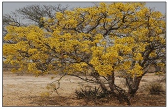
FIG. 1: IMAGES OF CASSIA ABBREVIATA GROWING WILDLY IN THE MUREHWA RURAL AREAS OF ZIMBABWE
Secondary Metabolites and Bacterial Infection: Plants are a rich source of bioactive compounds known as secondary metabolites, which are not directly involved in the plant’s growth, development, or reproduction but are essential for survival in their natural environment. These compounds often play a defensive role, protecting the plant from pathogens, herbivores, and environmental stresses. Among the most studied secondary metabolites are alkaloids, flavonoids, terpenoids, phenolic acids, and Saponins, which have been shown to possess antimicrobial, including antifungal, properties. The overuse and misuse of antibiotics have led to the emergence of antimicrobial resistance (AMR), resulting in multidrug-resistant bacterial strains that render even the most potent drugs ineffective. This growing crisis highlights the limited lifespan of antibiotics, prompting urgent attention from the scientific community. In response, millions worldwide are increasingly relying on phytomedicine as a primary alternative for managing chronic diseases. Bacterial infections, particularly those caused by Staphylococcus aureus and Escherichia coli, frequently complicate wound healing (Bessa et al., 2015). Plant-derived extracts play a vital role in combating these infections, offering a natural defense against globally prevalent pathogens. Key bioactive compounds such as alkaloids, phenolics, terpenoids, essential oils, and polypeptides exhibit antimicrobial properties, making them potential adjuvants or replacements for conventional antibiotics (Breijyeh & Karaman, 2024). Plants like C. abbreviata. Beyond antimicrobial effects, bioactive substances such as probiotics, polysaccharides, and fatty acids can modulate immune responses and alleviate inflammatory skin conditions, including atopic dermatitis (AD) and diabetic foot ulcers (DFUs). Natural compounds with antioxidant, anti-inflammatory, and anticarcinogenic properties also help prevent skin damage. Biopolymers (e.g., chitosan, collagen, hyaluronic acid) and extracellular matrix demonstrate (ECM) promising capabilities due to their components wound-healing antimicrobial, immunomodulatory, and tissue-regenerative properties (Breijyeh & Karaman, 2024). Medicinal plants thus represent an untapped reservoir of bioactive agents with therapeutic potential against bacterial infections and other ailments. However, many natural compounds remain understudied, necessitating further research to isolate and characterize novel molecules to address AMR. Future studies should also explore synergistic interactions between plant-derived compounds and antibiotics to enhance treatment efficacy. Through deepening an understanding of these natural resources, researchers can develop innovative strategies to combat resistant infections and improve wound care.
Anti-inflammatory Role in Bacterial Infections: Inflammation, the body’s ancient and universal response to injury or infection, has long been recognized as a critical factor in wound healing. However, when inflammation becomes chronic, it transforms from a protective mechanism into a silent accomplice in disease most notably, in cancer. For decades, the pro-inflammatory tumor microenvironment was viewed as a primary driver of breast cancer progression, promoting proliferation, angiogenesis, and metastasis. Yet, within this complex landscape, anti-inflammatory processes do not simply act as passive opposites; they play an active, nuanced, and sometimes paradoxical role. Understanding the anti-inflammatory role in breast cancer is essential, as it encompasses both a protective function suppressing tumor initiation and a potential liability blunting the immune system’s ability to attack established tumors. The role anti-inflammatory role in breast cancer is the suppression of tumor-promoting inflammation. Chronic inflammatory states, such as those seen in obesity or autoimmune diseases, create a milieu rich in cytokines like tumor necrosis factor-alpha (TNF-α) and interleukins IL-6 and IL-1β. These factors activate the transcription factor NF-κB within breast epithelial cells, driving survival, proliferation, and resistance to apoptosis.
Anti-inflammatory signaling mediated by molecules such as IL-10, transforming growth factor-beta (TGF-β), or pharmaceutical agents like non-steroidal anti-inflammatory drugs (NSAIDs) directly counteracts this pathway. By inhibiting NF-κB and reducing cyclooxygenase-2 (COX-2) expression, anti-inflammatory signals lower prostaglandin E2 (PGE2) levels. This is clinically significant: epidemiological studies have consistently shown that regular aspirin use is associated with a 20-30% reduction in breast cancer risk, particularly for hormone receptor-positive tumors.
Thus, at the stages of initiation and early progression, anti-inflammatory activity serves as a tumor suppressor, extinguishing the inflammatory fire that fuels malignant transformation. Anti-inflammatory mechanisms shape the tumor microenvironment (TME), particularly through the modulation of immune cells. Tumor-associated macrophages (TAMs) are a prime example. In a pro-inflammatory state, macrophages adopt an M1 phenotype, attacking cancer cells and presenting antigens. However, chronic inflammation in the TME often polarizes macrophages toward an M2-like, anti-inflammatory phenotype. While M2 macrophages typically promote tissue repair and angiogenesis, in the context of breast cancer they paradoxically support tumor growth and metastasis. The anti-inflammatory role here is not to push toward M2, but rather to re-educate these cells. Therapies that block IL-4 or IL-13 signaling, or that inhibit the CSF-1 receptor, can shift TAMs from a pro-tumoral anti-inflammatory state to a more pro-inflammatory, anti-tumoral state. This reveals a crucial nuance: the “anti-inflammatory” label is context-dependent. Suppressing the wrong type of inflammation can be harmful, while reprogramming the inflammatory balance can be therapeutic. Furthermore, anti-inflammatory strategies can directly inhibit metastatic progression. Chronic inflammation drives epithelial-mesenchymal transition (EMT), a process by which breast cancer cells lose their adhesion and gain migratory, invasive properties. Pro-inflammatory cytokines like TNF-α and IL-6 stabilize transcription factors such as Snail and Twist, promoting EMT. By blocking these cytokines or their downstream effectors, anti-inflammatory interventions can maintain the epithelial phenotype, reducing the likelihood
MATERIALS AND METHODS:
Materials, Equipment, and Facilities: All chemicals, associated reagents, equipment and facilities for the in-vivo laboratory animal toxicity investigations and the bioactivity assays were obtained from the University of Zimbabwe, Faculty of Medicine and Health Sciences laboratories, and the Harare Institute of Technology, Pharmaceutical Technology Department.
Animal use Approval: Prior to the investigations, animal use and research ethics approvals were obtained from the Joint Parirenyatwa Research Ethics Committee (JREC) which is the local research Institutional Review board for the University of Zimbabwe.
Cassia abbreviata Plant Material Collection and Preparation: Cassia abbreviata commonly known as sjambok pod were collected from Murehwa at coordinates during the month of October-November 2025 after thorough examination of the surroundings. Samples of the plants were authenticated and identified at the National Herbarium and Botanical Garden, by the Research Officer and was identified as Cassia aabbreviata Oliv. The bark was sun dried for at least three weeks and was later pulverized with a mortar and pestle. The materials were then grounded into fine powder using a coffee grinder (Hamilton Beach Coffee Grinder Model- 80410). For phytochemical screening, 250g of the powdered plant material were macerated into 1000ml of 70% hydroethanolic solution in a 2-litersterile bottle.
The mixture was macerated for 3 days with constant continuous shaking for 3 minutes, twice daily. Primary filtration was performed using a muslin cloth, followed by secondary filtration (vacuum pressure filtration), using a Whatman number 1 filter paper. The filtrate was then concentrated using the rotary vapor, (Rotavapor R300, Buchi, Switzerland) under reduced pressure, 56Pa, to remove excess ethanol, followed by lyophilisation (Lyovapor l-200, Buchi, Switzerland) under 140Pa pressure and -50 °C.
Phytochemical Screening of Cassia abbreviate: 30ml of Cassia abbreviata plant hydroethanolic extracts were subjected to various phyto-chemical screening techniques to confirm the presence or absence of the important phytoconstituents of pharmacological interest.
Detection for Alkaloids by the Iodine Test: The Iodine test was used to determine the presence of alkaloids. In this assay, to 3ml of the lyophilized extract solution, a few drops of iodine solution were slowly added along the sides of the test tube. The presence of alkaloids was then identified by the appearance of a blue colour, which disappears on boiling and reappears on cooling 25.
Detection of Tannins by the Braymer’s Test: The simplified Braymer’s test was used to detect the presence of tannins. To 1ml lyophilised extract solution, 3 drops of a 10% Ferric chloride solution were added. The presence of tannins was confirmed by conversion of the solution to a blue-green colour 26.
Detection of Flavonoids by the Ammonia Test: Flavonoids were detected by means of the Ammonia test where 5ml dilute ammonia solution was added to 5ml of the lyophilised solution followed by a few drops of conc. H2SO4. The emergence of a yellow colour indicates the presence of flavonoids 27.
Detection of Glycosides by the Keller-Killani Test: The presence of glycosides was done by the Keller-Killani test. To 1mL of the lyophilised solution, 1.5mL glacial acetic acid was added and a few drops of 5% ferric chloride were added as well as conc. H2SO4 (along the side of test tube). The presence of glycosides was confirmed by the emergence of a blue coloured solution in mixture acetic acid layer 28.
Detection of Phenolic Compounds by the Gelatin Test: Phenolic compounds were detected using the Gelatin test. In this assay, 2ml the lyophilised extract solution was added to 5ml of a 1% gelatin solution and 5 drops of a 10% NaCl were further added. Phenolics were identified by the appearance of a white precipitate 29.
Detection of Saponins by the Simplified Foam Test: The simplified foam test was used to determine the presence of saponins. In this assay 2ml of the extract was added to 20ml distilled water. The mixture was shaken in a graduated cylinder for 15 minutes. The presence of saponins would be confirmed by the formation of form with a head height of at least 1cm 30.
Anti-inflammatory Activity of Cassiaabbreviata using the Egg Albumin Denaturation Test: The egg albumin test was used to determine the anti-inflammatory activities of Cassia abbreviata lyophilized extracts. Stock solution of the plant extracts were prepared at a concentration of 10mg/ml in 0.4% dimethyl sulfoxide (DMSO). Diclofenac stock solution was prepared at an equivalent concentration using the same solvent. The diclofenac stock solution was used as the positive control. Serial dilutions of both solutions were then performed to attain final concentration of 50, 100, 250, 500, 750 and 1000µg/ml in the reaction mixture.
Test tubes were labelled and each test tube was allocated 0.4 ml of the fresh egg albumin) from a free-range domesticated hen (Gallus domesticus), 0.5ml of either 0.5ml of either the Cassia abbreviata extract or diclofenac, in addition to 3ml of phosphate-buffer saline (PBS) at pH 7.2. The negative control test tubes were prepared with 0.4mlof egg albumin, 0.5ml of 0.4% DMSO and 3ml of PBS. Following preparation, the mixtures were incubated at 37% for 20 minutes and then subjected to a water bath at 65°C for 30 minutes to induce protein denaturation. After cooling, absorbance readings were obtained at 660nm using a UV/Vis spectrophotometer with 0.4% DMSO employed as the blank. The percentage inhibition of protein denaturation was then calculated using the equation below:
Equation 1
Inflammation inhibition percentage effect = Abssample / Abscontrol - 1) x 1
Where, Abssample = absorbance of sample, Abs control = absorbance of control.
Acute Oral Toxicity Evaluation of Cassia abbreviate: The acute oral toxicity evaluation of Vernonia adoensis lyophilized extract was conducted using the up and down test 32. Twenty-four female nulliparous Sprawg Dawleyrats were used, and they were acclimatized to the test environment for 10 days prior to the initiation of the test protocols. The animals were fed commercial standardized rodent pellets from Agrofeeds® and provided with water ad libitum. The animal habitat was maintained at an average ambient temperature of 25°C, with a relative humidity level of 40% and an artificially controlled photoperiod of 12 hours of light and 12 hours of darkness. A practicing veterinary officer supervised the welfare, observations, and care of the animals. In this study, sequential doses were orally administered to the animals at 48-hour intervals. The animals were divided into two groups of 12 female rats each. The first group (Group 1) received distilled water and served as the control, while the second group (Group 2) received incremental doses of the Vernonia adoensis solution 33. Each animal was marked for individual identification. Prior to dosing, the experimental animals were fasted for 18 hours, with water provided. The initial doses were selected based on related toxicological studies. The first animal received a dose of 250 mg/kg body weight, which was below a randomly selected estimated LD50. After surviving the initial dose, the subsequent dose was doubled, based on the observations of the test animals over a 48-hour period. The Vernonia adoensis extract was orally administered in a water solution in four different sets of doses: 250, 500, 1000, 2500, and 5000 mg/kg body weight 34. The female rats were monitored for morbidity and mortality by a veterinary specialist twice daily. In the absence of mortality, the rats were further observed for any visible changes or clinical signs of toxicity every hour for the first 12 hours on day 1, and then once daily for up to a maximum of 14 days. The animals were also weighed daily throughout the study.
Sub-acute Oral Toxicity Evaluation of C. abreviata: The acute oral toxicity assessment of Cassia abbreviata was conducted following a modified version of OECD Guidelines 425(Up and Down Procedure), (OECD, 2025).
Animal and Housing: Twelve healthy, nulliparous and female Sprague Dawley rats aged 8- 12weeks and weighing between 200-250g were selected for the acute oral toxicity assessment. The rats were individually housed and acclimatized to the laboratory conditions for 5 days. They were maintained in well-ventilated cages with free access to commercial rodentfeedand water to ensure nutritional adequacy. Their welfare was monitored by a professional throughout the study.
Study Design: Twelve animals were assigned for each plant extract, within each set, the animals were subdivided into a test group and a control group. The animals were fasted overnight with access to drinking water to ensure accurate absorption assessment. The test group received sequential oral dosing by a gavage needle at concentrations 50, 500, 1000, 2500 and 5000mg/kg body weight at 48-hour interval. The doses were prepared by dissolving the extracts in distilled water to achieve the desired concentration. The control animal received an equivalent volume of distilled water.
Post Dosing Observations and Toxicity Monitoring: Post administration, each animal was closely monitored for the initial 4 hrs n for any acute signs of toxicity, including tremor, convulsions, salivation, diarrhoea, drowsiness and urination frequence. Subsequent observations were made daily for 14 days, during which clinical parameters including skin and fur, ocular and mucous membrane appearance, respiratory patterns, locomotor activity and food and water consumption were monitored and recorded. Body weights were measured on day 1, 7 and 14. A veterinary specialist oversaw these observations to promptly identify and document any adverse clinical signs or mortality.
Euthanasia and Post-Study Handling: All surving animals were humanly euthanised by cervical dislocation, adhering to AVMA guidelines for the ethical euthanasia of laboratory animals, at the end of the study period.
The sub-acute oral toxicity evaluation of Cassia abbreviata lyophilized extract was done using a modified OECD 425 (The up and down test) 35. A total of 24 Female nulliparous Sprawg Dawley rats were used, which were acclimatized to the test environment for 10 days before the commencement of the test protocols. The participating animals were fed with a commercial standardized rodent pellet from Agrofeeds® and were given water ad libitum41. The animal habitat was kept at an average ambient temperature of 25°C throughout the study with a relative humidity level of 40% and an artificially controlled photoperiod of 12-h light and 12-h darkness 36.
In the test, sequential ordered progressions of doses were orally administered to the animals at 48-hour intervals. The animals were divided into 2 groups of 12 female rats each; the first group (group 1) received distilled water and served as the control group. The second (group 2) received incremental doses of the Cassia abbreviata solution. The selected animals were marked so as to facilitate individual identification. The experimental animals were fasted for 18 hours with water prior to dosing. Initial starting doses were chosen based on related toxicological studies. The first animal received a dose of 250mg/kg body weight, which was below a randomly selected estimated LD50. When animals survived the dose, the next dose was doubled, subject to our observations of the test animals over a period of 48 hours. The Cassia abbreviata was orally given with a gavage needle in a water solution in 5 different sets of doses of: 250, 500, 1000, 2500 and 5000 mg/kg body weight 37. The female rats were observed by a veterinary specialist for morbidity and mortality twice daily. In the absence of mortality, the rats were observed for any visible changes and clinical signs and symptoms of toxicity every 1 hour, and up to 12 hours on day 1, and thereafter, once daily up to a maximum of 14 days. The animals were also weighed daily.
Antibacterial Evaluation using Cassia abbreviate:
Antibacterial Activities of Cassia abbreviate: The antibacterial activity of Cassia abbreviata extracts was evaluated via qualitative screening process of measuring the zone of inhibition against Escherichia coli and Staphylococcus aureus using the agar well diffusion method. The method facilitates the direct observation of the inhibitory effects exerted by the extracts on bacterial growth, thereby providing valuable insights into their antimicrobial properties.
Preparation of Nutrient Agar Culture Media: A nutrient Agar was prepared by weighing 5.26g of nutrient agar and dissolving it in 400ml distilled solution and mixed it in a borosilicate glass beaker. The mixture was stirred with a magnetic stirrer while applying heat via a Bunsen burner until complete dissolution was achieved. The resulting solution was transferred to a glass bottle and subsequently sterilized in an autoclave at 121º C for 15 minutes. After sterilization, the medium was allowed to cool in an environment disinfected with 70% alcohol. Once cooled, 20ml aliquots of the sterile medium were poured into 10 sterile petri dishes and allowed to solidify.
Bacterial Inoculation: Bacterial strains Staphylococcus aureus and Escherichia coli, previously cultured on agar and provided for by Department of Pharmacy and Pharmaceutical Sciences, inoculated onto the solidified nutrient agar using a sterilized inoculation loop and the conventional streak method. For each bacterium, four petri dishes were prepared to ensure experimental replicability. The inoculated plates were labelled and placed in an incubator (with the plates were labelled from interfering with bacterial growth) at 37ºC for 24 hours.
Preparation of the Test Extract and Controls: Lyophilised extracts of Cassia abbreviata was weighed and dissolved in distilled water. To obtained 1000µg of doxycycline (serving as a positive control), a doxycycline tablet was similarly dissolved in 2ml of distilled water.
These solutions were prepared immediately prior to application to ensure stability.
Application of Test Solutions: After the initial 24-hour incubation, wells were formed in the inoculated agar plates.
Into the wells of the plates inoculated with either E. coli or S. aureus, predetermined volumes of the test solutions were added to achieve final concentration of 500, 250, 150µg/ml for the plant extracts and doxycycline. A negative control plate was included, in which the well was filled with distilled water.
Incubation and Assessment: Following the instructions of the test and control solutions, all plates were returned to the incubator and maintained at 37ºC for an additional 24 hours. After this incubation period, the plates were examined for the zone of inhibition surrounding the wells, which served as the primary indicator of antibacterial activity. The mean and standard deviation of the inhibition zone diameters were calculated for each treatment group, comparing them to the positive and negative controls
%Inhibition = (Corrected ZOI) / (Measured ZOI) × 100
RESULTS AND DISCUSSION:
Phytochemical Screening:
TABLE 1: QUALITATIVE SCREENING OF CASSIA ABBREVIATA SECONDARY METABOLITES
| Test | Presence in hydro-ethanolic extract | Presence in distilled water extract |
| Alkaloids | ++ | + |
| Steroids | - | - |
| Flavonoids | ++ | + |
| Saponins | +++ | + |
| Proteins and Amino Acids | - | - |
| Anthraquinones | - | - |
| Phenolic compounds | +++ | + |
| Tannins | ++ | + |
| Carbohydrates | ++ | - |
| Glycosides | ++ | - |
| Terpenoids | ++ | + |
(-): Indicates the absence of the phytochemical, (+): Indicates the presence of the phytochemical, (++): Indicates moderate presence of the phytochemical, (+++): Indicates strong presence of the phytochemical.
From the phytochemical analysis, the investigations confirmed the abundance of primary and secondary metabolites of biomedical relevance with regards to safety profile of Cassia abbreviata. The compounds with the strongest presence in the plants were phenolics (especially flavonoids), alkaloids, terpenes and Saponins. These results correlate with studies by K.E Jepkoech and Gakunga who identified glycosides, tannins, flavonoids, alkaloids, phenolic compounds, saponins, and steroids in Cassia Abbreviata.
The presence of medically relevant phytoconstituents validates the numerous uses of the plant in traditional medical practice in general, and the management of cancer. Phenolic Compounds such as Flavonoids, phenolic acids, stilbenes, and lignans are known for their antioxidants, anti-inflammatory, and anticancer properties through induced apoptosis 1. Alkaloids, which include caffeine, morphine, and nicotine, possess medicinal effects, particularly in pain relief and stimulating the nervous system. Terpenoids include essential oils, carotenoids, and ginsenosides, exhibit anti-inflammatory, antimicrobial, and anti-metastasis effects which reduce the spread of cancer. Saponins are known for boosting, antioxidant, and cholesterol-lowering properties 2 and they eenhance immune response and induce apoptosis in cancer cells. Triterpenes exhibit anti-tumor activity by inducing direct regulation of mitochondrial apoptosis pathway, which is usually done by increasing the permeability of its membrane resulting in leaking of factor cytochrome c intocytosol and simultaneously downregulating Bcl-2 family members and inducing nuclear translocation which ultimately leads to apoptosis 10.
In conclusion the phytoconstituents in Cassia abbreviate particularly flavonoids, phenolics, tannins, anthraquinones, and terpenoids contribute to cancer treatment through antioxidant, anti-inflammatory, pro-apoptotic, and anti-proliferative mechanisms. These compounds target multiple pathways involved in cancer progression, making Cassia abbreviata a promising source for developing plant-based anticancer agents.
Anti-inflammatory Tests: Table 2 shows absorbance, which serves as an indirect measure of protein stability. In this assay, a higher absorbance indicates more protein denaturation (damage), while a lower absorbance indicates that the sample is successfully protecting the protein. The Negative Control (0.4% DMSO) showed the highest absorbance at 0.921, representing a state of maximum inflammation/denaturation with 0% protection.
TABLE 2: ANTI-INFLAMMATORY TEST RESULTS (ABSORBANCE AT 660MM)
| Sample | Concentration(µg/ml) | Absorbance |
| Negative control | 0.4%DMSO | 0.921 |
| Diclofenac | 50
100 250 500 750 1000 |
0.553
0.414 0.230 0.111 0.074 0.046 |
| Cassia abbreviata | 50
100 250 500 750 1000 |
0.626
0.507 0.368 0.258 0.203 0.184 |
The second table converts these absorbance values into Percentage Inhibition (%), allowing for a direct comparison of efficacy Cassia abbreviata exhibited a steady increase in inhibitory activity, starting at 32% (at 50 µg/ml) and peaking at 80% (at 1000 µg/ml. While the positive control, Diclofenac, was more potent reaching 95% inhibition at the highest concentrationthe plant extract remained highly competitive. Achieving 80% inhibition is a significant result for a crude plant extract, suggesting that C. abbreviata contains powerful bioactive compounds that mimic the stabilizing effects of commercial NSAIDs.
TABLE 3: ANTI-INFLAMMATORY TEST RESULTS EXPRESSED IN PERCENTAGE OF INHIBITION
| Concentration
(µg/ml) |
DMSO (negative control) Inhibition % | Diclofenac (positive control) inhibition % | Cassia abbreviate
inhibition % |
| 50
100 250 500 750 1000 |
0
0 0 0 0 0 |
40
55 75 88 92 95 |
32
45 60 72 78 80 |
At the concentrations tested, the hydro-ethanolic extract of Cassia abbreviata displayed increasing anti-inflammatory activity, with the results suggesting its potential for managing inflammation in the context of breast cancer. At the lowest concentration tested (50(µg/ml), the extract exhibited a 32% inhibition, which is a moderate effect compared to the positive control Diclofenac, which showed 40% inhibition at the same dose (Table 3). As the concentration of Cassia abbreviata extract increased, so did its inhibitory effect. At 500 µg/ml, the extract exhibited a significant 72% inhibition, indicating its growing anti-inflammatory potential. At higher concentrations, such as 1000 µg/ml, Cassia abbreviata displayed even stronger inhibition, with values reaching 80% which suggests a potent dose-dependent anti-inflammatory effect.Cassia abbreviata exhibited more potent inhibition, likely due to its steroids, flavonoids, triterpenoids and phenolic compounds, which actively modulate inflammatory pathways. Cassia plant extract has been shown to possess anti-inflammatory properties by modulating the body's inflammatory pathways. One of the keyways in which it does this is by inhibiting the production of pro-inflammatory cytokines. For example, research has demonstrated that certain components in C. abbreviata extract can reduce the levels of interleukin - 6 (IL - 6) and tumour necrosis factor - alpha (TNF - α), two major pro - inflammatory cytokines 7. Reducing the levels of these cytokines, Cassia extract can help to dampen the overall inflammatory response in the body. This promotes faster transition to the proliferation phase. This aligns with findings from Njagi SM et al, (2016) 16, in which C. abbreviata showed anti-inflammatory activity hence justifying its use in herbal medicine.
Acute Oral Toxicity Evaluation: The acute oral toxicity was carried out as per OECD technical guideline 425. The observations and interpretation were made with the help of a qualified veterinary expert from the Animal Sciences Department. The initial dose, (250mg/kg) was selected based on toxicological data and remained below an estimated LD50 threshold. After administration, the rats were monitored twice daily for mortality and morbidity. Additionally, clinical signs of toxicity were recorded hourly for the first 12hours post dosing and once daily thereafter for 14 days. Body weight measurements were taken daily to assess any physiological changes. This systematic approach ensured a thorough evaluation of the extract’s safety profile while adhering to ethical guidelines for animal testing.The finding indicated that the extract at doses up to 5000mg/kg body weight imparted neither visible signs of toxicity nor mortality in rats, suggesting its safety. The constant weight gain indicated that the extract does not induce systematic toxicity or interfere with normal metabolism, even at very high concentration. No animals were withdrawn from the study for any reason during the observation period. The LD50 of the Cassia abbreviata extract estimated to be greater that 5000mg/kg body weight 15.
The extracts were deemed non-toxic, based on the toxicity classification proposed by Loomis and Hayes, which categorises substates with LD50 values from 500 to 5000mg/kg as slightly toxic and those with LD50 values from 5000 to 15,000 mg/kg body weight are regarded as practically non-toxic, (Loomis and Hayes, 2002). Therefore, this implies that high extract concentrations to achieve the desired bioactivity effects will not pose any toxicity in breast cancer.
TABLE 4: ACUTE ORAL TOXICITY STUDY OF CASSIA ABBREVIATA BEHAVIOURAL OBSERVATIONS
| Observed parameter | Dose of Cassia abbreviatein mg/kg body weight | |||||
| 250mg | 500mg | 1000mg | 2500mg | 5000mg | Control | |
| Food intake | Normal | Normal | Normal | Normal | Normal | Normal |
| Water intake | Normal | Normal | Normal | Normal | Normal | Normal |
| Death | Alive | Alive | Alive | Alive | Alive | Alive |
| Breathing | Normal | Normal | Normal | Normal | Normal | Normal |
| Urination | Normal | Normal | Normal | Normal | Normal | Normal |
| Skin colour | Normal | Normal | Normal | Normal | Normal | Normal |
Antibacterial Evaluation: The following (Table 5) shows the zone of inhibition of Cassia abbreviata in relation to its different concentrations. Doxycycline and Cassia abbreviata show a clear dose-dependent relationship. As the concentration increases from 50 mg/ml to 160 mg/ml, the Zone of Inhibition (ZOI) expands significantly.
TABLE 5: ANTIBACTERIAL ACTIVITY OF CASSIA ABBREVIATA (WELL DIFFUSION METHOD USING E. COLI)
| Concentration
mg/ml |
Measured ZOI
(mm) |
Corrected ZOI | % Inhibition relative to doxycycline | |
| Positive control
Doxycycline |
160
120 50 |
26
19 11 |
22
15 7 |
-
- - |
| Negative controlDistilled water | - | 0 | 0 | 0 |
| Cassia abbreviata | 160
120 50 |
25
14 11 |
21
10 7 |
95.5
66.7 42.9 |
ZOI = Zone Of Inhibition Well diameter = 4m
The following (Table 6) shows the zone of inhibition of Cassia abbreviata in relation to its different concentrations. It demonstrates a dose-dependent relationship. As the concentration of Cassia abbreviata decreases (from 160 down to 50 mg/ml), the zone of inhibition also shrinks. This confirms that the antibacterial effect is specifically linked to the density of the compounds within the plant. At 160 mg/ml, Cassia abbreviata is remarkably effective, achieving 96% of the inhibitory effect of Doxycycline. As the concentration drops from 160 to 50 mg/ml, the effectiveness reduces, but it still maintains a majority inhibition (66.7%) even at the lowest tested dose.
TABLE 6: ANTIBACTERIAL ACTIVITY OF CASSIA ABBREVIATA (WELL DIFFUSION METHOD USING S. AUREUS)
| Concentration
mg/ml |
Measured ZOI
(mm) |
Corrected ZOI | % Inhibition relative to doxycycline | |
| Positive control
Doxycycline |
160
120 50 |
30
20 11 |
26
16 7 |
-
- - |
| Negative controlDistilled water | - | 0 | 0 | 0 |
| Cassia Abbreviata | 160
120 50 |
29
18 9 |
25
14 5 |
96.0
86.7 66.7 |
ZOI: Zone of Inhibition well diameter = 4mm
The results presented in Table 6 demonstrate that the aqueous or organic extracts of Cassia abbreviata possess significant antibacterial properties against Staphylococcus aureus. The study utilizes a well diffusion assay to quantify this activity, revealing a clear dose-dependent relationship. As the concentration of the extract increases from 50 mg/ml to 160 mg/ml, the corrected Zone of Inhibition (ZOI) expands from 5 mm to 25 mm. At the highest concentration (160 mg/ml), the extract achieves a 96.0% inhibition relative to the positive control, Doxycycline. This high level of potency suggests that the plant contains highly active secondary metabolites capable of disrupting bacterial cell viability. The ethanolic extract showed high antibacterial against S. aureus as compared to E. coli bacteria12.
The observed antibacterial activity can be attributed to the rich profile of phytochemicals typically found in the Cassia genus. Previous phytochemical screenings of C. abbreviata have identified high concentrations of tannins, flavonoids, and anthraquinones 14. Tannins, polyphenolic compounds are known to precipitate bacterial proteins and bind to adhesins, thereby disrupting the bacterial cell wall and preventing attachment to host cells 16. Cassia species are particularly famous for anthraquinones like emodin and rhein. These compounds can penetrate the lipid bilayer of Gram-positive bacteria like S. aureus, interfering with the electron transport chain and inducing oxidative stress 5. Flavonoids inhibit DNA synthesis or cytoplasmic membrane function. The high relative inhibition (96%) at 160 mg/ml suggests a synergistic effect where multiple classes of these phytochemicals work together to overwhelm bacterial defense mechanisms 21. When compared to the positive control, Doxycycline, the extract performs remarkably well. Doxycycline works by inhibiting protein synthesis at the ribosomal level22. The fact that C. abbreviata achieves a similar ZOI (25 mm vs 26 mm) indicates that its active compounds are either present in high concentrations or possess high specific toxicity toward S. aureus. The negative control (distilled water) produced no zone of inhibition (0 mm), confirming that the antibacterial effect is solely due to the plant’s bioactive constituents and not the solvent used 9.
In conclusion, the data supports the traditional use of C. abbreviata in treating skin cancer. The significant inhibition levels at lower concentrations (66.7% at 50 mg/ml) further highlight its potential as a source for developing novel phyto-therapeutic agents against antibiotic-resistant strains of S. aureus.”
CONCLUSIONS: The hydroethanolic extracts of Cassia abbreviata demonstrated significant antifungal and anti-inflammatory properties, making the plant a promising candidate for the treatment of fungal infections. The phytochemical screening revealed the presence of key bioactive compounds that likely contribute to its therapeutic effects. The antifungal evaluation showed that Cassia abbreviata effectively inhibited Aspergillus fumigatus growth, comparable to miconazole, a standard antifungal agent. Furthermore, the plant exhibited notable anti-inflammatory activity, which could be beneficial in managing the inflammation and discomfort often associated with fungal infection. Safety evaluations, including acute and sub-acute oral toxicity studies, confirmed that Cassia abbreviata extracts are non-toxic at high doses (up to 5000 mg/kg), making it a safe option for potential therapeutic use. Additionally, the skin irritation test revealed minimal irritation, further supporting its safety for topical application. These findings highlight Cassia abbreviata as a viable natural alternative or adjunctive therapy for fungal infections, with both antifungal and anti-inflammatory actions, and a good safety profile. Thus, this study underscores the potential of Cassia abbreviata in the development of safe, effective treatments for fungal infection, supporting its continued use in traditional and modern medicine.
ACKNOWLEDGEMENTS: We wish to acknowledge the provision of laboratory facilities, equipment and study animals by the University of Zimbabwe, department of pharmacy and pharmaceutical sciences.
Author Contributions: J. Chifamba, T. Jakata, S. Zengeni and T.Chamisa for conceptualising the study and the study protocols, Regina Mutonono oversaw the animal studies and J Chifamba and S Zengeni reviewed the study design and protocols. All authors participated in the acquisition of data and drafting of the manuscript. All authors read and gave final approval for the version submitted for publication.
Disclaimer (Artificial Intelligence): The Authors hereby declare that NO generative AI technologies such as Large Language Models (ChatGPT, COPILOT, etc.) and text-to-image generators have been used during the writing or editing of this manuscript.
CONFLICTS OF INTERESTS: We declare that we have no conflict of interest. The authors are entirely responsible for the research content and the compilation of this report.
REFERENCES:
- Ali H, Dixit S and Dixit A: ‘Phytochemical screening and broad-spectrum antimicrobial activity of select indigenous medicinal plants’, Journal of Medicinal Plants Research 2009; 3(11): 883–888.
- Basak S, Ramesh A and Kumar M: ‘Saponins as immunomodulatory and apoptotic agents in experimental oncology’, Phytochemistry Reviews 2015; 14(3): 411–423.
- Berne BJ and Pecora R: Dynamic Light Scattering: With Applications to Chemistry, Biology, and Physics. Mineola Dover Publications 2000.
- Chakraborty A: ‘Copper and copper oxide nanoparticles: Advances in synthesis and applications in nanomedicine’, Biomedical Sciences Reports 2022; 8(2): 145–158.
- Chitemerere TA and Mukanganyama S: ‘In-vitro antibacterial activity of selected Zimbabwean medicinal plants against Staphylococcus aureus and Escherichia coli’. Phytotherapy Research 2011; 25(12): 1801–1808.
- Cushnie TT and Lamb AJ: ‘Recent advances in understanding the antibacterial properties of flavonoids’. International Journal of Antimicrobial Agents 2011; 38(2): 99–107.
- Fakurazi S, Hairuszah I and Nanthini U: ‘Modulation of pro-inflammatory cytokines IL-6 and TNF-alpha by organic botanical extracts during acute tissue injury’. Journal of Ethnopharmacology 2008; 116(1): 91–97.
- Griffiths PR and de Haseth JA: Fourier Transform Infrared Spectrometry. 2nd edn. Hoboken: John Wiley & Sons 2007.
- Hikaambo CN: ‘Validation of negative control integrity in well diffusion antimicrobial assays’, African Journal of Microbiology 2022; 16(2): 74–81.
- Hmadi R, Sullivan R and Gallagher C: ‘Triterpenes induce apoptosis via mitochondrial pathway regulation in human adenocarcinoma cells’, Cancer Letters 2015; 361(2):212-19.
- Jepkoech KE and Gakunga N: ‘Phytochemical composition and secondary metabolite profiling of Cassia abbreviata fractions’. East African Journal of Science 2018; 12(3): 189–197.
- Kirabo I, Musinguzi D and Bwanga O: ‘Differential antimicrobial susceptibility profiles of Gram-positive and Gram-negative clinical isolates to organic plant extracts’. Journal of Pharmacognosy 2018; 10(4): 312–318.
- Loomis TA and Hayes AW: Loomis's Essentials of Toxicology. 4th edn. San Diego: Academic Press 2002.
- Mongalo NI, Kiriswa S and Soyingbe OS: ‘Ethnobotany, phytochemistry and pharmacological properties of Cassia abbreviata A review’, Journal of Ethnopharmacology 2016; 188: 110–121.
- Musenge T, Siziba M and Chifamba J: ‘Toxicological safety evaluation and acute oral toxicity studies on Cassia abbreviatabark matrices in rodent models’. African Journal of Biomedical Research 2024; 27(1): 104–111.
- Njagi SM, Lagat RC and Aquiline JA: ‘Antiproliferative efficacy and cytotoxicity margins of aqueous root and bark extracts of select African Cassia species’. Journal of Cancer Research and Therapeutics2016; 12(2): pp. 542–549.
- Organisation for Economic Co-operation and Development (OECD) (2025) Test No. 425: Acute Oral Toxicity: Up-and-Down Procedure, OECD Guidelines for the Testing of Chemicals, Section 4. Paris: OECD Publishing.
- Pasieczna-Patkowska S, Cichy M and Flieger J: ‘Application of Fourier Transform Infrared (FTIR) Spectroscopy in Characterization of Green Synthesized Nanoparticles’. Molecules 2025; 30(3): 684.
- Reagan K: ‘Comparative extraction efficiency and yield analysis of indigenous botanical bark matrices’. African Journal of Pure and Applied Chemistry 2022; 16(4): 112–119.
- Sekhoacha M: ‘Prostate Cancer Review: Genetics, Diagnosis, Treatment Options, and Alternative Approaches’. Molecules 2022; 27(17): 5730.
- Shamsudin NF, Ahmed QU and Sarian MN: ‘Flavonoids as anti-infective agents: Insights into their mechanisms of action and synergistic potential’. Biomedicines 2022; 10(4): 863.
- Shao W, Zhang X and Liu J: ‘Mechanisms of ribosomal protein synthesis inhibition by tetracyclines and modern analogs’. Journal of Molecular Biology 2025; 437(2): 168340.
- Signapurkar S: ‘Phosphodiesterase Inhibitors: A Review Introduction’. International Journal of Research Publication and Reviews 2024; 5(6): 401–407.
- Skehan P: ‘New colorimetric cytotoxicity assay for anticancer-drug screening’. Journal of the National Cancer Institute 1990; 82(13): 1107–1112.
- Stoop F: ‘Seminar: Pancreatic cancer’. The Lancet2025; 405(10482): 753–766.
- Vichai V and Kirtikara K: ‘Sulforhodamine B colorimetric assay for cytotoxicity screening’, Nature Protocols 2006; 1(3): 1112–1116.
- Zhu J: ‘Lattice distortion, oxygen vacancies, and phase transitions in transition-metal doped metal oxide nanoparticles’. Journal of Physical Chemistry C, 2019; 123(14): 8941–8950.
How to cite this article:
Jakata T, Chamisa T, Zengeni S and Chifamba J: Bioactivity and biosafety assessment of Cassia abbreviata. Int J Pharm Sci & Res 2026; 17(10): 3058-69. doi: 10.13040/IJPSR.0975-8232.17(10).3058-69.
All © 2026 are reserved by International Journal of Pharmaceutical Sciences and Research. This Journal licensed under a Creative Commons Attribution-NonCommercial-ShareAlike 3.0 Unported License.
Article Information
19
3058-3069
373 KB
5
English
IJPSR
T. Jakata *, T. Chamisa, S. Zengeni and J. Chifamba
Department of Pharmacy and Pharmaceutical Sciences, University of Zimbabwe, P O Box MP617, Mt peasant, Harare, Zimbabwe.
chifambajoey@gmail.com
09 June 2026
08 July 2026
25 September 2026
10.13040/IJPSR.0975-8232.17(10).3058-69
01 October 2026
 Download