




BIOSYNTHESIS OF MAGNESIUM OXIDE NANOPARTICLES MEDIATED BY FICUS SYCOMORUS FOR THE PRELIMINARY EVALUATION OF WART-RELEVANT BIOLOGICAL ENDPOINTS
HTML Full TextBIOSYNTHESIS OF MAGNESIUM OXIDE NANOPARTICLES MEDIATED BY FICUS SYCOMORUS FOR THE PRELIMINARY EVALUATION OF WART-RELEVANT BIOLOGICAL ENDPOINTS
B. Chinhongo *, Joey Chifamba and Shingirai Zengeni
Department of Pharmacy and Pharmaceutical Sciences, Faculty of Medicine and Health Sciences, University of Zimbabwe, P. O. Box MP167, Mt Pleasant, Harare, Zimbabwe.
ABSTRACT: Cutaneous warts are common benign proliferative lesions caused by human papillomavirus and remain a therapeutic challenge because of recurrence, cost, and limited accessibility of some conventional treatments. This study evaluated the phytochemical profile, preliminary anti-inflammatory activity, and feasibility of green synthesis of magnesium oxide nanoparticles using hydroethanolic leaf extract of Ficus sycomorus. The extract was subjected to qualitative phytochemical screening, total phenolic content estimation, in-vitro anti-inflammatory assessment using the egg albumin denaturation assay, and preliminary nanoparticle biosynthesis and characterization. Phytochemical screening indicated the presence of alkaloids, flavonoids, tannins, saponins, glycosides, and phenolic compounds, while total phenolic content was estimated at 2.64 mg TAE/g extract. In the egg albumin denaturation assay, the crude extract exhibited concentration-dependent inhibition of protein denaturation, increasing from 26% at 50 µg/mL to 80% at 1000 µg/mL, compared with 35–83% for diclofenac. Green synthesis mediated by Ficus sycomorus extract produced approximately 387.52 mg of magnesium-containing precipitate. FTIR analysis suggested the involvement of plant phytochemicals in particle formation or stabilization, while UV–Vis spectroscopy showed an absorption peak at 299.4 nm, providing preliminary optical evidence of particle formation. However, confirmatory characterization using XRD, SEM/TEM, EDX, DLS, PDI, and zeta potential analysis is required to establish MgO phase formation, particle size, morphology, crystallinity, purity, and colloidal stability. Overall, the findings suggest that Ficus sycomorus leaf extract contains bioactive phytoconstituents with preliminary anti-inflammatory activity and may support green synthesis of magnesium-containing nanomaterials. Further antiviral, keratolytic, cytotoxicity, wart-specific efficacy, and dermatological safety studies are required before any therapeutic claims for wart management can be made.
Keywords: Ficus sycomorus, Magnesium oxide nanoparticles, Green synthesis, Phytochemical screening, Anti-inflammatory activity, Protein denaturation assay, Hydroethanolic leaf extract, Plant-based therapeutics
INTRODUCTION: Human papillomavirus (HPV) associated warts are common dermatological conditions in Zimbabwe 1.
Conventional treatments, including laser therapy and cryotherapy, may be costly, require repeated application, and are associated with recurrence 2.
These limitations support the need for affordable and locally relevant therapeutic approaches. Green-synthesized nanoparticles prepared using medicinal plants such as Ficus sycomorus may provide an environmentally friendly platform for preliminary biological investigation; however, their direct efficacy in wart management remains unconfirmed 3.
Warts Management: Wart treatment approaches can broadly be classified as patient-applied therapies and physician-administered procedures. Patient-applied options include salicylic acid, podophyllotoxin, topical 5-fluorouracil, tretinoin, and imiquimod 4. Some topical agents, including diphencyprone, dinitrochlorobenzene, and squaric acid, act by inducing contact dermatitis, which may support wart clearance through immune stimulation. However, their use is limited by pruritus, local irritation, and safety concerns. Other less commonly used topical agents include formalin and glutaraldehyde. Physician-administered treatments include laser surgery, blunt dissection, electrodesiccation, and cryotherapy 5.
Herbal Management of Warts: Several medicinal plants, including Euphorbia hirta and Solanum dulcamara, have been reported to possess traditional or experimental relevance in wart management. In some regions, latex from the fig tree (Ficus carica) is used traditionally for wart treatment. Ficin, a proteolytic enzyme present in fig latex, has been suggested to contribute to this effect. Garlic (Allium sativum) has also been used traditionally for skin conditions, and allicin has been proposed to interfere with HPV-related processes. Although these reports support the ethnomedicinal relevance of plant-derived preparations, robust pharmacological and clinical evidence is still required before therapeutic claims can be made 5.
Ficus sycomorus: Ficus sycomorus, commonly known as fig-mulberry, is a semi-deciduous tree belonging to the family Moraceae. It commonly reaches 20–21 m in height and may grow taller under favorable conditions. The species has a broad crown, spreading branches, and heart-shaped leaves. Flowering and fruiting may occur throughout the year, with peak fruiting commonly reported between July and December 6.
The bark color varies from green to yellow or orange, and the tree is commonly found in savannah areas, near rivers, streams, and sites with a high-water table. Traditionally, the latex, bark, and other parts of F. sycomorus have been used for burns, ulcers, inflammation, ringworm, sore throat, respiratory complaints, diarrhoea, pain, and other conditions. These ethnomedicinal uses provide a rationale for preliminary phytochemical and biological evaluation 7.
Magnesium Nanoparticles and Warts Treatment: Magnesium oxide nanoparticles (MgO NPs) synthesized using plant extracts have attracted interest because of their physicochemical properties and biological activities. Green synthesis methods using extracts from plants such as Carica papaya, Azadirachta indica, and Mangifera indica have been reported to produce MgO-based nanomaterials with antibacterial activity against organisms such as Escherichia coli, Staphylococcus aureus, and Pseudomonas aeruginosa 8.
These effects have been associated with surface reactivity, oxidative stress generation, and membrane-disruptive mechanisms. However, such antibacterial findings cannot be directly extrapolated to HPV-associated warts without antiviral, keratolytic, cytotoxicity, and dermatological efficacy studies. In-vitro studies however suggest MgONPs are potentially candidates for the development of novel antiviral preparations 9.
Nanotechnology: Nanotechnology is an innovative discipline centered on the manipulation of matter at the atomic and molecular levels. Nanotechnology, with its applications in health, agriculture, and water purification, presents distinctive qualities that can tackle significant issues like health disparities and food security 10.
Nanotechnology has emerged as one of the most valuable and beneficial technological advancements for humanity. Nanotechnology encompasses the integration, manipulation, and regulation of molecules and atoms to create systems, devices, structures, materials, and components at the nanoscale level. Nanotechnology operates on a nanoscale range of 1 nm to 100 nm 11.
Main Aim: To biosynthesize and preliminarily characterize magnesium oxide nanoparticles mediated by Ficus sycomorus leaf extract and to evaluate selected biological endpoints relevant to inflammation.
Specific Objectives:
- To perform qualitative phytochemical screening of the lyophilized crude leaf extract of Ficus sycomorus.
- To estimate the total phenolic content of the hydroethanolic leaf extract.
- To assess the in vitro anti-inflammatory activity of Ficus sycomorus leaf extract using the egg albumin denaturation assay.
- To biosynthesize and preliminarily characterize MgO-based nanoparticles mediated by Ficus sycomorus leaf extract.
MATERIALS AND METHODS: All chemicals, equipment, reagents, and laboratory facilities used for the in-vitro and bioactivity assays were obtained from the Department of Pharmacy and Pharmaceutical Sciences, Faculty of Medicine and Health Sciences, University of Zimbabwe. Authorization to collect and use the plant material for research purposes was obtained from the National Herbarium and Botanic Garden of Zimbabwe. A copy of the authorization/ authentication letter from the National Herbarium and Botanic Garden of Zimbabwe is provided as supporting documentation.
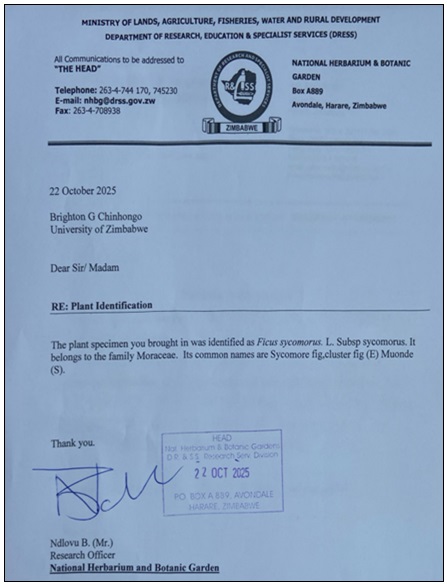
FIG. 1: BOTANICAL VERIFICATION OF FICUS SYCOMORUS
Ethical approval was obtained from the Joint Research Ethics Committee (JREC) (JREC Ref: 472/2025) dated 4th November 2025. No human participants were involved in this study. Permission to use the departmental laboratories was obtained from the Department of Pharmacy and Pharmaceutical Sciences.
Ficus sycomorus Plant Material Collection and Preparation: Ficus sycomorus leaves, commonly known as fig-mulberry leaves, were collected from Mazowe, Zimbabwe, in October 2025 after inspection of the collection site. The plant material was authenticated and identified as Ficus sycomorus by a research officer at the National Herbarium and Botanic Garden of Zimbabwe. No voucher specimen number was assigned for this study.
The leaves were sun-dried for at least three weeks and then pulverized using a mortar and pestle. The dried material was further ground into a fine powder using a coffee grinder (Hamilton Beach Coffee Grinder, Model 80410). For phytochemical screening and extraction, 530 g of powdered plant material was macerated in 1000 mL of 70% hydroethanolic solution in a sterile bottle. The mixture was macerated for three days with continuous shaking. Primary filtration was performed using muslin cloth, followed by secondary filtration under vacuum using Whatman No. 1 filter paper. The filtrate was concentrated using a rotary evaporator (Rotavapor R-300, Buchi, Switzerland) under reduced pressure to remove excess ethanol.
Approximately 300 mL of crude liquid hydroethanolic extract was obtained and used for subsequent experimental procedures. To estimate dry extract yield, a representative 20 mL aliquot of the crude liquid extract was lyophilized to constant weight. The mass of lyophilized extract obtained from this aliquot was used to estimate the total dry extract content of the 300 mL crude extract and to calculate percentage yield relative to the initial dried plant material.
Phytochemical Screening of Ficus sycomorus: A 30 mL aliquot of the Ficus sycomorus hydroethanolic leaf extract was subjected to qualitative phytochemical screening to determine the presence or absence of selected phytoconstituents of pharmacological interest.
Detection of Flavonoids by Ammonia Test: Two millilitres of Ficus sycomorus extract solution were mixed with 2 mL of dilute ammonia solution, followed by a few drops of concentrated sulphuric acid. Development of a yellow color that became colorless indicated the presence of flavonoids 12.
Detection of Alkaloids by the Wagner’s Test: A few drops of Wagner’s reagent were added along the side of a test tube containing 2 mL of extract. Formation of a brown precipitate indicated the presence of alkaloids 13.
Detection of Tannins by the Ferric Chloride Test: Four drops of 10% ferric chloride solution were added to 3 mL of Ficus sycomorus extract. Formation of a greenish-black color indicated the presence of tannins 14.
Detection of Phenolic Compounds by the Gelatine Test: A few drops of 5% ferric chloride solution were added to 2 mL of Ficus sycomorus extract. Formation of a bluish-black color indicated the presence of phenolic compounds 12.
Detection of Saponins by Simplified Foam Test: Five milliliters of distilled water were added to 2 mL of extract, and the mixture was shaken in a graduated cylinder for 10 min. Formation of persistent foam with a height of at least 6 cm indicated the presence of saponins 15.
Total Phenolic Content Assay: Total phenolic content is commonly determined by comparing the response of plant extracts with a phenolic standard 16. In this study, because the Folin–Ciocalteu reagent was unavailable, total phenolic content was estimated using a modified direct UV spectrophotometric method with tannic acid as the calibration standard 17. Phenolic compounds absorb ultraviolet radiation due to their aromatic rings and hydroxyl substituents; therefore, sample absorbance was measured at 270 nm and compared with a tannic acid calibration curve 18. This modified method provides an estimate of total phenolic constituents rather than the conventional Folin–Ciocalteu reducing-capacity assessment. Results were expressed as tannic acid equivalents (TAE).
A 1 mg/mL tannic acid stock solution was prepared by dissolving 100 mg of tannic acid in 100 mL of methanol. Serial dilutions were prepared by transferring 0, 1, 2, 3, 4, and 5 mL of the stock solution into labelled test tubes and making each volume up to 10 mL, producing an approximate calibration range of 0–500 µg/mL. The absorbance of each tannic acid standard was measured at 270 nm using a UV–Visible spectrophotometer, and a calibration curve of absorbance versus concentration was constructed.
The Ficus sycomorus hydroethanolic extract was diluted in methanol at a ratio of 1 mL extract to10 mL solvent, and absorbance was measured at 270 nm. The concentration of phenolic compounds in the extract was determined from the tannic acid calibration curve and expressed as milligrams of tannic acid equivalents per gram of extract (mg TAE/g extract). All measurements were performed in triplicate, and mean absorbance values were used to estimate total phenolic content. The formula used for estimation was C = C1 × v / m where C is the total phenolic content in mg/g, is the concentration of tannic acid obtained from the calibration curve, V is the volume of extract solution in milliliters, and m is the mass of extract in grams. The extract solution yielded approximately 1.86 g of lyophilized extract per 20 mL of bulk hydroethanolic extract.
Ficus sycomorus Anti-inflammatory Test (Egg Albumin Assay): The egg albumin anti-inflammatory test is based on the principle of inhibition of protein denaturation. During inflammation, proteins may become denatured due to heat, chemicals, or other stress conditions, leading to the production of auto-antigens that contribute to inflammatory reactions. Substances with anti-inflammatory properties are capable of stabilizing proteins and preventing or reducing their denaturation. In this test, egg albumin is subjected to heat-induced denaturation in the presence and absence of the test sample. The degree of turbidity or coagulation produced is measured spectrophotometrically. A lower level of protein denaturation in the presence of the test sample indicates anti-inflammatory activity. Stock solutions of the plant extract and diclofenac were prepared at a concentration of 10 mg/mL in 0.4% dimethyl sulfoxide (DMSO). Diclofenac was used as the positive control.
Serial dilutions of both solutions were prepared to obtain final concentrations of 50, 100, 250, 500, 750, and 1000 µg/mL in the reaction mixture. Each test tube contained 0.4 mL of fresh egg albumin, 0.5 mL of either Ficus sycomorus extract or diclofenac solution, and 3 mL of phosphate-buffered saline (PBS). Negative-control tubes contained 0.4 mL of egg albumin, 0.5 mL of 0.4% DMSO, and 3 mL of PBS. The mixtures were incubated at 37 °C for 20 min and then heated in a water bath at 65 °C for 30 min to induce protein denaturation. After cooling, absorbance was measured at 660 nm using a UV–Visible spectrophotometer, with 0.4% DMSO used as the blank. Percentage inhibition of protein denaturation was calculated using the following equation:
Inhibition% = (Abs(control) - Abs(sample) / (Abs(control) × 100
Biosynthesis of MgO-Based Nanoparticles: The biosynthesis of MgO-based nanoparticles was attempted through the reaction of magnesium nitrate with phytochemical constituents present in the plant extract. These constituents, including flavonoids, phenolic compounds, alkaloids, and tannins, may act as reducing, capping, and stabilizing agents under alkaline reaction conditions.
When magnesium nitrate solution was mixed with the plant extract under controlled temperature, pH, and stirring conditions, visible turbidity and precipitate formation were used as preliminary indicators of particle formation. The resulting precipitate was isolated, purified, and dried before characterization. Because confirmatory techniques such as X-ray diffraction and electron microscopy were not performed, the material is described cautiously as MgO-based or magnesium-containing nanoparticles.
Materials and Equipment:
- Ficus sycomorus leaves hydroethanolic extract (100 ml)
- Magnesium nitrate crystals (10 g)
- Two (2) 100ml volumetric flasks
- One (250ml) Volumetric flask
- Sodium hydroxide solution for pH adjustment
- Dropper
- Filter paper x3
Apparatus:
- Analytical Balance
- pH meter
- Sonicator/ Water Bath
The experiment was carried out in triplicate. In the first experiment, 1 g of magnesium nitrate and 1 g of Ficus sycomorus hydroethanolic extract were accurately weighed using an analytical balance. Each was transferred into separate 100 ml volumetric flasks and 50 ml of distilled water was added to each flask.
The Ficus sycomorus solution was stirred in a water bath at 50 °C for 15 min and then filtered into a 250 mL volumetric flask. The 0.13 M magnesium nitrate solution was added to the filtered extract, and the pH was adjusted to 10.5–12.0. The resulting mixture was stirred in a water bath at 50 °C for 1 h, during which a cloudy precipitate formed. The mixture was then left at room temperature for 12–24 h to allow the reaction to continue. The mixture was centrifuged for 30 min, after which the supernatant was discarded and the pellet was collected and dried in an oven at 100 °C for 6 h. The process was repeated using 2 g of magnesium nitrate with 5 g of Ficus sycomorus extract and then 5 g of magnesium nitrate with 11 g of Ficus sycomorus extract to improve precipitate yield.
Characterization: The synthesized magnesium-containing nanoparticles were characterized using Fourier-transform infrared spectroscopy (FTIR) and ultraviolet–visible spectroscopy (UV–Vis) to assess functional groups and optical absorption behavior, respectively.
FTIR:
Principle: Fourier-transform infrared spectroscopy (FTIR) is an analytical technique used to identify functional groups and chemical bonds in a sample by measuring absorption of infrared radiation at characteristic wave numbers. In this study, FTIR was used to identify functional groups in the plant extract and to infer possible involvement of biomolecules in nanoparticle formation and stabilization.
Procedure: The diamond platform of the FTIR spectrophotometer was cleaned using distilled water and methanol to remove contaminants. The dried nanoparticle sample was placed on the diamond platform to cover the surface. Spectra were recorded over the range of approximately 4000–400 cm⁻¹. The spectra were exported from the instrument software, and absorption peaks were analysed to identify functional groups present on the surface of the synthesized material. The same procedure was repeated for the lyophilized Ficus sycomorus leaf extract for comparison.
UV-VIS:
Principle: Ultraviolet–visible spectroscopy (UV–Vis) is an analytical technique used to assess the optical absorption behaviour of dispersed particles over ultraviolet and visible wavelengths. In this study, UV–Vis analysis was used as a preliminary tool to observe absorption features of the synthesized magnesium-containing material. Because MgO nanoparticles do not exhibit surface plasmon resonance in the same manner as noble metal nanoparticles, UV–Vis results were interpreted cautiously and were not used as sole confirmation of MgO nanoparticle formation.
Procedure: A small quantity of the synthesized magnesium-containing material was dispersed in distilled water to prepare a colloidal suspension. The suspension was mixed thoroughly to promote uniform dispersion. Distilled water was used as the blank for UV–Vis spectrophotometer calibration. The prepared suspension was transferred into a clean quartz cuvette, and the absorbance spectrum was recorded over a wavelength range of approximately 200–800 nm. The obtained spectra were saved and analysed to identify absorption features of the synthesized material.
RESULTS AND DISCUSSION:
Plant Extraction: Hydroethanolic leaf extract of Ficus sycomorus was successfully prepared by maceration. Approximately 300 mL of crude liquid (hydroethanolic) extract was obtained from 530 g of dried powdered leaf material. Lyophilization of a representative 20 mL aliquot of this crude liquid extract yielded approximately 1.86 g of dry extract. Based on the total crude extract volume of 300 mL, the estimated total dry extract content was 27.9 g. Relative to the initial 530 g of dried powdered leaf material, the estimated extraction yield was approximately 5.26%. The relatively low yield may be attributed to the use of leaves only, as other plant parts may contain higher levels of extractable secondary metabolites. Leaves were selected to minimize destructive harvesting of plant material.
Phytochemical Screening and Total Phenolic Content Assay: Qualitative phytochemical screening indicated the presence of secondary metabolites of potential biomedical relevance in the hydroethanolic leaf extract of Ficus sycomorus. The detected phytochemical classes included alkaloids, flavonoids, saponins, phenolic compounds, glycosides, and tannins.
TABLE 1: QUALITATIVE PHYTOCHEMICAL SCREENING RESULTS OF FICUS SYCOMORUS HYDROETHANOLIC LEAF EXTRACT
| Phytochemical Class | Test Used | Observed Reaction | Result |
| Flavonoids | Ammonia test | Yellow color developed after addition of ammonia and became colorless after addition of concentrated sulphuric acid. | Present |
| Alkaloids | Wagner’s test | Brown precipitate formed after addition of Wagner’s reagent. | Present |
| Tannins | Ferric chloride test | Greenish-black color developed after addition of ferric chloride solution. | Present |
| Phenolic compounds | Ferric chloride test | Bluish-black color developed after addition of ferric chloride solution. | Present |
| Saponins | Foam test | Persistent foam of at least 6 cm was observed after shaking with distilled water. | Present |
| Glycosides | Acetic acid–ferric chloride–sulphuric acid test | Blue color developed after addition of glacial acetic acid, ferric chloride, and concentrated sulphuric acid. | Present |
The phytochemical screening was qualitative and was based on visible color change, precipitate formation, or foam persistence according to the respective screening tests. These findings provide preliminary evidence of phytoconstituent classes in the extract; however, confirmatory quantitative assays or chromatographic profiling would be required to determine the exact identity and concentration of the detected compounds. The estimated total phenolic content of the hydroethanolic extract was 2.64 mg TAE/g extract. This value was compared with previous studies reporting total phenolic content in different plant parts, although direct comparison should be made cautiously because extraction methods, standards, plant parts, and reporting units differ among studies 19. Further quantitative assays and chromatographic profiling are recommended to identify and quantify the specific phenolic, and related compounds present in the extract.
Anti-inflammatory Evaluations: The in-vitro anti-inflammatory activity of Ficus sycomorus leaf extract was evaluated using the egg albumin denaturation assay. This assay estimates the ability of a test substance to inhibit heat-induced protein denaturation, a process associated with inflammatory responses. The extract showed a concentration-dependent increase in percentage inhibition, reaching 80% inhibition at 1000 µg/mL. Diclofenac, used as the positive control, showed 83% inhibition at the same concentration. Although the extract demonstrated notable activity in this assay, the result should be interpreted as preliminary and should not be considered evidence of wart-specific efficacy. The table below shows the raw spectrophotometric readings (absorbance) for the samples tested, which serves as the primary data source used to evaluate the anti-inflammatory potential of Ficus sycomorus.
TABLE 2: ANTI-INFLAMMATORY ASSAY RESULTS EXPRESSED AS ABSORBANCE AT 660 NM
| Sample | Concentration (µg/mL) | Absorbance |
| Negative control | 0.4%DMSO | 0.821 |
| Diclofenac | 50
100 250 500 750 1000 |
0.533
0.404 0.340 0.221 0.216 0.136 |
| Ficus sycomorus | 50
100 250 500 750 1000 |
0.606
0.557 0.368 0.238 0.223 0.164 |
Table 2 presents the calculated percentage inhibition values derived from the absorbance readings. Percentage inhibition represents the extent to which each sample reduced protein denaturation relative to the negative control.
TABLE 3: ANTI-INFLAMMATORY ASSAY RESULTS EXPRESSED AS PERCENTAGE INHIBITION
| Concentration
(µg/ml) |
DMSO(negative control) Inhibition % | Diclofenac (positive control) inhibition % | Ficus sycomorus inhibition % |
| 50
100 250 500 750 1000 |
0
0 0 0 0 0 |
35
51 62 73 74 83 |
26
32 55 71 73 80 |
The plot compares the percentage inhibition of diclofenac and Ficus sycomorus extract across the tested concentration range.
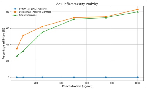
FIG. 2: ANTI-INFLAMMATORY ACTIVITY EXPRESSED AS PERCENTAGE INHIBITION VERSUS CONCENTRATION
Biosynthesis and Characterization of Nanoparticles: The first two nanoparticle synthesis attempts did not yield visible precipitate. In the third attempt, 5 g of magnesium nitrate dissolved in 30 mL of distilled water was added to a filtered solution containing 11 g of Ficus sycomorus hydroethanolic leaf extract in 50 mL of distilled water, resulting in precipitate formation. This observation suggests that higher precursor and extract concentrations may be required to improve yield. Further optimization studies are needed to establish reproducible conditions for the synthesis of MgO-based nanoparticles mediated by Ficus sycomorus leaf extract. The final yield was 387.52 mg, which was insufficient for extended safety, dosage, and formulation evaluations.
The FTIR spectra for the lyophilized extract and the synthesized magnesium-containing material are presented below.
For the extract, eleven peaks were observed, as shown below.
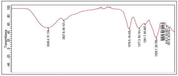
FIG. 3: FTIR SPECTROGRAM FOR THE LYOPHILIZED EXTRACT
For the synthesized magnesium-containing material, the following spectrum was obtained.
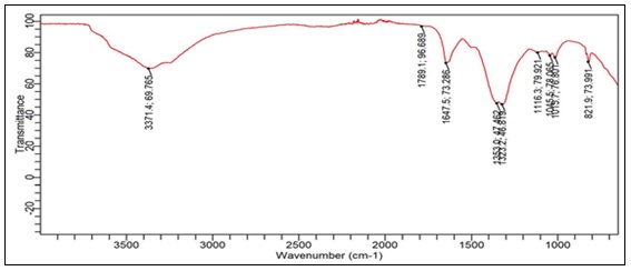
FIG. 4: IMAGE ILLUSTRATING THE FTIR SPECTROGRAM FOR THE BIOSYNTHESIZED MgONPs
In the FTIR spectrum, a broad peak at approximately 3371 cm⁻¹ was observed, which may be attributed to O–H stretching vibrations associated with alcohols and phenolic compounds. The peak at approximately 1647 cm⁻¹ may correspond to C=O stretching, aromatic C=C vibrations, or amide-related functional groups. These observations suggest possible adsorption of phytochemical constituents onto the surface of the synthesized magnesium-containing material.
Peaks around 1352 cm⁻¹ may be associated with phenolic O–H bending and C–O stretching. Changes observed between 1116 and 1055 cm⁻¹ may correspond to C–O stretching in alcohols, ethers, and glycosidic linkages, indicating possible interactions between polysaccharides or phenolic compounds and the synthesized material. Overall, the FTIR findings suggest the involvement of Ficus sycomorus phytochemicals in capping or stabilizing the synthesized magnesium-containing particles; however, FTIR alone is insufficient to confirm MgO nanoparticle formation, crystallinity, morphology, size, or purity.
The image below shows the UV-Vis spectrogram derived from the UV-Vis Spectometry.
The UV–Vis spectrum of the synthesized magnesium-containing material showed a distinct absorption peak at 299.4 nm. This absorption feature indicates the presence of UV-active magnesium-containing particles or phytochemical-stabilized material in the dispersion. However, MgO nanoparticles do not exhibit surface plasmon resonance in the same manner as noble metal nanoparticles; therefore, the observed peak should not be interpreted as a plasmonic band.
The UV–Vis result provides preliminary optical evidence of particle formation, but further characterization using X-ray diffraction, scanning or transmission electron microscopy, energy-dispersive X-ray spectroscopy, dynamic light scattering, polydispersity index, and zeta potential analysis is required to confirm MgO phase formation, particle size, morphology, crystallinity, purity, and colloidal stability.
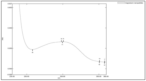
FIG. 5: UV-VIS SPECTROGRAM FOR BIOSYNTHESIZED MgONP CANDIDATE MATERIAL
ACKNOWLEDGEMENTS: The authors acknowledge the Department of Pharmacy and Pharmaceutical Sciences, University of Zimbabwe, for providing laboratory facilities and technical support. The authors also extend their appreciation to the Department of Chemistry at the Medicines Control Authority of Zimbabwe (MCAZ) for additional analytical assistance, and further acknowledge the technical staff within the department for their support and training on the analytical equipment used in this research. The National Herbarium and Botanic Garden of Zimbabwe is gratefully acknowledged for plant authentication, and the Joint Research Ethics Committee (JREC) is acknowledged for granting ethical approval for this research.
Authors’ Contributions: This work was carried out in collaboration among all authors. Author BC led the study conception, coordination, design, methodology, data collection, and preparation of the original manuscript draft. Authors JC and SZ contributed to the conceptual framework, supervised the study, and reviewed the manuscript. All authors reviewed and approved the final manuscript.
CONFLICT OF INTEREST: The authors declare no conflicts of interest regarding this work.
REFERENCES:
- M. R. M. T. M. F. M. K. M. B. P. J. C. Z. Manyere NR: Human papillomavirus genotype distribution in genital warts among women in Harare-Zimbabwe. J Obstet Gynaecol 2019.
- Widschwendter A: Recurrence of genitals warts in pre-HPV vaccine era after laser treatment. Arch Gynecol Obstet 2019; 300(3): 661–668, doi: 10.1007/s00404-019-05242-5.
- Alholy T and Khaddam W: “Biosynthesis of magnesium oxide nanoparticles using Opunitaficus indica and their antifungal effect against Aspergillus niger. Journal of Research in Pharmacy 2023; 27(3): 1188–1201, doi: 10.29228/jrp.408.
- Bruggink SC: Cryotherapy with liquid nitrogen versus topical salicylic acid application for cutaneous warts in primary care: randomized controlled trial. Can Med Assoc. J 2010; 182(15): 1624–1630, doi: 10.1503/cmaj.092194.
- Gogoi HP, Ruby JJ, Ruby JJ and Professor A: “Treatment and management of warts-a comprehensive review. 2025.
- Hossain MA: A review on Ficus sycomorus: A potential indigenous medicinal plant in Oman. Elsevier B.V. 2019; 01, doi: 10.1016/j.jksus.2018.07.002.
- AL Rahman F Magbool F, Elnima EI, S. ME and Hussein SEO: Phytochemical and antimicrobial screening of stem bark and leaves extracts from Ficus sycomorus 2017; [Online]. Available: www.wjpmr.com
- Chen H, Chen X, Chen Y, Tang L, Shi WJ and Ou YH: L-type association between magnesium intake and human papillomavirus infection in US adult women: based on NHANES 2003–2016 data. Front Nutr 2025; 12, doi: 10.3389/fnut.2025.1594489.
- Nasim I: Antioxidant & amp; anti inflammatory activity of magnesium oxide nanoparticles - an in-vitro Int J Dent Oral Sci 2021; 2913–2917, doi: 10.19070/2377-8075-21000568.
- Bayda S, Adeel M, Tuccinardi T, Cordani M and Rizzolio F: The history of nanoscience and nanotechnology: from chemical–physical applications to nanomedicine. Molecules 2019; 25(1): 112, doi: 10.3390/molecules25010112.
- Muparadzi N: Nanotechnology: a review on the application of nano-materials in water purification and nano-medicine in the Zimbabwean context. Journal of Materials Science Research and Reviews 2025; 8(2): 347–359, doi: 10.9734/jmsrr/2025/v8i2409.
- Altemimi A, Lakhssassi N, Baharlouei A, Watson DG and Lightfoot DA: Phytochemicals: Extraction, isolation, and identification of bioactive compounds from plant extracts 2017; 01, MDPI AG. doi: 10.3390/plants6040042.
- Dey P: Analysis of alkaloids (indole alkaloids, isoquinoline alkaloids, tropane alkaloids). In Recent Advances in Natural Products Analysis, Elsevier 2020; 505–567. doi: 10.1016/B978-0-12-816455-6.00015-9.
- Shaikh JR and Patil M: Qualitative tests for preliminary phytochemical screening: An overview. Int J Chem Stud 2020; 8(2): 603–608, doi: 10.22271/chemi.2020.v8.i2i.8834.
- Kareru J, Keriko M, Gachanja AN and Kenji GM: Direct detection of triterpenoid saponins in medicinal plants. 2008. [Online]. Available: www.africanethnomedicines.net
- Abugri DA and Mcelhenney WH: Extraction of total phenolic and flavonoids from edible wild and cultivated medicinal mushrooms as affected by different solvents. 2013. [Online]. Available: http://scholarsresearchlibrary.com/archive.html
- Pérez M, Dominguez-López I and Lamuela-Raventós RM: The chemistry behind the folin–ciocalteu method for the estimation of (poly) phenol content in food: total phenolic intake in a Mediterranean dietary pattern. J Agric Food Chem 2023; 71(46): 17543–17553, doi: 10.1021/acs.jafc.3c04022.
- Siddiqui N, Rauf A, Latif A and Mahmood Z: “Spectrophotometric determination of the total phenolic content, spectral and fluorescence study of the herbal Unani drug Gul-e-Zoofa (Nepetabracteata Benth). J Taibah Univ Med Sci 2017; 12(4): 360–363, doi: 10.1016/j.jtumed.2016.11.006.
- Al-matani SK, Al-Wahaibi RNS and Hossain MA: In-vitro evaluation of the total phenolic and flavonoid contents and the antimicrobial and cytotoxicity activities of crude fruit extracts with different polarities from Ficus sycomorus. Pacific Science Review A: Natural Science and Engineering 2015; 17(3): 103–108, doi: 10.1016/J.PSRA.2016.02.002.
How to cite this article:
Chinhongo B, Chifamba J and Zengeni S: Biosynthesis of magnesium oxide nanoparticles mediated by Ficus sycomorus for the preliminary evaluation of wart-relevant biological endpoints. Int J Pharm Sci & Res 2026; 17(10): 3135-45. doi: 10.13040/IJPSR.0975-8232.17(10).3135-45.
All © 2026 are reserved by International Journal of Pharmaceutical Sciences and Research. This Journal licensed under a Creative Commons Attribution-NonCommercial-ShareAlike 3.0 Unported License.
Article Information
26
3135-3145
1805 KB
5
English
IJPSR
B. Chinhongo *, Joey Chifamba and Shingirai Zengeni
Department of Pharmacy and Pharmaceutical Sciences, Faculty of Medicine and Health Sciences, University of Zimbabwe, P. O. Box MP167, Mt Pleasant, Harare, Zimbabwe.
bgchinhongo@protonmail.com
20 May 2026
17 June 2026
26 June 2026
10.13040/IJPSR.0975-8232.17(10).3135-45
01 October 2026
 Download