




CHITOSAN AND CARBOXYMETHYL CHITOSAN (CMC)-COATED MAGNETITE NANOPARTICLES FOR DRUG DELIVERY TO CANCER CELLS: EFFECT OF MOLECULAR WEIGHT ON DRUG RELEASE
HTML Full TextCHITOSAN AND CARBOXYMETHYL CHITOSAN (CMC)-COATED MAGNETITE NANOPARTICLES FOR DRUG DELIVERY TO CANCER CELLS: EFFECT OF MOLECULAR WEIGHT ON DRUG RELEASE
Johar K. Al Khalifah, O. M. Lemine, Alia Mustafa, Khaled Greish and Kheireddine El-Boubbou *
Department of Chemistry, College of Science, University of Bahrain, Sakhir 32038, Bahrain.
ABSTRACT: Novel effective delivery systems for chemotherapeutic drugs are of utmost demand in nanomedicine. Herein, we prepared magnetite (Fe₃O₄) nanoparticles (NPs) coated with either low molecular (LM) or high molecular (HM) weight Chitosan or Carboxymethyl Chitosan (CMC) and assessed their drug loading and release efficiencies. Chitosan/CMC-coated MNPs were fully characterized by various physiochemical techniques including TEM, XRD, DLS, FTIR, and VSM. These techniques clearly elucidated the nanostructure, morphology, surface charge, and magnetization of all functional MNPs. We then loaded them with the anticancer drug Dox to achieve biocompatible, biodegradable, and pH-responsive drug nanocarriers. Drug release was evaluated at both neutral and acidic pH to mimic endosomal/lysosomal conditions. HM Chitosan-NPs or CMC-NPs showed faster and more sustained controlled release (~ 90 - 99%) at acidic pH, while LM NPs exhibited slightly lower drug release (~ 60 - 65%). Notably, almost negligible release was found at neutral physiological pH for all MNP formulations, which is crucial for successful pH-triggered delivery system. The delivery of Dox using CMC-NPs was then evaluated in human monocytic leukemia THP-1 cancer cells. Remarkably, while the unloaded CMC-NPs were non-toxic to the tested cell lines, Dox@CMC-MNPs were found to be highly potent causing apoptotic cell death to cancer cells (IC50 = 16 µg/mL), as evident from live confocal images. These results show great promise for the utilization of Chitosan/CMC-MNPs for potential in-vivo cancer theranostics.
Keywords: Magnetite nanoparticles, Chitosan; Carboxymethyl chitosan; Doxorubicin, pH-responsive drug delivery, Cancer theranostics
INTRODUCTION: Magnetic iron oxide nanoparticles (MNPs) have been widely used for biomedical applications including imaging, biosensing, catalysis, magneto-guidance, and drug delivery 1-3.
Among the various MNPs, magnetite (Fe₃O₄) NPs are the most widely investigated, due to their low distinctive features including low toxicity, easy magnetic separation, stability, cost-effectiveness, simple synthesis, and versatile surface functionalization 4.
In particular, the preparation of colloidal polymer-coated MNPs for biomedical applications remains challenging, primarily due to issues related to stability, biocompatibility, toxicity, and achieving consistent reproducible material properties 5. For such applications, it is desirable to modify the surface of iron oxides with a protective coating layer, as the NP performance is closely linked to their optimized structures, sizes, magnetic properties, and enhanced stabilities. Many polymers are well known to coat MNP surfaces including but not limited to polyethylene glycol (PEG), polyacrylic acid (PAA), polyvinylpyrrolidone (PVP), Hyaluronic Acid (HA), Starch, Chitosan etc. The addition of a biocompatible complexing polymer provides protection, enhances colloidal stability, and reduces agglomeration in the NP formulation, thereby preventing degradation or oxidation of the core material over time. Specifically, polysaccharides such as HA, Starch, Chitosan, and their derivatives are amphiphilic, water-soluble, biocompatible, and non-toxic polymers that have been utilized for drug delivery to cancer cells 6-9.
Chitin, poly (β-(1→4)-N-acetyl-D-glucosamine), is a naturally occurring polysaccharide of significant importance, first identified in 1884 10. This biopolymer is synthesized by a wide range of living organisms and is considered the second most abundant natural polymer after cellulose. In nature, chitin exists as highly ordered crystalline microfibrils that serve as structural components in arthropod exoskeletons and in the cell walls of fungi and yeast 11. Despite its widespread natural occurrence, crab and shrimp shells remain the primary commercial sources of chitin Fig. 1A. In industrial processing, chitin is extracted from crustaceans through acid treatment. When the degree of deacetylation reaches ~ 50%, the material becomes soluble in aqueous acidic media and is referred to as chitosan. Chitosan is, thus, the N-deacetylated derivative of chitin, although this N-deacetylation is almost never complete 12. Chitosan becomes soluble through protonation of the –NH₂ group at the C-2 position of the D-glucosamine repeat unit, converting the polysaccharide into a polyelectrolyte in acidic media. As one of the pseudo-natural cationic polymers, chitosan exhibits unique properties that highlight its wide range of applications. Recently, chitosan has attracted considerable attention due to its promising properties for pharmaceutical applications, particularly its biodegradability, biocompatibility, and ability to enable controlled drug release 13-15. Furthermore, chitosan exhibits mucoadhesive and penetration-enhancing properties, making it especially attractive for drug delivery 14, 16. The cationic nature of the polymer allows interaction with negatively charged biological membranes, thereby enhancing drug absorption and retention. Although numerous efforts have been reported to prepare functional chitosan derivatives through chemical modifications, solubilities in aqueous solvents and some binary solvent systems are still a major concern which underscores a limitation in their reactivity and processability. Chemically-modified chitosan structures resulting in improved solubility have been reported by many workers 17. For instance, carboxymethyl chitosan (CMC) has been widely studied due to its ease of synthesis, amphiphilic nature, and increased solubility 18-20. Chitosan exhibits low solubility at physiological pH (~7.0), which limits its use in systems requiring higher solubility or controlled drug release. Chemical modification of the hydroxyl and amino groups in the chitosan chains with carboxymethyl moieties can significantly alter its properties Fig. 1B. The water solubility of CMC across different pH conditions is determined by the degree of carboxymethylation. Another limitation of chitosan is its rapid water absorption and high swelling in aqueous media, which can lead to accelerated drug release 21. Thus, chitosan and its derivatives are considered suitable functional materials due to their excellent characteristics, including biocompatibility, biodegradability, non-toxicity, high nitrogen content, and chelation/adsorption capabilities. They are well-suited for the fabrication of films, tablets, gels, and nanotechnology delivery systems, offering prospects for commercialization and industrial-scale production.
Because Chitosan- and CMC-based formulations exhibit enhanced solubility in aqueous media, they represent an attractive alternative platform and good potential for the development of controlled-release drug delivery systems 22. In fact, Chitosan and CMC-NPs serve as useful drug delivery carriers in cancer chemotherapy 23, 24. For instance, Doxorubicin (Dox) is frequently used as a frontline chemotherapeutic drug against a variety of cancers including breast, ovary, bladder, thyroid, acute lymphoblastic leukemia (ALL), and acute monocytic leukemia (AML). However, there are major pitfalls of Dox, including serious side-effects such as cardiotoxicity, myelosuppression, extravasation during administration, and unselective delivery 25, 26. Consequently, liposomal formulation of Dox (Doxil) has been FDA approved for the treatment of ovarian cancer in patients who have failed conventional chemotherapy 27. This liposomal nanoformulation encapsulates the drug in tiny, fat-like particles (liposomes) to improve tumor delivery, reduce heart toxicity (cardiotoxicity), and prolong circulation via PEGylation, exploiting the EPR (enhanced permeability & retention) effect for better cancer treatment with fewer side effects than free drug 28. These formulations use pH gradients for stable loading and are a key nanotechnology advance, with newer versions (i.e. TLD) aiming for even smaller sizes and better tumor penetration 29. Chitosan NPs have been also used for successful and promising Dox delivery to MCF-7 and Dox-resistant MCF-7 breast cancer cells, because of their biocompatibility, high drug encapsulation efficiency, and target specificity 30. Chitosan and CMC-based NPs form spherical structures where the positively charged Chitosan binds to negatively charged iron-based NPs capable of carrying therapeutic molecules. In these systems, drugs like Dox can be either physically entrapped, attached, or encapsulated within the chitosan through complexation, allowing the drug to be held securely within the structure. The hydrophilic chitosan stabilizes the NPs in aqueous environments and protects the drug from premature degradation. This strategy of complexing macromolecules with chitosan is well-suited for the design of controlled-release drug delivery systems. While the majority of research utilizing chitosan and iron oxide NPs have focused on solid tumors, a sparce of literature has examined their utilization in hematological malignancies, specifically acute leukemias. Leukemia constitutes a large group of hematological malignancies that are ominous for their rapid proliferation and rapid clinical deterioration. Drug delivery for this specific subset of disease remains unmet and demands urgent clinical need. To the best of our knowledge, no prior study has comprehensively investigated how the molecular weight of Chitosan/CMC-coated MNPs influences drug delivery performance, with a particular focus on leukemia cell models.
In this study, different molecular weights (Mws) of Chitosan-or CMC-coated MNPs were prepared by KHB methodology and then loaded with the anticancer drug Dox to achieve a biocompatible, biodegradable, and pH-responsive drug nanocarrier. The as-prepared functionalized MNPs were examined by various electronic and spectroscopic techniques, including TEM, XRD, FTIR, and VSM, which provided detailed insights into the structure, morphology, and magnetization of the chitosan/CMC-coated MNPs. Drug release profiles were then evaluated at both neutral and acidic pH for Chitosan-MNPs and CMC-MNPs of different Mws. Finally, cellular internalization and cytotoxic effects of CMC-MNPs and Dox@CMC-MNPs were investigated on THP-1 leukemic cancer cells, indicating that they are promising nanocarriers as pH-triggered delivery system. It is to be noted that this work represents a systematic investigation of addressing the effect of different Mws of Chitosan and chemically-modified Chitosan MNP formulations on the drug release profiles, making this exploration a novel contribution in the field. The potential promise of using such unique Chitosanylated MNPs will be a great addition to the drug delivery toolbox.
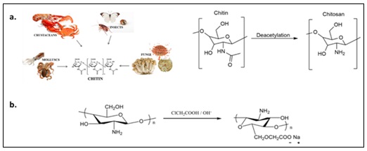
FIG. 1: (A) THIS FIGURE SHOWS THE PREPARATION OF CHITOSAN FROM CHITIN, (B) PREPARATION OF CARBOXYMETHYL CHITOSAN (CMC) FROM CHITOSAN
MATERIALS AND METHODS: All reagents and solvents were obtained from commercial suppliers and used as received, without further purification. Iron (III) chloride hexahydrate (FeCl3 6H2O) (98-102%), iron (II) sulfate heptahydrate (FeSO4 7H2O) (≥ 99%), Chitosan (Low Mw (LM) and High Mw (HM)), Doxorubicin Ready-Made (Dox, 2 mg/mL), monochloroacetic acid, ethanol (C2H5OH) (Assay 96%), 2-Propanol (C3H8O) (Assay 99.8%), and 28% ammonium hydroxide (NH4OH) were all purchased from Sigma-Aldrich. All reactions were carried out under an inert nitrogen atmosphere. For biological assays, phosphate buffered saline (PBS), Roswell Park Memorial Institute medium (RPMI 1640), fetal bovine serum (FBS), Hoechst 33342 stain, L-glutamine, and Penicillin/Streptomycin (PS) were all purchasedfrom Invitrogen. MTT (thiazolyl bluetetrazolium bromide) was purchased from Bioworld, USA. Cancer cell lines were purchased from the American Type Culture Collection (ATCC) and grown in RPMI 1640 medium supplemented with 10% FBS and 1% PS. Human cancer cells used in this study are THP-1 human monocytic cell lines derived from an acute monocytic leukemia patient.
Characterization: X-ray diffraction (XRD) was performed using Rigaku Uitima IV equipped with Cu-Kα radiation source (0.15418 nm) with angle ranging from 20⁰ to 80⁰ and the crystal structure parameters were obtained through Rietveld analysis. The morphology and high-resolution structural features of the samples were investigated using a Talos L120C G2 transmission electron microscope (TEM) (Thermo Fisher Scientific, USA)at an accelerating voltage of120 kV.Prior to imaging, the nanoparticle samples were dispersed in water, sonicated for 5 minutes, and drop-casted onto carbon-coated copper grids (TEM grid from EMS-Electron Microscopy Science, USA). High-resolution TEM (HR-TEM) was subsequently performed to examine the crystalline structure of the nanoparticle cores at the atomic scale. Clear lattice fringes were resolved, enabling measurement of interplanar (d-spacing) values, which were used to assess crystallinity and phase characteristics.Dynamic light scattering (DLS) measurements were assessed on Malvern Zetasizer Nano ZS instrument. Fourier transform infrared (FTIR) spectra (400–4000 cm-1) were recorded as KBr pellet forms using Shimadzu IRAffinity-1. Magnetic characterizations were performed using vibrating sample magnetometer (VSM) with 1.8 T magnets at ambient temperature. UV-vis absorption spectra were measured using ORION AQUAMATE 8000 UV-vis spectrophotometer in quartz cuvette cell over the wavelength range 200-800 nm.
Preparation of Carboxymethyl Chitosan (CMC): CMC was prepared following a previous procedure, with slight modification 31. First, either LM or HM Chitosan (0.5 g) was dissolved in 20% NaOH (2 g NaOH + 10 ml distilled water) in a 100 ml beaker and stirred for 15 minutes. Monochloroacetic acid (5.29mmol, 0.5 g) was then added in batches to the solution with continuous stirring for two hours in a water bath at 40 °C. After cooling the solution to room temperature, it was neutralized with 2% acetic acid to obtain a neutral medium. An excess of 70% ethanol (20 ml) was poured into the neutral solution and then filtered, followed by extensive washing with distilled water and methanol to get rid of the non-reactive substances. Finally, the desired product “CMC” was obtained as a solution to be utilized in the preparation of coated NPs.
Chitosan/CMC-coated Fe3O4 NPs: All polymer-coated MNPs were prepared using our previously reported Ko-precipitation Hydrolytic Basic “KHB” method. FeCl3.6H2O (0.3 g) was mixed with 2 mL of Chitosan solution (0.2 g Chitosan for every 10 ml water), followed by addition of FeSO4.7H2O (0.2 g) and stirred for few minutes at 80°C under nitrogen gas. NH4OH (~ 3 mL) was slowly added until pH = 12-14, where the solution turned from brown to black-brick colored depicting the formation of magnetite NPs and stirring was continued for an additional 3 hrs. The MNP suspensions were subsequently purified by centrifugation (4500 rpm, 5 min), washed multiple times with isopropanol, ethanol, and water, and finally dried at 80 °Cto produce ~ 150 mg (~ 90 % yield) of Chitosan-coated MNPs. For the preparation of CMC-coated NPs, similar procedure was followed using 3 ml of the as-prepared CMC precursor yielding ~ 140 mg of CMC-MNPs.
Drug Loading and Release: A total of 1 mg of Chitosan/CMC-coatedMNPs was loaded with Dox by incubating them with 1 mL of Dox solution (20 µg/mL), followed by continuous shaking for 24 hrs to ensure maximum drug loading. The drug-loading and release behavior of Dox-loaded Chitosan/CMC-coated NPs was then evaluated using UV-vis spectroscopy. First, the characteristic peaks of free Dox were recorded as a reference and calibration curve of Dox was generated. Drug loading was confirmed by the disappearance of the red color from the supernatant and reduction of Dox absorbance in the UV-vis spectrum at 480 nm. The drug loading efficiency (content) was calculated using the following equation:
Loading efficiency = WL/W0 × 100
Where WL is the amount of Dox loaded onto MNPs and W0 is theamount of Dox in the initial solution. The amount of Dox loaded onto the MNPs was determined by calculating the difference between the initial Dox concentration and the concentration remaining in the supernatant.
For the release study, the loaded MNPs were dispersed separately in two acetate buffer media: an acidic (pH ~ 4.5) buffer to simulate endosomal/lysosomal conditions, and neutral (pH ~ 7.4) to assess release under physiological conditions at room temperature.
Samples were shaken on a rotary shaker for selected periods of time and the Dox-loaded MNPs were isolated from the solution using hand-held magnet where aliquots were analyzed by UV-vis spectroscopy. Three replicates for each release experiment were performed and the results are plotted as mean ± S.D.
Cell Viability Assays: Non-adherent THP-1monocytic cancer cells were used as a representative cell type for leukemia. The cells were plated in flat-bottom 96-well plates ata density of ~ 104 cells/well in 100 µL of their respective growth medium. Serial dilutions of the various NP formulations (0.5 – 50μg/mL) were added to cell plates (200 μL per well) in triplicates in a cell culture medium (RPMI supplemented with 10% FBS and 1% PS). Wells at the edges were left empty to minimize edge effects. After 48 hrs of incubation at 37 °C with 5% CO₂, the medium was removed, and cells were washed with PBS. Cell viability was assessed using the MTT assay. Briefly, 20 μL of MTT solution (5 mg/mL) was added to each well and incubated for 4 hrs at 37 °C. The supernatant was then discarded, and the resulting formazan crystals were dissolved in DMSO. Absorbance was measured at 590 nm using an iMark microplate reader. The percentage of viable cells was calculated by comparing the absorbance of treated wells to that of untreated control wells, which were set at 100% viability. IC₅₀ values were determined from dose-response curves generated using polynomial dose-response fitting.
Live Confocal Microscopy Imaging: Non-adherent THP-1 monocytic leukemia cells were suspended in the respective media in 8-well dish and were exposed to Dox@Chitosan-MNPs (10 µg/mL NPs) and further incubated for different periods of times (i.e. 6 and 48 hrs). Hoechst 33342 stain was then added. The cells were allowed to settle down for 20 min before microscopic visualization. To mimic physiological conditions, nofixation of cells was conducted.
Preparation and Characterization of Chitosan/CMC-MNPs: Chitosan/CMC-coated MNPs with relatively uniform and homogeneous sizes were synthesized using our previously reported Ko-precipitation Hydrolytic Basic (KHB) approach, 32, 33 as depicted in Fig. 2. The approach relies on sequential basic hydrolytic in situ co-precipitation (Ko-precipitation) of inexpensive iron salts (Fe3+: Fe2+; 2:1) compartmentalized by the stabilizing polymers (i.e. Chitosan/CMC) in the presence of a base (i.e. NH4OH). This allowed the formation of stabilized aqueous colloidal dispersions of Chitosan/CMC-coated MNPs. Delightfully, the synthetic methodology for the MNPs demonstrated good reproducibility, as repeated syntheses consistently yielded comparable percentage yields (~ 90 – 95 %) and good aqueous dispersity.
It is to be noted that two different chitosan samples were used: (i) LM Chitosan with an average Mw range of 50–190 kDa and a degree of deacetylation (DDA) of ~ 75-85 %, and (ii) HM Chitosan presenting an average Mw of 310–375 kDa and a DDA of ≥ 75%. TEM, XRD, DLS, FTIR, and VSM analyses confirmed the structure, morphology, surface charge, and magnetic properties of the as-synthesized MNPs. TEM images revealed that Chitosan/CMC-coated MNPs exhibited a uniform, quasi-spherical morphology Fig. 3, with average particle sizes of approximately 8–10 nm.HR-TEM image Fig. 3F revealed a well-defined lattice fringe with an interplanar spacing (d-spacing) of ~ 0.445 nm, corresponding to the most prominent (111) crystallographic plane. To further elucidate the crystalline structure and phase composition of the as-synthesized MNPs, X-ray diffraction (XRD) analysis was performed.
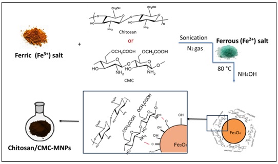
FIG. 2: SCHEMATIC ILLUSTRATION FOR THE PREPARATION OF CHITOSAN OR CMC-COATED MNPS USING KO-PRECIPITATION HYDROLYTIC BASIC (KHB) METHOD
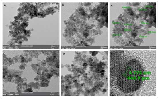
FIG. 3: TEM IMAGES AT DIFFERENT MAGNIFICATIONS OF (A-C) CHITOSAN-COATED MNPS AND (D-E) CMC-COATED MNPS SHOWING UNIFORM AVERAGE SIZES ~ 10 NM. (F) HR-TEM IMAGE WITH AN AVERGAE INTERPLANAR DISTANCE (D-SPACING) ~ 0.4449 NM, DEPCITING THE EXCELLENT CRYSTALLINTY OBTAINED FOR THE AS-PREPARED MAGNETITE NPS
The XRD patterns indicated that all samples possessed well-crystallized, single-phase inverse spinel structures Fig. 4. Each spectrum exhibited well-defined peaks, indicative of the crystalline nature of the samples. Main peaks were identified at (31), (37), (44), (54), (57), (63), and (75) 2Ɵcorresponding to (220), (311), (400), (422), (511), (440), and (533) which are related to the spinel structure of pure Fe3O4 cubic spinel phases (JCPDS # 01-088-0315). No additional peaks were observed, suggesting that no other iron oxide phases are formed. Inverse spinel magnetite has a face-centered cubic (FCC) crystalline lattice where oxygen and metal ions are positioned on the tetrahedral (A) and octahedral sites (B), where the tetrahedral A sites are occupied exclusively by ferric ions (Fe3+), while the octahedral B sites contain an equal mixture of ferric (Fe3+) and ferrous (Fe2+) ions in a 1:1 ratio 34. Rietveld refinement of the XRD patterns confirmed the formation of pure Fe₃O₄ cubic inverse spinel phases with space group Fd3m, in good agreement with JCPDS #01-088-0315. In the Rietveld refinement, the quality of agreement between the experimentally observed X-ray diffraction patterns and the calculated patterns is evaluated using several statistical parameters, including the profile R-factor (Rp), weighted profile R-factor (Rwp), expected R-factor (Re), goodness-of-fit (S), and chi-square (χ2). The goodness-of-fit (S) is calculated as the ratio between Rwp and Re (S= Rwp / Re) and indicates how closely the refinement approaches the statistically expected agreement. A value of S close to 1 indicates that the calculated model adequately describes the experimental data. χ2 is the square of the goodness-of-fit (χ2=S2) and is commonly used as an overall indicator of refinement quality; values approaching unity indicate a statistically acceptable refinement. Herein, the Rietveld refinements exhibited S values close to unity (1.08–1.11) and χ2 values near 1 (1.17–1.24), indicating excellent agreement between the observed and calculated diffraction patterns. Importantly, Rietveld analysis perfectly corroborated XRD and TEM results with lattice parameters (a = b = c = 8.36/7) for all samples and crystallite sizes between 7 and 10 nm Table 1.
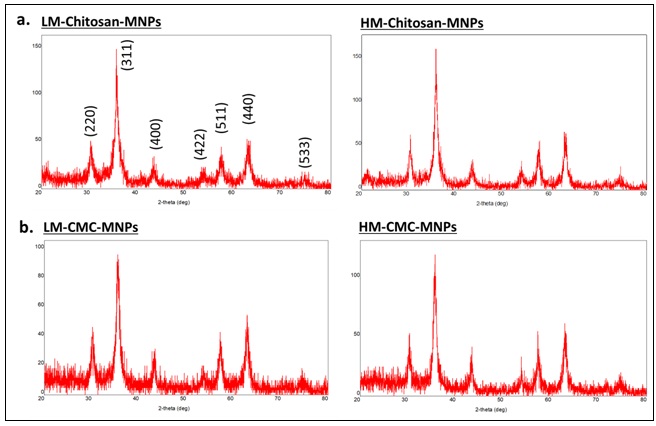
FIG. 4: XRD DIFFRACTION PATTERNS OF THE VARIOUS MW (LM AND LM) CHITOSAN/CMC-COATED MNPS, A) CHITOSAN-MNPS AND B) CMC-MNPS, CLEARLY SHOWING THE FORMATION OF PURE CRYSTALLINE PHASES OF FE3O4
Next, DLS measurements of aqueous dispersions of Chitosan/CMC-MNPs were recorded. While TEM measures the core sizes of the MNPs, DLS records the hydrodynamic radii in water. Chitosan-MNPs revealed hydrodynamic sizes (DH) between 275 and 434 nm with uniform and narrow size distributions as evident from the sharp peaks and low polydispersity index (PDI) values for all samples Fig. 5A. It is to be noted that the hydrodynamic sizes of CMC-MNPs (~275 - 301 nm, PDI = 0.423 – 0.497) were found to be slightly smaller than Chitosan-MNPs (365 - 434 nm, PDI = 0.633 - 1). HW Chitosan-MNPs (PDI = 1) shows higher PDI than LW Chitosan-MNPs (PDI = 0.633), while HW CMC-MNPs (PDI = 0.497) and LW CMC-MNPs (PDI = 0.423) illustrated slightly better PDI. This is likely attributable to the successful carboxymethyl substitution of chitosan, which resulted in MNPs with a narrow size distribution.
All the functionalized MNPs exhibited good stability in aqueous dispersion, even after prolonged storage. No significant changes in particle size were observed over time, further confirming their excellent colloidal stability.Zeta potential measurements indicated positive charge surfaces for all samples (ξ ≥ + 5 mV), further confirming coating with the positively charged Chitosan/CMC Fig. 5B. Notably, the average surface charges of Chitosan-MNPs were found to be slightly higher (ξ = +12 and 18 mV) than CMC-MNPs (ξ = + 6 and 8 mV), due to carboxymethylation of Chitosan. Moreover, zeta potential of bare Fe3O4 MNPs was found to be equal to ξ = - 12.5 mV, further confirming Chitosan/CMC coating the magnetite surfaces. FTIR analysis was then conducted to validate the successful coating of Chitosan/CMC onto MNPs Fig. 6. The presence of iron oxide (Fe3O4) core is clearly evident by the Fe−O stretching bands appearing at ~ 560 - 620 cm−1. The broad peaks at ~ 3400 cm−1 are ascribed to the O-H and N-H stretching vibrations of hydroxyl and amino groups on MNPs. Symmetric and asymmetric C-H stretching vibrations clearly appear at ~2850 cm⁻¹ and 2920 cm⁻¹. The presence of a strong peak at ~ ~1150 cm-1 is a characteristic signature of the C-O-C stretching vibrations. Another major peak is the stretching vibration of C=O carbonyl evident at ~ 1635 cm−1, which coincides with the N-H and O-H bending vibrations occurring ~1600-1650 cm-1.[35, 36]Peaks at 1601 cm−1 (due to symmetric axial deformation of –COOH) and 1392 cm−1 (due to asymmetric axial deformation of –COOH) confirm the existence of carboxymethyl groups on chitosan 37. All these functional vibrations provide clear evidence of the successful functionalization of Fe3O4 MNPs with Chitosan or CMC, suggesting strong interactions and stability of the chitosan/CMC-MNPs probablyvia electrostatic/ hydrogen bonding interactions between amino, hydroxyl, and carboxyl groups of chitosan or CMC and the hydroxyl groups on MNP surfaces.
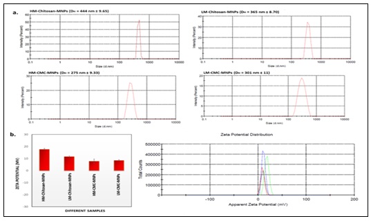
FIG. 5: DLS MEASUREMENTS FOR THE DIFFERENT MWS OF CHITOSAN/CMC-COATED MNPS DISPERSED IN WATER. A) REPRESENTATIVE SIZE DISTRIBUTION PLOTS; B) AVERAGE ZETA POTENTIAL (Ξ) SURFACE CHARGES REPRESENTED AS BAR GRAPHS ALONG WITH A REPRESENTATIVE ZETA POTENTIAL DISTRIBUTION FOR THE DIFFERENT CHITOSAN/CMC-COATED MNPS. ERROR BARS REPRESENT THREE INDEPENDENT MEASUREMENTS REPORTED AS AVERAGE MEANS ± S.D.
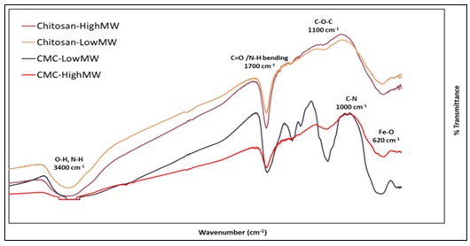
FIG. 6: FTIR FOR THE DIFFERENT AS-PREPARED CHITOSAN/CMC-COATED MNPS. THE FTIR SPECTRA CLEARLY SHOW THE SUCCESSFUL CHITOSAN AND CMC COATING THE IRON OXIDE CORE
Magnetic Properties: Magnetically-responsive MNPs are composed of elements possessing magnetic properties such as iron, nickel, cobalt, or metal oxides.Consequently, there is a strong demand for the preparation of stabilized polymer-coated MNPs with high saturation magnetization (Ms) using simple, scalable, cost-effective, and reproducible methods. The preparation of colloidal and well-dispersed MNPs with controlled particle sizes and biocompatible polymer coatings have recently revealed an intriguing capability to mediate drug delivery applications.Therefore, designing MNPs that have optimal magnetization for magnetic-guided drug delivery applications is highly demanded. The significance stems from preparation of high-performance water-dispersible magneto-responsive NPs and tuning their physicochemical parameters.
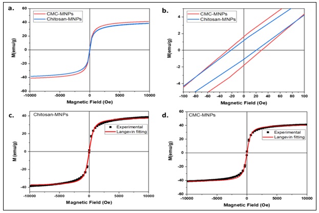
FIG. 7: FIELD-DEPENDENT MAGNETIZATION HYSTERESIS LOOPS AT ROOM TEMPERATURE FOR (A,B) CHITOSAN-MNPS AND CMC-MNPS AND (C,D) LAW OF APPROACH TO SATURATION (LAS) FITTINGS
To determine the magnetic behavior for the as-prepared Chitosan/CMC-coated MNP samples, field-dependent magnetizations were conducted. As depicted in Fig. 7, the hysteresis loops (M-H) as a function of applied magnetic field at room temperature are recorded. From the magnetization curves, the saturation magnetization (Ms), coercivity (Hc), and remanence (Mr) values were extracted and are summarized in Table 2. It can be observed that the MNPs show super paramagnetic characteristics with very small Hc(< 30 Oe) and almost negligible Mr. The Ms magnetization obtained for the Chitosan-MNPs and CMC-MNPs were found to be equal to 38.77 and 41.25 emu/g, respectively. The magnetic measurements revealed relatively high saturation magnetizations, reflecting a high degree of magnetic ordering and crystallinity in the samples. To further assess their distinctive magnetic behavior, the effective anisotropy constant (Keff) was calculated, as it is a key parameter directly influencing magnetization. The law of approach to saturation (LAS) was employed to determine Keff, using the following equation applied to the experimental magnetization data.
M(H) = Ms (1-b/H2)
Where b is a parameter which is deduced from the fitting of experimental magnetization with the above equation. Keff is then determined by:
Keff = μ0 Ms √(15b/4)
Fitting the equation to the experimental magnetization data showed that all M–H curves were well described by the LAS model, yielding very high Kₑff values~ 1.2 - 1.3 ×10⁵ erg/cm3 Fig. 6B.
TABLE 1: RIETVELD XRD ANALYSIS OF THE DIFFERENT AS-PREPARED CHITOSAN/CMC-MNPS
| Sample | Phase Composition (%) | Crystallite size (nm) | Microstrain
(%) |
Lattice parameters (Å) | Fit parameters |
| LM-CMC-MNPs | Fe3O4 100%(7) | 8.51 (7)
|
0.36(3)
|
a= b=c= 8.357(6)
α=β=γ=90 |
Rwp=44.27 Rp=33.55 Re=40.23
S=1.0997 χ2=1.2094 |
| HM-CMC-MNPs | Fe3O4 100%(5) | 8.82(6)
|
0.11(3)
|
a= b=c= 8.362(4)
α=β=γ=90 |
Rwp=42.69 Rp=32.23 Re=39.41
S=1.0827 χ2=1.1722 |
| LM-Chitosan-MNPs | Fe3O4 100%(6) | 6.72(2)
|
0.425(18)
|
a= b=c= 8.361(7)
α=β=γ=90 |
Rwp=41.81 Rp=31.11 Re=37.54
S=1.1130 χ2=1.2388 |
| HM-Chitosan-MNPs | Fe3O4 100%(4) | 8.99 (13)
|
0.303(6)
|
a= b=c= 8.369(17)
α=β=γ=90 |
Rwp=41.32 Rp=30.83 Re=37.91
S=1.0892 χ2=1.1864 |
Rwp: Weighted profile R-factor; Re: Expected R-factor; Rp: Profile R-factor; S: goodness of fit (S=Rwp/Re); χ2=S2
TABLE 2: MAGNETIC PARAMETERS OBTAINED FROM M–H CURVES AND LAW OF APPROACH TO SATURATION (LAS) FOR CHITOSAN/CMC-MNPS
| Samples | Hc (Oe) | Mr (emu/g) | Mr/Ms | Keff | Saturation Ms (emu/g) | |
| Experimental | Lange in fitting | |||||
| Chitosan -MNPs | 29.7 | 1.64 | 0.04 | 1.20 x 10⁵ | 38.77 | 38.37 |
| CMC-MNPs | 20.9 | 1.14 | 0.02 | 1.29 x 10⁵ | 41.25 | 41.68 |
Drug Loading and Release: Significant pH variations exist at the systemic, tissue, and sub-cellular levels 38. This pH gradient has been utilized as an endogenous trigger for designing nanocarriers capable of selectively releasing their payloads in acidic tumor environments 29, 39, 41. For example, the extracellular milieu of tumor tissues is more acidic (pH 6.2–6.9) than that of normal tissues and systemic blood pH. Additionally, the acidic conditions within intracellular compartments such as endosomes and lysosomes (pH 4.0–6.5) have been exploited to design pH-responsive nanocarriers that release their cargo intracellularly 38, 40. pH-responsive nanocarriers are typically constructed from polymers that undergo transitions between hydrophilic (swollen) and hydrophobic (collapsed) states in response to protonation and deprotonation of labile functional groups, such as amino and carboxylic acid groups, under varying pH conditions. This behavior provides a mechanism for pH-triggered payload release. Polymers containing amine groups have been extensively investigated for such applications, as protonation enhances polymer hydrophilicity 42, 43. Furthermore, amine groups can effectively neutralize the acidic endosomal environment viaprotonation, raising the endosomal pH. This process facilitates ionic influx into the endosome, causing it to swell and rupture, ultimately releasing the internalized NPs - a phenomenon known as the proton-sponge effect 44. For instance, smart pH and thermoresponsive N,O-Carboxymethyl Chitosan-conjugated nanogels, possessing enhanced swelling behavior at lower pH, exhibited improved therapeutic efficacy of Dox in MCF-7 and MDA-MB-231 breast cancer cells 45.
To explore the possibility of Chitosan/CMC-coated MNPs as pH-responsive drug nanocarriers Fig. 8A, we first loaded them with the anticancer drug Doxorubicin (Dox). 1 mL of Dox solution (20 µg/mL) was incubated with the respective MNPs, followed by continuous shaking for 24 hrs to ensure maximum entrapment. Drug loading content of Dox@Chitosan/CMC-coated NPs was evaluated by UV–vis spectroscopy which was confirmed by the complete disappearance of the red color in the supernatant along with a decrease in Dox absorbance. Almost quantitative amount of drug (i.e. 100% loading efficiencies) was successfully loaded onto all samples of Chitosan/CMC-coated MNPs (i.e. 20 µg Dox/mg MNPs), as evident from absorption spectroscopy Fig. 8B. These results were reproducible when using different batches of Chitosan/CMC-coated MNPs (1 mg/mL). Dox can be loaded into Chitosan/CMC-coated MNPs primarily through electrostatic interactions, hydrogen bonding, and Van der Waals interactions taking advantage of Chitosan's structure and Dox's properties. This physio-adsorption of Dox onto the coated MNPs offers the advantage of preserving both the structure of the NPs and the conjugated Dox. MNPs in which drug molecules are covalently conjugated to their surfaces typically show low drug entrapment efficiencies and limited drug release due to the covalent bonding. In addition, the proper orientation of the drug’s active functional groups is critical for biological activity.Thus, we anticipated that simple physio-adsorption interactions would be a more suitable approachto preserve the activity of the drug on the MNPs.
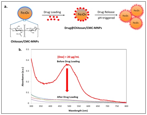
FIG. 8: (A) SCHEMATIC REPRESENTATION FOR THE DRUG LOADING AND PH-DEPENDENT DRUG RELEASE; B) UV-VIS SPECTRA BEFORE AND AFTER LOADING OF DOX ONTO THE DIFFERENT MNPS
With Dox@Chitosan/CMC-MNPs in hand, we subsequently examined their in-vitro drug release profiles Fig. 9. One notable feature of chitosan is its pH-dependent solubility, which can be leveraged for targeted drug administration 46. Importantly, very few reports 47, if any explored the effects of different Mws of chitosan on drug release behavior. To conduct the release studies, all loaded MNPs were dispersed separately in two media: an acidic buffer (pH ~ 4.5) to simulate endosomal/lysosomal conditions, and neutral (pH ~ 7.4) conditions to assess release under physiological conditions. At selected time points, samples were analyzed by UV-vis spectroscopy to monitor the re-appearance of the characteristic Dox peaks, indicating the extent of drug release in each medium. We compared between LM and HM Chitosan- and CMC-coated MNPs in drug releasing experiments Fig. 9A. The release of drug was measured with time until 24 hrs under pH 4.5 and pH 7.4 for all the samples. For all formulations, at acidic medium (pH = 4.5), higher and faster drug release was observed compared to neutral pH = 7.4. Among the samples, HM-Chitosan MNPs exhibited the greatest release at acidic pH, giving almost quantitative release within 24 hrs, while the HM-CMC showed slightly lower release (~ 90 %) Fig. 9B. In contrast, LM-Chitosan and CMC MNPs showed lower drug release behaviour (~ 60 - 65%) at acidic pH compared to HM-Chitosan/CMC-MNPs Fig. 9C.
Remarkably, drug release profiles at neutral pH were markedly low (≤ 20 %) for all formulations, indicating the stability of Dox-loaded MNPs at neutral physiological pH. This enhanced release at acidic pH is attributed to decreased interactions between Dox and Chitosan bonds, which lead to faster diffusion of the anticancer drug. The initial burst phase of Dox release is ascribed to Dox located at the surface of the particles, while the remainder of the unreleased Dox is sustainably released as it is assumed to be entrapped within the Chitosan/CMC-MNPs. Chitosan and CMC are sensitive to pH, where the bonding between Dox and Chitosan/CMC will be protonated in acidic medium, leading to releasing the drug in a controlled sustained fashion.In endosomes and lysosomes (pH ~ 4 – 6.5), protonation of amine groups increases polymer hydrophilicity causing NP swelling, disassembly, and consequent drug release. In general, CMC was found to be slightly more stable than chitosan, that means CMC show more controlled and a bit slower release profiles, where the carboxymethyl group is anticipated to interact with the drug stronger. This controlled yet sustained release of Dox is advantageous for achieving cytotoxic intracellular concentrations, in both in-vitro and in-vivo cancer therapy, consistent with previous observations 48. Together, these findings indicate that the physically bound Dox molecules in our system can be released rapidly, efficiently, and in a sustained manner, highlighting our Chitosan/CMC-MNPs as promising nanocarriers for imaging, drug delivery, and therapeutics.
One of the key benefits of using nano-based drug delivery systems is their ability to provide more targeted and less toxic solutions, effectively addressing the challenges of poor specificity and the dose-limiting toxicities often associated with conventional anticancer drugs. Thus, to better understand and determine the intracellular uptake of MNPs in cancer cells, live confocal laser scanning microscopy was employed. For these experiments, we chose the optimal HMCMC-MNP formulation, as it has excellent water solubility and colloidal dispersions, smallest sizes, positive surface charges, and suitable in-vitro drug release profiles.
A representative human monocytic leukemic cancer cell line (THP-1) was treated with Dox@CMC-MNPs and imaged at various time points Fig. 10. Notably, confocal images demonstrated that Dox is effectively delivered to the cytoplasm within relatively short time frames of 6 hrs Fig. 10A. Apoptotic features observed after 48 hrs of incubation with Dox-loaded MNPs clearly reveal morphological changes associated with effective nanoparticle-mediated drug delivery. As the NPs penetrate the cells and release Dox, the cells exhibit characteristic signs of apoptosis, including cell shrinkage, membrane blebbing, and the formation of irregular cellular fragments Fig. 10B. We then proceeded to evaluate, quantitively, the cytotoxic potentials of CMC-MNPs and Dox@CMC-MNPs against this cell line using thiazolyl blue tetrazolium bromide (MTT) cell viability assay. While unloaded CMC-MNPs exhibited no cytotoxicity toward the cells (IC50 > 200 µg/mL), even at the highest NP concentrations tested, Dox@CMC-MNPs showed significant toxicity against THP-1 leukemic cells, killing the cancer cells with an IC50 = 16 ± 2.2 µg/mL Fig. 10C. Although free Dox, at equivalent concentrations, exhibited more pronounced cytotoxicity against THP-1 cells than Dox@CMC-MNPs, the noted cytotoxicity of our formulation could prove superior for in-vivo situations. Dox is known to have initial rapid distribution to internal organs within 5 minutes. This rapid distribution can account for the systemic toxicity of the drug and to lesser exposure of leukemic cells to the drug in circulation. The current formulation is expected to have longer half-life in circulation, innate to its hydrodynamic size. This, in turn, can ensure longer exposure and selectivity to cancer cells that will internalize the Dox-loaded CMC-NPs through phagocytosis/pinocytosis that is common to most hematological malignancy cells. We anticipate that the observed pronounced potency of Dox@CMC-MNPs primarily results from the selective and differential uptake of the NPs by cells, followed by the intracellular release of Dox over time in response to biochemical changes, particularly the pH-triggered conditions within endosomes and lysosomes. The drug payload subsequently undergoes sustained translocation to the nucleus, where it exerts its cytotoxic effect. Markedly, the Dox@CMC-MNP nanocarriers described here offer inherent advantages, as it minimizes the undesirable diffusion-associated side effects of the free drug and enables more selective and targeted drug delivery.
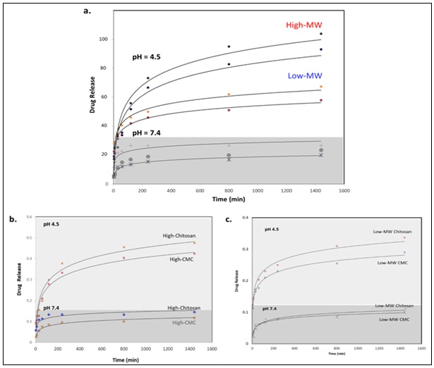
FIG. 9: THIS FIGURE SHOWS A) DOX RELEASE PROFILES FOR HM AND LM CHITOSAN/CMC-COATED MNPS UNDER ACIDIC AND NEUTRAL MEDIA. B) DOX RELEASE FROM HM CHITOSAN AND CMC MNPS UNDER ACIDIC AND NEUTRAL PH. C) DOX RELEASE FROM LM CHITOSAN AND CMC MNPS UNDER ACIDIC AND NEUTRAL pH. THE RESULTS SHOW HIGHER SUSTAINED DRUG RELEASE OF HM-CHITOSAN OR CMC COMPARED TO LM-CHITOSAN OR CMC AT ACID PH CONDITIONS, WHILE NONE OF THE FORMULATIONS EXHIBIT SIGNIFICANT RELEASE AT NEUTRAL pH
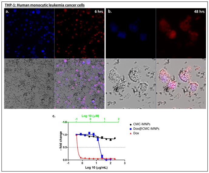
FIG. 10: CONFOCAL MICROSCOPY IMAGES OF THP-1 CELLS AFTER INCUBATION WITH DOX@CMC-MNPS (10 ΜG/ML MNPS; 0.37 ΜM DOX) FOR A) 6 HRS AND B) 48 HRS. (A,B) BLUE HOECHST CHANNEL SHOWING POSITIONS OF THE NUCLEI, RED DOX CHANNEL, AND OVERALL OVERLAY WITH TRANSMITTED LIGHT (TL). THE IMAGES CLEARLY SHOW SUCCESSFUL DELIVERY OF DOX TO THE CYTOPLASM, CAUSING APOPTOSIS WITH TYPICAL APOPTOTIC FEATURES INCLUDING CONDENSATION, SHRINKAGE, AND MEMBRANE BLEBBING. C) MTT CELL PROLIFERATION CYTOTOXICITY ASSAY FOR UNLOADED CMC-MNPS, DOX@CMC-MNPS, AND FREE DOX ON THP-1 LEUKAEMIA CELLS. X-AXIS: NP CONCENTRATIONS EXPRESSED IN µG/ML; Y-AXIS: ABSORBANCE AT 450 NM. EXPERIMENTS WERE CARRIED OUT IN TRIPLICATES AND AVERAGE RESULTS ARE PLOTTED
CONCLUSION: In conclusion, different Chitosan/CMC-coated MNPs with varying MWs were successfully prepared and utilized as nanocarriers for drug delivery to cancer cells. Carboxymethylation of chitosan proved to be effective in enhancing aqueous solubility while maintaining sustained and controlled drug-release profiles. By using HM or LM Chitosan or CMC-MNPs, pH-triggered Dox release was evaluated at both acidic and neutral pH conditions. In our system, a relatively fast initial burst release over the first 2 hrs was observed, followed by a more controlled slower drug release, with almost quantitative discharge at acidic pH = 4.5, but almost no significant release at the physiological neutral pH. HM Chitosan/CMC-MNPs showed slightly higher drug release profiles compared to LM counterparts.
This behaviour is consistent with the increased protonation of functional groups at low pH. Confocal microscopy and viability cytotoxicity assays revealed pronounced killing of THP-1 cells using Dox@CMC-MNPs, causing apoptotic cell death, but not when unloaded CMC-MNPs were utilized. This sustained release of Dox is distinctive and likely advantageous for achieving cytotoxic intracellular concentrations, benefiting both in-vitro and in-vivo cancer therapy. Therefore, drug-loaded chitosan-based nanocarriers may offer broad applicability and considerable potential for the development of controlled-release formulations and leukaemia management, warranting further in-vivo testing.
ACKNOWLEDGMENTS: This work was funded and supportedby the Deanship of Postgraduate and Scientific Research at University of Bahrain (UoB) (Project No. 10). The authors would like to thank the continuous support of the Chemistry Department, College of Science at UoB. Special and utmost thanks to Mr. Manohar Reddy from Electron Microscopy Department, EXPRESSMED Diagnostics and Research laboratories for conducting TEM and HR-TEM images.
Author Contributions: K.H.B. conceived, designed, and conceptualized the study. J.K.A. prepared all the Chitosan- and CMC-coated MNPs and performed the drug loading and release experiments. O.M.L. conducted magnetization and calculated magnetic parameters. A.M. carried out XRD and Rietveld analyses. K.G. helped with performing the MTT and confocal microscopy data as well as DLS measurements. K.H.B. and J.K.A. analyzed the experimental data and wrote the manuscript. All authors revised and approved the manuscript.
Data Availability: The authors confirm that the data supporting the findings of this study are available within the article. Further inquiries can be directed to the corresponding author, upon request.
Ethics Declaration: Not applicable
CONFLICTS OF INTERESTS: The authors declare no competing financial and/or non-financial interests in relation to the work described.
REFERENCES:
- El-Boubbou K: Magnetic iron oxide nanoparticles as drug carriers: clinical relevance. Nanomedicine 2018; 13(8): 953-971.
- Wu W, Jiang CZ and Roy VAL: Designed synthesis and surface engineering strategies of magnetic iron oxide nanoparticles for biomedical applications. Nanoscale 2016; 8(47): 19421-19474.
- Wahajuddin M and Arora S: Superparamagnetic iron oxide nanoparticles: magnetic nanoplatforms as drug carriers. Int J Nanomedicine 2012; 7: 3445-3471.
- El-Boubbou K: Magnetic iron oxide nanoparticles as drug carriers: preparation, conjugation and delivery. Nanomedicine 2018; 13(8): 929-952.
- Li K, Nejadnik H and Daldrup-Link HE: Next-generation superparamagnetic iron oxide nanoparticles for cancer theranostics, Drug Discovery Today 2017; 22(9): 1421-1429.
- El-Dakdouki MH, El-Boubbou K, Zhu DC and Huang X: A simple method for the synthesis of hyaluronic acid coated magnetic nanoparticles for highly efficient cell labelling and in-vivo imaging, RSC Adv 2011; 1: 1449-1452.
- El-Dakdouki MH, Zhu DC, El-Boubbou K, Kamat M, Chen J, Li W and Huang X: Development of multifunctional hyaluronan-coated nanoparticles for imaging and drug delivery to cancer cells. Biomacromolecules 2012; 13: 1144-51.
- Grewal AK and Salar RK: Chitosan nanoparticle delivery systems: An effective approach to enhancing efficacy and safety of anticancer drugs. Nano Trans Med 2024; 3: 100040.
- Troncoso OP and Torres FG: Non-conventional starch nanoparticles for drug delivery applications. Medical Devices & Sensors 2020; 3(6): e10111.
- Rinaudo M: Chitin and chitosan: Properties and applications. Prog Polym Sci 2006; 31(7): 603-632.
- Kumar RMNV: A review of chitin and chitosan applications. React Funct Polym 2000; 46(1): 1-27.
- Aranaz I, Alcántara AR, Civera MC, Arias C, Elorza B, Heras Caballero A and Acosta N: Chitosan: An overview of its properties and applications. Polymers 2021; 13(19).
- Rodrigues S, Dionísio M, López CR and Grenha A: Biocompatibility of chitosan carriers with application in drug delivery. J Funct Biomater 2012; 615-641.
- TM MW, Lau WM and Khutoryanskiy VV: Chitosan and its derivatives for application in mucoadhesive drug delivery systems. Polymers 2018; 10(3).
- Ali A and Ahmed S: A review on chitosan and its nanocomposites in drug delivery. Int J Biol Macromol 2018; 109: 273-286.
- Stefanache A, Lungu II, Anton N, Damir D, Gutu C, Olaru I, Plesea Condratovici A, Duceac M, Constantin M, Calin G, Duceac LD and Boev M: Chitosan nanoparticle-based drug delivery systems: advances, challenges, and future perspectives. Polymers 2025; 1453.
- Elgadir MA, Uddin MS, Ferdosh S, Adam A, Chowdhury AJK and Sarker MZI: Impact of chitosan composites and chitosan nanoparticle composites on various drug delivery systems: A review. J Food Drug Anal 2015; 23(4): 619-629.
- Shariatinia Z: Carboxymethyl chitosan: Properties and biomedical applications. Int J Biol Macromol 2018; 120: 1406-1419.
- Mourya VK, Inamdara N and Ashutosh Tiwari N: Carboxymethyl chitosan and its applications. Adv Mater Lett 2010; 1(1): 11-33.
- Geng Y, Xue H, Zhang Z, Panayi AC, Knoedler S, Zhou W, Mi B and Liu G: Recent advances in carboxymethyl chitosan-based materials for biomedical applications. Carbohydrate Polymers 2023; 305: 120555.
- Sonia TA and Sharma CP: Chitosan and its derivatives for drug delivery perspective, in: R. Jayakumar, M. Prabaharan, R.A.A. Muzzarelli (Eds.), Chitosan for Biomaterials I. Advances in Polymer Science, Springer Berlin Heidelberg, Berlin, Heidelberg 2011; 23-53.
- Sangnim T, Dheer D, Jangra N, Huanbutta K, Puri V and Sharma A: Chitosan in oral drug delivery formulations: a review. Pharmaceutics 2023; 15(9).
- Park JH, Saravanakumar G, Kim K and Kwon IC: Targeted delivery of low molecular drugs using chitosan and its derivatives. Adv Drug Deliv Rev 2010; 62(1): 28-41.
- Yadav M, Kaushik B, Rao GK, Srivastava CM and Vaya D: Advances and challenges in the use of chitosan and its derivatives in biomedical fields: A review. Carbohydr Polym Technol Appl 2023; 5: 100323.
- Tacar O, Sriamornsak P and Dass CR: Doxorubicin: an update on anticancer molecular action, toxicity and novel drug delivery systems. J Pharm Pharmacol 2013; 65(2): 157-170.
- Belger C, Abrahams C, Imamdin A and Lecour S: Doxorubicin-induced cardiotoxicity and risk factors. Int J Cardiol Heart Vasc 2024; 50: 101332.
- Subramani K, Hosseinkhani H, Khraisat A, Hosseinkhani M and Pathak Y: Targeting nanoparticles as drug delivery systems for cancer treatment. Current Nanoscience 2009; 5(2): 135-140.
- Bai X, Smith ZL, Wang Y, Butterworth S and Tirella A: Sustained drug release from smart nanoparticles in cancer therapy: a comprehensive review. Micromachines 2022; 13(10).
- Tsirogianni AG, Chountoulesi M and Pippa N: Recent advances in ph-responsive liposomes: lessons learnt and new directions in nanomedicine development. Materials 2025; 4295.
- Unsoy G, Khodadust R, Yalcin S, Mutlu P and Gunduz U: Synthesis of Doxorubicin loaded magnetic chitosan nanoparticles for pH responsive targeted drug delivery. Eur J Pharm Sci 2014; 62: 243-250.
- Saber LF and Al-Kadhimi AAHA: Preparation of Schiff bases of hydrazide prepared from carboxymethyl-Chitosan. AIP Conf Proc 2023; 2839(1).
- El-Boubbou K, Lemine OM, Ali R, Huwaizi SM, Al-Humaid S and AlKushi A: Evaluating magnetic and thermal effects of various Polymerylated magnetic iron oxide nanoparticles for combined chemo-hyperthermia. New Journal of Chemistry 2022; 46(12): 5489-5504.
- El-Boubbou K: Acid-stabilized iron-based metal oxide colloidal nanoparticles, and methods thereof, US Patent, 2020; US10629339B2.
- Nguyen MD, Tran HV, Xu S and Lee TR: Fe3O4 nanoparticles: structures, synthesis, magnetic properties, surface functionalization, and emerging applications. Appl Sci 2021; 11301.
- Petcharoen K and Sirivat A: Synthesis and characterization of magnetite nanoparticles via the chemical co-precipitation method, Materials Science and Engineering: B 2012; 177(5): 421-427.
- Lori N, Eydokia P, Vassilis Z, Anna P, Eleana H and Christos NP: Magnetic nanoparticles in medical diagnostic applications: synthesis, characterization and proteins conjugation. Current Nanoscience 2016; 12(4): 455-468.
- de Abreu FR and Campana-Filho SP: Characteristics and properties of carboxymethylchitosan. Carbohydrate Polymers 2009; 75(2): 214-221.
- Lim EK, Huh YM, Yang J, Lee K, Suh JS and Haam S: pH-triggered drug-releasing magnetic nanoparticles for cancer therapy guided by molecular imaging by MRI. Adv Mater 2011; 23(21): 2436-2442.
- Zhao Z, Huang D, Yin Z, Chi X, Wang X and Gao J: Magnetite nanoparticles as smart carriers to manipulate the cytotoxicity of anticancer drugs: magnetic control and pH-responsive release. J Mater Chem 2012; 22(31): 15717-15725.
- Banerjee SS and Chen DH: Multifunctional pH-sensitive magnetic nanoparticles for simultaneous imaging, sensing and targeted intracellular anticancer drug delivery. Nanotechnology 2008; 19(50): 505104.
- Zhu L, Wang D, Wei X, Zhu, X Li J, Tu C, Su Y, Wu J, Zhu B and Yan D: Multifunctional pH-sensitive superparamagnetic iron-oxide nanocomposites for targeted drug delivery and MR imaging. Journal of controlled release. Official Journal of the Controlled Release Society 2013; 169(3): 228-38.
- Nunziata G, Pollonio D, Lacroce E and Rossi F: Smart pH-Responsive polymers in biomedical Applications: Nanoparticles, hydrogels, and emerging hybrid platforms, Mater. Today Chem 2025; 49: 103063.
- Obireddy SR and Lai WF: ROS-Generating amine-functionalized magnetic nanoparticles coupled with carboxymethyl chitosan for pH-responsive release of doxorubicin. Int J Nanomedicine 2022; 17: 589-601.
- Lu ZR and Sun D: Mechanism of pH-sensitive amphiphilic endosomal escape of ionizable lipid nanoparticles for cytosolic nucleic acid delivery. Pharm Res 2025; 42(7): 1065-1077.
- Verma NK, Purohit MP, Equbal D, Dhiman N, Singh A, Kar AK, Shankar J, Tehlan S and Patnaik S: Targeted smart pH and thermoresponsive n,o-carboxymethyl chitosan conjugated nanogels for enhanced therapeutic efficacy of doxorubicin in MCF-7 breast cancer cells. Bioconjug Chem 2016; 27(11): 2605-2619.
- Cavallaro G, Micciulla S, Chiappisi L and Lazzara G: Chitosan-based smart hybrid materials: a physico-chemical perspective. J Mater Chem B 2021; 9(3): 594-611.
- Yang HC and Hon MH: The effect of the molecular weight of chitosan nanoparticles and its application on drug delivery. Microchemical Journal 2009; 92(1): 87-91.
- Radwan AM, El-Atrash A, Elkholy M, Nawar NF, Tousson E and Ghoneim AI: In-vivo antitumor activity of doxorubicin loaded on chitosan functionalized Pb2Mn2Fe12O22 magnetic nanoparticles. Scientific Reports 2025; 15(1): 34313.
How to cite this article:
Khalifah JKA, Lemine OM, Mustafa A, Greish K and El-Boubbou K: Chitosan and carboxymethyl chitosan (CMC)-coated magnetite nanoparticles for drug delivery to cancer cells: effect of molecular weight on drug release. Int J Pharm Sci & Res 2026; 17(11): 3473-88. doi: 10.13040/IJPSR.0975-8232.17(11).3473-88.
All © 2026 are reserved by International Journal of Pharmaceutical Sciences and Research. This Journal licensed under a Creative Commons Attribution-NonCommercial-ShareAlike 3.0 Unported License.
Article Information
15
3473-3488
2492 KB
8
English
IJPSR
Johar K. Al Khalifah, O. M. Lemine, Alia Mustafa, Khaled Greish and Kheireddine El-Boubbou *
Department of Chemistry, College of Science, University of Bahrain, Sakhir 32038, Bahrain.
khboubbou@uob.edu.bh
12 June 2026
14 July 2026
25 July 2026
10.13040/IJPSR.0975-8232.17(11).3473-88
01 November 2026
 Download