




COMPILATION OF YOUNG PATIENTS WITH RARE PRESENTATION UNDERGOING RADICAL MULTIVISCERAL SURGICAL RESECTIONS AND RECONSTRUCTION
HTML Full TextCOMPILATION OF YOUNG PATIENTS WITH RARE PRESENTATION UNDERGOING RADICAL MULTIVISCERAL SURGICAL RESECTIONS AND RECONSTRUCTION
Gouthaman Shanmugasundaram *, Sivasundari Maharajan, R. Shrivathsan, Salapathy Shanmugam, Dharanya Thanumalayan, Ramakrishnan Gurukailasam, A. K. Khader Hussain and R. Ranjith
Department of Surgical Oncology, Apollo Speciality Hospitals, Vanagaram, Chennai, Tamil Nadu, India.
ABSTRACT: Background: Multivisceral surgical resections (MVR) are highly specialised procedures involving the surgical resection of multiple organs or tissue groups within the thorax, abdominal and pelvic cavities. Here we describe a series of patients who underwent complex major mutivisceral resections unique in their type in the world literature. Materials and Methods: Case 1: Patient underwent Subtotal Sternectomy with en-bloc 3rd-6th partial rib resection with sternal reconstruction by Sandwich technique (2 layers of Prolene mesh with PMMA cement interposition) & Pectoralis Myocutaneous flap. Postoperative histopathology was suggestive of Epithelioid Malignant Peripheral Nerve Sheath tumor arising in a Schwannoma. Case 2: Patient underwent enbloc radical resection of the retroperitoneal tumor with right nephrectomy, subtotal cholecystectomy, partial right hemidiaphragm resection with vascular dissection following 5 cycles neoadjuvant chemotherapy. Postoperative histopathology showed features suggestive of Extra skeletal Ewing’s sarcoma, Grade III. Case 3: Patient underwent left radical nephrectomy with left colectomy with para –aortic lymph node dissection with pancreatic and extensive retroperitoneal dissection with colocolic anastomosis and omentectomy. Postoperative histopathology was suggestive of Fumarate hydratase deficient renal cell carcinoma infiltrating the perinephric fat, Gerota's fascia and left adrenal with high grade. Results: Case 1: Postoperatively patient received adjuvant chemotherapy and radiation therapy. Patient is disease free for the past 2 years and is on regular followup. Case 2: Patient received adjuvant chemotherapy and radiation therapy at native place. Patient developed unsalvageable local recurrence 8 months later and succumbed to the disease. Case 3: Patient has completed adjuvant chemotherapy. Patient is disease free and on regular followup. Conclusion: Multivisceral surgical resections (MVR) as part of extensive major surgical oncology resection procedures is feasible with least peri-operative morbidity for rare tumors in complex locations with meticulous technical expertise. The radicality and completeness of surgical resection determines the short-term and long-term oncological outcomes.
Keywords: Sternectomy, eMPNST, En Bloc resection, Ewing’s sarcoma, FHRCC
INTRODUCTION: Multivisceral surgical resections (MVR) are highly specialised procedures involving the surgical resection of multiple organs or tissue groups within the thorax, abdominal and pelvic cavities 1. In view of the extensive nature of the major surgical oncology resection procedures, there is a higher risk of significant peri-operative complications and increased risk of peri-operative mortality 2-4. Completeness of surgical resection to R0 margin is the prerequisite for long- and short-term oncological results. This aggressive surgical concept is accompanied by pre- and postoperative systemic therapy comprising of chemotherapy, and radiotherapy 5. In younger patients more aggressive surgical approaches are considered following neoadjuvant treatment. The involvement of multiple surgical speciality surgeons and the extensive number of organ or tissue groups resected illustrate the complexity of the surgical approaches. This underscores the need to co-ordinate the surgical procedures by the surgical oncology team to plan the resection safely 1. Here we describe a series of patients who underwent complex major mutivisceral resections unique in their type in the world literature.
MATERIALS AND METHODS:
Case 1: A 38 year old lady presented with complaints of Swelling in lower sternum of 1 month duration associated with pain of 2 weeks duration. She had history of shortness of breath. Patient had no history loss of weight/appetite. Patient had Hypothyroidism as a co-morbid illness. On examination, a diffuse swelling in lower sternum of size approximately 5x5cm was palpable, about 5cm inferior to Manubrium sternum. No significant axillary nodes or swelling elsewhere in the body. Examination of other systems were within normal. CT Chest showed an ill-defined lytic destruction in lower body of sternum associated with a large heterogeneous lesion showing enhancing solid areas, eccentric cystic/ necrotic areas and few calcific specks in anterior mediastinum indenting the heart. CT-guided Tru-cut biopsy of the tumor showed spindle cells arranged in sheets with oval to spindle shaped hyperchromatic nuclei suggestive of high-grade Sarcoma, Neuroblastoma or malignant Thymoma. Immunohistochemistry was done, in which S100 was strongly positive and Ki-67 was positive in many cells. Metastatic workup by PET-CT showed metabolically active, heterogeneously enhancing mass measuring 10x6.7x7.6cm in anterior mediastinum with lytic destruction of sternum with soft tissue component with no evidence of pulmonary nodules or active disease elsewhere. Patient underwent Subtotal Sternectomy with en-bloc 3rd-6th partial rib resection with sternal reconstruction by Sandwich technique (2 layers of Prolene mesh with PMMA cement interposition) & Pectoralis Myocutaneous flap under GA with Thoracic epidural Fig. 1.
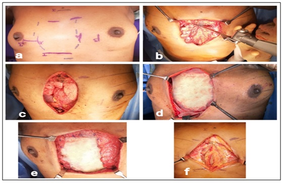
FIG. 1: INTRA-OPERATIVE IMAGES SHOWING (A) PRE-OPERATIVE PLANNING AND MARKING OF MARGINS WITH LOWER STERNUM SHOWING A VISIBLE MASS. (B) PARTIAL STERNECTOMY WITH RESECTION OF 3RD TO 6TH RIBS USING BONE SAW. (C) POST- EN-BLOC RESECTION OF LOWER STERNUM WITH 3RD TO 6TH RIBS ALONG WITH UNDERLYING TUMOUR, REVEALING THE HEART, BOTH LUNGS AND DIAPHRAGM. (D) DEFECT CLOSURE BY SANDWICH TECHNIQUE (2 LAYERS OF PROLENE MESH WITH PMMA BONE CEMENT INTERSPERSED BETWEEN THE LAYERS). (E) PECTORALIS MAJOR MOBILIZATION AND ANCHORING TO MESH. (F) MOBILISATION OF TISSUES JUST BEFORE RESECTION.
Patient tolerated the procedure well and the Post-op period was uneventful. Postoperative histopathology was suggestive of Epithelioid Malignant Peripheral Nerve Sheath tumor arising in a Schwannoma. Microscopy revealed an encapsulated neoplasm with tumor periphery composed of cells with wavy and buckled nuclei arranged in alternating hypo cellular and hyper cellular areas with focal vague Verocay body formation Fig. 2. Immunohistochemistry (IHC) showed tumor cells diffusely positive for Vimentin, S-100, SOX-10, focal positivity for CD56 and GFAP (in Schwannoma areas), loss of INI-1 in tumor nuclei at hyper cellular areas and tumor cells negative for Pan cytokeratin, EMA, HMB-45, Synaptophysin and Chromogranin. H3K27me 3 was retained in the tumor nuclei. MIB-1 proliferation index was 25-30% (diffusely increased in hyper cellular areas) Fig. 3.
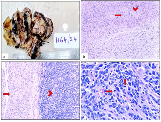
FIG. 2: (A) GROSS APPEARANCE OF THE ENCAPSULATED TUMOR WITH MYXOID AREAS IN THE PERIPHERY AND CENTRAL FLESHY AND NECROTIC AREAS. (B) CLASSICAL ANTONI A AND ANTONI B AREAS OF SCHWWANNOMA WITH VEROCAY BODY FORMATION (ARROW) AND FEW HYALINISED VESSELS (ARROW HEADS). H&E STAIN, 10X. (C) TRANSITION AREAS WITH CLASSICAL SCHWANNOMA ON LEFT (ARROW) AND MALIGNANT TRANSFORMATION COMPOSED OF MARKEDLY CELLULAR AND PLEOMORPHIC TUMOR CELLS ON RIGHT (ARROW HEAD). H&E STAIN, 10X. (D) EPITHELIOID TUMOR CELLS WITH PLEOMORPHIC ROUND NUCLEI AND EOSINOPHILIC NUCLEOLI AND BRISK MITOSIS (ARROW). H&E STAIN, 40X.
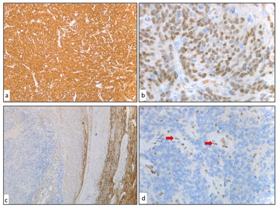
FIG. 3: (A) TUMOR CELLS WITH DIFFUSE S-100 AND (B) SOX-10 POSITIVITY. (C) GFAP POSITIVE IN THE PERIPHERAL SCHWANNOMA AREAS AND NEGATIVE IN THE EPITHELIOID MPNST AREAS. (D) LOSS OF INI-1 IN TUMOR CELLS WITH RETAINED NUCLEAR EXPRESSION IN VASCULAR ENDOTHELIAL CELLS AND STROMAL CELLS (ARROW).
Case 2: A 35 year old gentleman presented with right hypochondrial pain and was found to have a large right retroperitoneal mass detected at Bangladesh. He underwent exploratory laparotomy and partial excision of mass at the native place. Histopathology suggested undifferentiated pleomorphic sarcoma with positive Vimentin and CD99. He received 5 cycles of chemotherapy (AIM regimen -Adriamycin, Ifosfamide, Mesna). Patient had persistent, progressive large retroperitoneal mass and presented to us for further surgical management.
Examination revealed a large 30cmx25cm mass occupying whole of the right side of the abdomen in the right hypochondrium and right lumbar region extending to the right iliac fossa. MRI whole abdomen and pelvis showed 33.2cmx19.2cmx21.2 cm large fairly well defined thick walled cystic lesion with septation predominantly hyperintense, hypointense areas and focal extrinsic soft tissue component in the right retroperitoneum with significant extraneous compression/scalloping of the right kidney impinging the inferior surface of the right lobe of liver, gall bladder, IVC and pushing the right colon, duodenum along with head of pancreas medially to the left. PET CT showed no distant metastasis. He underwent enbloc radical resection of the retroperitoneal tumor with right nephrectomy, subtotalcholecystectomy, partial right hemidiaphragm resection with vascular dissection. Intraoperatively the large tumor was found pushing the right colon, duodenum and pancreas medially infiltrating the right kidney and adherent to right lobe of liver, gallbladder, right hemi diaphragm and IVC Fig. 4.
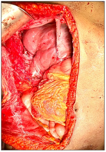
FIG. 4: POST RESECTION BED IN THE RIGHT HYPOCHONDRIUM SHOWING THE LIVER, DISTAL STOMACH WITH DUODENUM, RIGH COLON AND OMENTUM
Blood loss was 1000ml replaced with 4 units packed red blood cells. Postoperative histopathology showed poorly differentiated malignant neoplasm with round cell features. After IHC correlation the features were suggestive of extra skeletal Ewing’s sarcoma, Grade III. IHC was positive for Vimentin, focally positive for synaptophysin and diffusely positive for CD99, NKX2.2, FLI-1 with a Ki-67 of 25-30%. Tumor cells were negative for Pancytokeratin, CD45, Chromogranin A,CAM 5.2, desmin, Myo D1,WT-1, uroplakin, p63, p40, GATA-3, S-100, TLE1 and EMA Fig. 5.
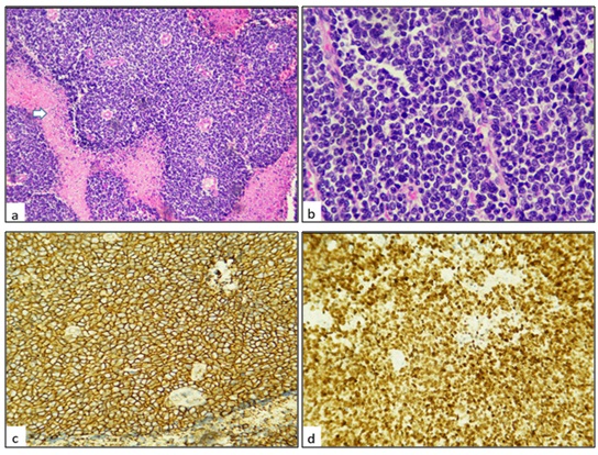
FIG. 5: A. HIGHLY CELLULAR MALIGNANT ROUND CELL TUMOUR WITH AREAS OF TUMOR NECROSIS (WHITE ARROW), 4X, H & E. B. ROUND CELLS HAVING HYPERCHROMATIC NUCLEI AND SCANT CYTOPLASM, 40X, H & E.C. IMMUNOHISTOCHEMISTRY FOR CD99 SHOWS DIFFUSE MEMBRANEOUS POSITIVITY, 10X.D. IMMUNOHISTOCHEMISTRY FOR NKX 2.2 SHOWS DIFFUSE NUCLEAR POSITIVITY, 10X.
Case 3: A 38 year old female presented with loss of appetite and loss of weight of more than 5 kilograms over 2-3 months. Patient had history of fever on and off. Patient received blood transfusion outside for anaemia. Past history of hysterectomy for uterine leiomyoma 9 years back. General examination revealed pallor. Abdominal examination showed a 20cm x 20cm mass palpable in the left lumbar region extending onto left hypochondrium and left iliac fossa. The mass was bimanually palpable and ballotable. Examination of other systems were within normal limits.USG whole abdomen and pelvis showed large heteroechoic lesion measuring 11.3cmx10.8cm x8.0cm noted in the lower and interpolar region of left kidney with increased vascularity encasing left hilum causing mass effect over pancreas superiorly and anteriorly. CT Whole abdomen and pelvis with contrast revealed a large infiltrative mass lesion infiltrating the mesocolon, adherent to pancreas, aorta and retroperitoneum. Left renal biopsy showed a low grade neoplasm with papillary pattern. IHC correlation showed features suggestive of cystic nephroma. TC 99m DTPA renal dynamic scintigraphy showed enlarged left kidney with mild to moderately impaired function and adequate clearance of radiotracer. Persistent relative photopenia in the interpolar to lower polar region of the enlarged left kidney. Normal sized right kidney with normal function. Whole body PET CT showed a large FDG avid heterogeneously enhancing exophytic mass lesion arising from the left kidney involving the renal pelvis and adjacent left ureter measuring 8.0 cm x 8.5cm x 10.5cm with focal loss of plane with the pancreas and spleen. Enlarged FDG avid retrocrural and para-aortic lymphnodes largest measuring 15mm x 15mm. Enlarged FDG avid omental nodules largest measuring 22mm in the infraumbilical region. No other FDG avid lesions in the rest of the body.
In view of the large size of the mass and infiltrative nature of the lesion, patient underwent left radical nephrectomy with left colectomy with para –aortic lymph node dissection with pancreatic and extensive retroperitoneal dissection with colocolic anastomosis and omentectomy Fig. 6.
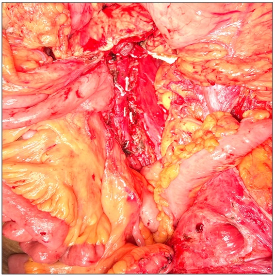
FIG. 6: POSTRESECTION BED IN THE LEFT LUMBAR REGION SHOWING THE PANCREAS, DUODENOJEJUNAL FLEXURE, ABDOMINAL AORTA WITH CLIPS AND COLOCOLIC ANASTOMOSIS
On POD 4, patient developed secondary haemorrhage and underwent emergency laparotomy with clot evacuation with controlling of ooze from the pancreatic bed and retroperitoneum. Patient was haemodynamically stable subsequently. 2 weeks later, patient had an abscess which was drained by CT guided aspiration and pigtail insertion. Postoperative histopathology was suggestive of Fumarate hydratase deficient renal cell carcinoma infiltrating the perinephric fat, Gerota's fascia and left adrenal with high grade. Macroscopically the tumor was involving the entire left kidney measuring 13.8cmx12.4cmx10.5cm. Microscopy showed renal parenchyma with an infiltrating solid cystic neoplasm composed of tumor cells arranged in mixed tubulopapillary, tubulocystic and glandular with focal cribriform pattern. The tumor cells had enlarged vesicular nuclei with single prominent inclusion like nucleoli surrounded by clear halo and abundant eosinophilic cytoplasm. Mitotic activity is brisk. The tumor extensively infiltrates the renal sinus fat and focally infiltrates the renal pelvis. Multiple small vessel lymphovascular Emboli were present with focal perineural invasion. 1 out of 36 lymph nodes harvested showed tumor deposits. On immunohistochemistry, the tumor cells were positive for Pancytokeratin, Vimentin, AMACR and PAX8 with focal positivity for high molecular weight cytokeratin. The tumor cells were negative for cytokeratin 7, cytokeratin 20, GATA3, p63, CD10, carbonic anhydrase and ALK-1. INI-1 expression was intact in the tumor nuclei. FH (fumarate hydratase) showed loss of staining within the tumor cells with positive internal control. Ki-67 proliferation index was 20% Fig. 7.
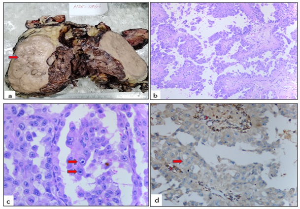
FIG. 7: A. CUT SURFACE OF KIDNEY SHOWS LARGE FLESHY MASS (ARROW) WITH AREAS OF HEMORRHAGE ALMOST REPLACING THE KIDNEY PARENCHYMA. B. SHOWS TUMOR CELLS PREDOMINANTLY ARRANGED PAPILLARY CORES, H&E, 100X. C. TUMOUR CELLS HAVING PROMINENT INCLUSION LIKE EOSINOPHILIC NUCLEOLI SURROUNDED BY CLEAR HALO (ARROWS), H&E, 400X. D. IMMUNOHISTOCHEMISTRY FOR FUMARATE HYDRATASE (FH) SHOWS LOSS OF STAINING WITHIN TUMOUR CYTOPLASM (ARROW), WHEREAS STROMAL CELLS SERVE AS INTERNAL CONTROL WITH GRANULAR CYTOPLASMIC POSITIVITY (ARROW HEAD), 400X MAGNIFICATION.
RESULTS:
Case 1: Postoperatively patient received adjuvant chemotherapy and radiation therapy. Patient is disease free for the past 2 years and is on regular followup.
Case 2: Patient received adjuvant chemotherapy and radiation therapy at native place. Patient developed unsalvageable local recurrence 8 months later and succumbed to the disease.
Case 3: Patient has completed adjuvant chemotherapy. Patient is disease free and on regular followup.
DISCUSSION: Malignant peripheral nerve sheath tumors (MPNSTs) are rare neoplasms and account for only 5% of all malignant soft-tissue sarcomasand 6, 7. The most common sites of MPNSTs occurrence are the torso, extremities and head and neck with anterior mediastinum being an extremely rare location 8. The majority of mediastinal neurogenic tumors arise in the posterior mediastinum, with only 3% found in the anterior mediastinum. Only few patients with MPNST of the anterior mediastinum have been reported in the world literature 9-12. Variants of MPNST include rhabdomyoblastic, glandular, and epithelioid MPNST 13. Epithelioid MPNST (EMPNST) accounts for 5% or less of all MPNSTs 14, 15. Only few cases of EMPNST are described in the English literature. EMPNS Tarise from the transformation of a pre-existing benign nerve sheath neoplasm which is typically a schwannoma rather than neurofibroma 16. These tumors most commonly affect adult patients (median age - 44years) although a wide age range (6 years to 80 years) and site distribution is observed. Men and women are equally affected.
The Epithelioid variant of MPNST is a distinct subtype that is characterized by predominantly epithelioid cytomorphology. EMPNST shows diffuse S-100 positivity and INI1 loss in two thirds of cases (67%) and is frequently multilobular. Conventional MPNST may sometimes show focally epithelioid morphology whereas, EMPNST is characterized by a predominance of epithelioid tumor cells showing a multilobular growth pattern with myxoid and/or fibrous stroma, with foci of spindled morphology. Most tumors showed moderate to severe cytologic atypia and the median mitotic rate is 5/ 10HPF. Tumors show aggressive biological behaviour with high risk of recurrence, metastasis, disease-related death regardless of histologic grade and anatomic depth of tumor 17. EMPNST arising in association with a schwannoma is a rare occurrence that has been reported occasionally in some cases being preceded by epithelioid malignant change in the schwannoma 14-16, 18, 19, 20.
Aggressive surgery is considered to improve prognosis of the MPNST 21, 22. The mainstay of management of localized EMPNST is complete surgical resection with or without neoadjuvant/adjuvant radiation. The role of chemotherapy in the management of localized disease remains to be defined 23. Our patient underwent radical surgery with complex reconstruction followed by adjuvant chemotherapy and radiotherapy.Our patient is the first female patient to have EMPNST arising from a schwannoma in the anterior mediastinum infiltrating the sternum treated by subtotal sternectomy (R0 resection) with reconstruction by sandwich technique(bone cement interspersed between prolene mesh) in the world literature. Ewing’s sarcoma (ES) is a rare malignant tumor described in 1921 by Ewing composed of small round cells 24. ESs have been reported throughout the human body and classified as ES of the bone, extraskeletal ES (EES), malignant small cell tumors of the chest wall (Askin tumor) and soft tissue-based primitive neuroectodermal tumors (PNET) 25, 26. EESs often occur in soft tissues in the paravertebral region, chest, extremities, and retroperitoneal regions 27. Most cases involve children, adolescents, and young adults with disease onset between the ages of four and 25 in > 90% of cases 28. More than 50% of ESs in adults are ESS developing in the trunk, intraabdominal tissues, retroperitoneum, and viscera 29-31. EES is a very rare sarcoma with an annual incidence of about 0.4/million with retroperitoneal EES contributing to ≈20–30% of those cases 32, 33. Only case reports and small case series exist for retroperitoneal EES in view of the rarity of the disease 34-40.
In view of the rapid growth of these tumors and tendency to develop widespread metastasis, they carry poor prognoses 41. On histopathology, ES-EWS appears similar to the classical EWS. It shows a uniform population of closely packed small round cells with indistinct nucleoli, arranged in sheets with round nuclei, high nuclear-cytoplasmic ratio and scant vacuolated cytoplasm due to the presence of glycogen responsible for the Periodic Acid Schiff (PAS) positivity 42, 43. In case of EWS, IHC reveals a strong, diffuse, membranous positivity with CD99. Vimentin, NSE and S-100 are also frequently expressed 44. Our case was positive for CD99, Vimentin and NKX2.2 and FLI-1.
For all Ewing’s Sarcoma Family of Tumors (ESFT), including retroperitoneal EES, initial treatment with vincristine, doxorubicin, cyclophosphamide, ifosfamide, and etoposide (VDC/IE) and subsequent multi-visceral resection should be considered the standard of care 45. Surgery and radiation are still critical to the management of EES and retroperitoneal EES 46. Our patient is the first young male patient with retroperitoneal EES in the world literature to have multivisceral resection of the retroperitoneal mass with enbloc right nephrectomy, partial diaphragmatic resection and subtotal cholecystectomy.
He completed adjuvant chemotherapy and radiotherapy at native place, but succumbed to the disease 8 months later due to unsalvageable local recurrence. Fumarate Hydratase deficient Renal cell carcinoma (FHdRCC) mainly affects younger patients and is a rare, aggressive subtype of type 2 papillary Renal Cell Carcinom (RCC) metastasing early from small solitary lesions. It is due to the inactivating mutation of the Fumarate Hydratase (FH) gene with a lifetime risk of 15% for FH mutation carriers to develop RCC 47. FHdRCC can be associated with an autosomal dominant hereditable syndrome, hereditary leiomyomatosis and renal cell cancer (HLRCC) syndrome with propensity to develop smooth muscle tumors of the skin and uterus. The WHO lists FHdRCC as a subtype of ‘HLRCC-associated RCC’ in their RCC classification. The general term FHdRCC includes both hereditary and known sporadic forms of cancer development 48. FH-deficient RCC is highly malignant and has the propensity to metastasize when the tumor volume is very small. The average age of onset of FHdRCC in Europe and the United States is 20–25 years earlier than that of sporadic RCC in the general population 49.
The main clinical manifestations include hematuria, osphyalgia and other nonspecific symptoms. The pathological features of the tumor are that of an invasive growth with invasion into the surrounding tissue. Microscopically, the tumor cells are arranged into papillary, solid, tubular or cystic patterns. The histological structure is similar to that of type II papillary renal cell carcinoma, collecting duct carcinoma and sarcomatoidcarcinoma 50. The two immunohistochemical biomarkers that show a high correlation with the diagnosis of FH-deficient RCC are FH and S-(2-succino)-cysteine (2SC) 51. Loss of Fumarate Hydratase combined with positivity with 2SC IHC is used in the diagnosis of these tumors. Muller et al, reported a sensitivity of 87.5% and a specificity of 100% for FH loss in the diagnosis of FH-deficient RCCs 52.
Surgical resection of the primary lesion remains an important treatment for organ-confined FH-RCC. The surgical strategy is also different from that of common kidney cancer because of FH-RCC’s aggressive nature and high risk of metastasis. The results of a cohort from West China Hospital of Sichuan University (West China Hospital) showed that the proportion of patients with tumor maximum diameter < 10 cm (≤ cT2a stage) had a postoperative pathological upgrade (≥ pT3a stage) as high as 50%. Literature also report a group of patients in North America presented with non-organ confined pathological escalation in 65% 48. Hence, radical nephrectomy is preferentially recommended for patients with a high preoperative clinical diagnosis of FH-RCC, regardless of the tumor size and partial nephrectomy with wide margins may be considered for patients with functional or anatomic isolated kidneys or with underlying renal dysfunction. While considering laparoscopic surgery, the possibility of channel implantation needs to be fully considered, the benefits and risks associated needs to be communicated to the patient, and the nephrectomy should be done strictly outside the renal fascia. There is a lack of reliable clinical evidence on performing hilar and retroperitoneal lymph node dissection for patients with FH-RCC. Since retroperitoneal lymph node metastasis often occurs early in this disease, the extent of dissection should be based on preoperative imaging changes and intraoperative conditions 53.
Our patient is the first young female patient in the world literature to have undergone radical nephrectomy with colectomy with para-aortic lymph node dissection in FH-RCC. Bevacizumab combined with erlotinib (the E-B regimen) is still recommended for treating FH RCC in the NCCN kidney cancer treatment guidelines and the 2021 Chinese Society of Clinical Oncology guidelines, in spite of the literature lacking effective systemic treatment options for FH-RCC 54, 55. Our patient has completed adjuvant therapy and is disease free at present.
CONCLUSION: Multivisceral surgical resections (MVR) as part of extensive major surgical oncology resection procedures is feasible with least peri-operative morbidity for rare tumors in complex locations with meticulous technical expertise. The radicality and completeness of surgical resection determines the short-term and long-term oncological outcomes.
ACKNOWLEDGEMENT: We would like to thank the surgical team from the cardiothoracic surgery, urologic surgery, orthopaedic surgery and thoracic oncology for the immense support in the effective management of our patients.
Funding: There is no funding involved in the conduct of the study.
Compilation of young patients with rare presentation undergoing radical multivisceral surgical resections and reconstruction.
CONFLICTS OF INTEREST: The authors declare no conflict of interest.
REFERENCES:
- KaiTaiDerek Yeung,RoumaysaaBouyardan,Desmond Barton,David Nicol,Sacheen Kumar,Ricky and Harminder Bhogal: Multidisciplinarymultivisceral resections involving the upper digestive tract: a United Kingdom tertiary cancer centre experience. Langenbeck's Archives of Surgery 2025; 410:224.
- Schizas D, Giannakodimos I, Mylonas KS, Kapetanakis EI, Papavgeri Aand Lianos GD: Multivisceral resection for locally advanced gastric cancer: a systematic review and evidence quality assessment. J Clin Med 2023; 12(23):7360.
- Cohen L, Alam F, Flood MP, Mohan H, Jain A, Guerra G, Heriot AGand Kong JCH: A systematic review of minimally invasive cytoreductive surgery and hyperthermic intraperitoneal chemotherapy in patients with peritoneal malignancy. ANZ J Surg2024; 94(7–8):1324–1332.
- Loos M,Al-Saeedi M,HinzU,Mehrabi A, Schneider Mand Berchtold C: Categorization of differing types of total pancreatectomy. JAMA Surg2022; 157(2):120–128.
- Nadiradze G, Yurttas C, Königsrainer Aand Horvath P: Significance of multivisceral resections in oncologic surgery: A systematic review of the literature. World J Meta-Anal 2019; 7(6): 269-289.
- Nielsen GP, Antonescu CR and Lothe RA: Malignant peripheral nerve sheath tumour. In: WHO Classification of Tumours of Soft Tissue and Bone. Fletcher CDM, Bridge JA, Hogendoorn PCD and Mertens F (eds). 4th edition. IARC Press, Lyon2013; 5: 187-189.
- Italiano A, Delva FandMathoulin-Pelissier S: Effect of adjuvant chemotherapy on survival in FNCLCC grade 3 soft tissue sarcomas: a multivariate analysis of the French Sarcoma Group Database. Ann Oncol 2010; 21: 2436-2441.
- Sugio K, Inoue Tand Inoue K: Neurogenic tumors of the mediastinum originated from the vagus nerve. Eur J Surg Oncol 1995; 21: 214-216.
- Ingels GW, Campbell DC, Giampetro AM, Kozub RE and Bentlage CH: Malignant schwannomas of the mediastinum. Report of two cases and review of the literature. Cancer 1971; 27: 1190-1201.
- Ducatman BS and Scheithauer BW: Malignant peripheral nerve sheath tumors with divergent differentiation. Cancer 1984; 54: 1049-1057.
- Otani Y, Morishita Yand Yoshida I: A malignant Triton tumor in the anterior mediastinum requiring emergency surgery: report of a case. Surg Today 1996; 26: 834-836.
- Zisis C, Fragoulis S, Rontogianni D, Stratakos G and Bellenis I: Malignant triton tumour of the anterior mediastinum as inci dental finding. Monaldi Arch Chest Dis 2006; 65: 222 224.
- ManganoniAM, FarisoglioC, LonatiA, ZorziF, TucciG, and PintonPGC: “Cutaneous epithelioid malignant schwannoma: review of the literature and case report.” Journal of Plastic, Reconstructive and Aesthetic Surgery, 2009; 62(9):318-321.
- Laskin WB, Weiss SW and Bratthauer GL: Epithelioid variant of malignant peripheral nerve sheath tumor (malignant epithelioid schwannoma). Am J SurgPathol 1991; 15(12): 1136–1145.
- McMenamin ME and Fletcher CD: Expanding the spectrum of malignant change in schwannomas: epithelioid malignant change, epithelioid malignant peripheral nerve sheath tumor, and epithelioid angiosarcoma: a study of 17 cases. Am J SurgPathol 2001; 25(1): 13–25.
- Carter JM, O’Hara Cand Dundas G: Epithelioid malignant peripheral nerve sheath tumor arising in a schwannoma, in a patient with “neuroblastoma-like” schwannomatosis and a novel germline SMARCB1 mutation. Am J SurgPathol 2012; 36(1): 154–160.
- Epithelioid Malignant Peripheral Nerve Sheath Tumor clinicopathologic analysis of 63 Cases. Vickie Y. Jo,Christopher D.M. Fletcher.Am J SurgPathol 2015;39:673–682.
- Woodruff JM, Selig AMand Crowley K: Schwannoma (neurilemoma) with malignant transformation. A rare, distinctive peripheral nerve tumor. Am J SurgPathol 1994;18:882–895.
- Rasbridge SA, Browse NLand Tighe JR: Malignant nerve sheath tumour arising in a benign ancient schwannoma. Histopathology 1989;14:525–528.
- Mikami Y, Hidaka Tand Akisada T: Malignant peripheral nerve sheath tumor arising in benign ancient schwannoma: a case report with an immunohistochemical study. Pathol Int 2000;50:156–161.
- DucatmanBS, ScheithauerBW and PiepgrasDG: “Ma lignant peripheral nerve sheath tumors. A clinicopathologic study of 120 cases.” Cancer1986;57(10):2006–2021.
- WaneboJE,MalikJM,VandenBergSR, J Wanebo H,DriesenN and PersingJA: “Malignant peripheral nerve sheath tumors: a clinicopathologic study of 28 cases.” Cancer1993; 71(4):1247–1253.
- Epithelioid malignant peripheral nerve sheath tumor arising in schwannoma.Nicholas Heatley, Eva Kolson Kokohaare, Dirk C Strauss, Magnus Hallin, Robin L Jones, Cyril Fisher and Khin Thway: Rare Tumors2020; 12: 1-4.
- Ewing J: The Classic: Diffuse endothelioma of bone. Proceedings of the New York Pathological Society. 1921;12: 17. Clin OrthopRelat Res 2006;450:25–27. https://doi.org/10.1097/01.blo.0000229311.36007.c7.
- Murphey MD, Senchak LT, Mambalam PK, Logie CI, Klassen-Fischer MKand Krans dorf MJ: From the radiologic pathology archives: ewing sarcoma family of tumors: radiologic-pathologic correlation. Radiographics 2013;33(3):803–31.
- Arpaci E, Yetisyigit T, Seker M, Uncu D, Uyeturk U, Oksuzoglu B, Demirci U, Coskun U, Kucukoner M, Isıkdogan A, Inanc M, Alkis NandOzkan M: Prog nostic factors and clinical outcome of patients with Ewing’s sarcoma family of tumors in adults: multicentric study of the Anatolian Society of Medical Oncology. Med Oncol 2013;30(1):469
- Iwamoto Y: Diagnosis and treatment of Ewing’s sarcoma. Jpn J Clin Oncol 2007;37(2):79–89.
- Muralidhar D, Vasugi GAand Sundaram S: Incidence and demographic Profile of Ewings Sarcoma: experience from a Tertiary Care Hospital. Cureus 2021;13(9):e18339.
- Ulusan S, Koc Z, Canpolat ETandColakoglu T: Radiological findings of primary retroperitoneal ewing sarcoma. Acta Radiol 2007;48(7):814–8.
- Ozaki Y, Miura Yand Koganemaru S: Ewing sarcoma of the liver with multilocular cystic mass formation: a case report. BMC Cancer 2015;15:16.
- Nedham FN, Nagaraj V, Darwish Aand Al-Abbasi TA: Retroperitoneal blue cell round tumor (ewing sarcoma in a 35 years old male)- case report. Int J Surg Case Rep 2022;94:107045.
- Murthy SS, Challa S, Raju K, Rajappa SJ, Fonseca D, Gundimeda SD, Rao BV, Ahmed F, Kodandapani Sand Nambaru L: Ewing sarcoma with emphasis on extra-skeletal ewing sarcoma: a decade’s experience from a single centre in India. Clin Pathol 2020; 13.
- Applebaum MA, Worch J, Matthay KK, Goldsby R, Neuhaus J, West DCand Dubois SG: Clinical Features and Outcomes in Patients with Extraskeletal Ewing Sarcoma. Cancer 2011; 117: 3027–3032.
- Thyavihally YB, Tongaonkar HB, Gupta S, Kurkure PA, Amare P, Muckaden MAand Desai SB: Primitive Neuroectodermal Tumor of the Kidney: A Single Institute Series of 16 Patients. Urology 2008; 71: 292–296.
- Marinova L: Retroperitoneal Primitive Neuroectodermal Tumour (PNET). A Case Report and Review of the Literature. Rep Pract Oncol Radiother 2009; 14: 221–224.
- Li J, Nie Fand Li Y: Extraosseous Ewing’s Sarcoma/Peripheral Primitive Neuroectodermal Tumour of the Kidney: A Case Report and Literature Review. BMC Urol 2022; 22: 197.
- Koch A, Frigo S, Lecointre L, Hummel M,Akladios CY, Bergerat JP, Noël Gand Wattiez A: Retroperitoneal Primitive Neuroectodermal Tumor (PNET): Case Report and Review of Literature. Eur J Gynaecol Oncol 2017; 38: 314–318.
- Genov P, Serbezova I, Georgieva D, Koleva Gand Hristova I: A Rare Case of Retroperitoneal Primitive Neuroectodermal Tumor (PNET). Urol Case Rep 2021; 35: 101554.
- Das S, Aggarwal G, Gupta Sand Midha D: Primary Renal Ewing’s Sarcoma in an Adult: An Enigma. Innov. Surg. Sci 2021; 6: 20200022.
- Chatterjee A, Patkar S, Purandare N, Mokal Sand Goel M: Management of abdominal ewing’s sarcoma: a single institute experience. Indian JSO 2021; 12: 571–580.
- Javalgi AP, Karigoudar MHandPalur K: Blue Cell Tumour at Unusual Site: Retroperitoneal Ewings Sarcoma. J Clin Diagn Res 2016;10(4): 19-20.
- Ulusan S, Koc Z, Canpolat ETandColakoglu T: Radiological Findings of Primary Retroperitoneal Ewing Sarcoma. Acta Radiol2007;7:814-18.
- Gill M, Gupta P, Pawar Pand Sen R: Malignant small round cell tumour masquerading as mesothelioma of peritoneum: diagnosed on immunohistochemistry. International J. of Healthcare and Biomedical Research 2014;2:83-86.
- Zhang Z, Yu H, Shi Yand Shi H: A case report on retroperitoneal PNET/EWS. Case Reports in Clinical Medicine 2013;2:366-69.
- Brennan B, Kirton L, Marec-Bérard P, Gaspar N, Laurence V, Martín-Broto J, Sastre A, Gelderblom H, Owens Cand Fenwick N: Comparison of two chemotherapy regimens in Patients with Newly Diagnosed Ewing Sarcoma (EE2012): An Open-Label, Randomised, Phase 3 Trial. Lancet 2022; 400: 1513–1521.
- Practical Management of Adult Ultra-Rare Primary Retroperitoneal Soft Tissue Sarcoma: A Focus on Perivascular Epithelioid Tumours and Extraosseous Ewing Sarcoma. Sameer S. Apte, Eyal Mor, Catherine Mitchell andDavidE. Gyorki. Curr Oncol 2023; 30: 5953–5972.
- Forde C, Lim DHK, Alwan Y, Burghel G, Butland Land Cleaver R: Hereditary leiomyomatosis and renal cell cancer: Clinical, molecular, and screening features in a cohort of 185 affected individuals. EurUrol Oncol 2020; 3(6):764–72.
- Lau HD, Chan E, Fan AC, Kunder CA, Williamson SRand Zhou M: A clinicopathologic and molecular analysis of fumarate hydratase-deficient renal cell carcinoma in 32 patients. Am J SurgPathol 2020; 44(1):98–110.
- Doehn C, Grunwald Vand Steiner T: The diagnosis, treatment, and fol low-up of renal cell carcinoma. DtschArztebl Int 2016;113(35–36):590–6.
- Muller M, Ferlicot Sand Guillaud-Bataille M: Reassessing the clinical spectrum associated with hereditary leiomyomatosis and renal cell carcinoma syndrome in French FH mutation carries. Clin Genet 2017;92(6):606–15.
- Fumarate hydratase-deficient renal cell carcinoma: a case report and review of the literature YantingLv, Lide Song and MengjunHu.Journal of Medical Case Reports 2023; 17:182.
- Muller M,Guillaud-Bataille Mand Salleron J: Pattern multiplicity and fumaratehydratase (FH)/S-(2-succino)-cysteine (2SC) staining but noteosinophilic nucleoli with perinucleolar halos differentiate hereditary leiomyomatosis and renal cell carcinoma-associated renal cell carcinomas from kidney tumors without FH gene alteration. Mod Pathol 2018;31(06):974–983.
- Yu Shen, Zhenhua Liu, Qiang Wei and Wei Xue: Consensus on clinical diagnosis and treatment of fumarate hydratase-deficient renal cell carcinoma.Holistic Integrative Oncology 2024; 3:7.
- .Carril-Ajuria L, Colomba Eand Cerbone L: Response to systemic therapy in fumarate hydratase-deficient renal cell carcinoma [J]. Eur J Cancer 2021;151:106–14.
- Yonese I, Ito Mand Takemura K: A case of metastatic hereditary leiomyomatosis and renal cell cancer syndrome-associated renal cell carcinoma treated with a sequence of axitinib and nivolumab following cytoreductive nephrectomy. J Kidney Cancer VHL 2020;7(2):6–10.
How to cite this article:
Shanmugasundaram G, Maharajan S, Shrivathsan R, Shanmugam S, Thanumalayan D, Gurukailasam R, Hussain AKK and Ranjith R: Compilation of young patients with rare presentation undergoing radical multivisceral surgical resections and reconstruction. Int J Pharm Sci & Res 2026; 17(1): 3179-89. doi: 10.13040/IJPSR.0975-8232.17(1).3179-89.
All © 2026 are reserved by International Journal of Pharmaceutical Sciences and Research. This Journal licensed under a Creative Commons Attribution-NonCommercial-ShareAlike 3.0 Unported License.
Article Information
30
3179-3189
4424 KB
4
English
IJPSR
Gouthaman Shanmugasundaram *, Sivasundari Maharajan, R. Shrivathsan, Salapathy Shanmugam, Dharanya Thanumalayan, Ramakrishnan Gurukailasam, A. K. Khader Hussain and R. Ranjith
Department of Surgical Oncology, Apollo Speciality Hospitals, Vanagaram, Chennai, Tamil Nadu, India.
gouthamonco1@gmail.com
09 June 2026
21 June 2026
25 September 2026
10.13040/IJPSR.0975-8232.17(10).3179-89
01 October 2026
 Download