




EFFICIENT VOLTAMMETRIC DETERMINATION OF NIFEDIPINE AT PENCIL GRAPHITE BLEND PASTE ELECTRODE
HTML Full TextEFFICIENT VOLTAMMETRIC DETERMINATION OF NIFEDIPINE AT PENCIL GRAPHITE BLEND PASTE ELECTRODE
Y. Madhu Kiran, M. Sunil Kumar and N. Y. Sreedhar *
Electroanalytical Lab, Department of Chemistry, Sri Venkateswara University, Tirupati, Andhra Pradesh, India.
ABSTRACT: Pencil graphite electrodes were quite useful in voltammetric studies of wide range of organic compounds with biological and industrial significance. In the present research, HB pencil graphite paste electrode with 15% pencil 8B grade was fabricated. Cyclic voltammetry was used to investigate the electrochemical reduction mechanism of nifedipine at the electrode. The peak due to 4e- and 4H+ reduction of 2-nitro group of the drug to hydroxyl amine is selected for the voltammetric method development. The pencil graphite blend paste electrode produced good and consistent reduction peaks of nifedipine by differential pulse voltammetry. The optimisation of pH, scan rate, accumulation time and potential and other parameters was carried out using DPV peak signals. A linear range of 10 – 100 nM for nifedipine was obtained with equation Ip(μA) = 0.098C(nM)+4.309 (correlation coefficient, R2 = 0.999) and detection limits of 6.0 nM and quantitation limits of 20 nM. By the present method, recoveries values are obtained in the range of 97.0-101.0 % for nifedipine tablets with RSD values ranging from 2.5-3.8%.
Keywords: Pencil graphite electrode, Nifedipine, Differential pulse voltammetry, Cyclic voltammetry, Tablets
INTRODUCTION: Nifedipine is mainly used for cardiovascular diseases and chest related pain management. A number of analytical methods based on UV, fluorescence, GC, HPLC and LC-MS methods were reported with good sensitivity and selectivity. UV derivative method was developed for nifedipine determination along with atenolol in dosage forms and validated with gas chromatography 1. A fluorescence sensor based on molecularly imprinted polymer - silane functionalized carbon dots was reported for nifedipine determination in serum and urine samples with good recoveries 2.
A 3D molecularly imprinted polymer based fluoroprobe with carbon quantum dots and polydopamine was used for the sensitive determination of Nifedipine 3. Further, Nifedipine was determined by rapid capillary gas chromatography employing electron capture detection, applied for plasma samples 4. HPTLC method was developed for nifedipine, for bulk drug and dose formulations 5. Micellar liquid chromatographic procedure was reported for the determination of nifedipine in serum and urine samples and photostability studies 6.
A multi-criteria decision making approach for simultaneous HPLC determination of atenolol and nifedipine was developed for content uniformity testing in capsules 7. Nifedipine was determined by SPE-HPLC-UV in plasma samples, for the pharmacokinetic studies 8. UPLC- MS/MS method was developed for nifedipine in plasma samples and applied for bioequivalence studies 9.
Nifedipine along with other 4 calcium channel antagonists was determined by UHPLC-MS/MS for therapeutic drug monitoring and pharmacokinetics in plasma samples 10. Voltammetry offers many advantages like simple, rapid and sensitive methods for the determination of drugs in different matrices. Differential pulse adsorptive stripping polarography procedure was developed and applied for the determination of nifedipine 11. Activated glassy carbon electrode for nifedipine was used by voltammetry and applied for tablet and capsule formulations 12. Further nifedipine was determined by square wave adsorptive stripping voltammetry by employing hanging mercury drop electrode and the method applied for pregnant woman plasma 13. Polyvinyl pyrrolidine modified carbon paste electrode was used in differential pulse voltammetry for the determination of nifedipine in tablet samples 14. Barium stannate perovskite needle shaped structure modified glassy carbon electrode was developed for simultaneous voltammetric determination of nifedipine and nitrofurantoin and the protocol was successfully applied for plasma, urine and serum samples 15.
A variety of electrodes for nifedipine were reported such as hanging mercury drop electrode and a mercury meniscus modified silver amalgam electrode 16, MWCNT modified glassy carbon electrode 17, β-cyclodextrin and MWCNT paste electrode 18, Ag nanoparticles modified glassy carbon electrode 19, MgO nanoplatelet modified screen-printed electrode with atenolol 20, zinc oxide with functional CNTs based electrode 21, silver phosphate/strontium phosphate nanoparticles (AgP/SrP NPs) modified glassy carbon electrode 22. Lately modified pencil graphite electrode was used for the determination of significant molecules 23-24. There is a dire necessity for a simple, cost-effective electrode for the direct, rapid and sensitive analysis of nifedipine, aptly addressed by the present research. Hence the aim of the work involves developing a pencil graphite blend paste electrode with HB and 8 B grades and then it is used for sensitive and selective voltammetric determination of nifedipine in two tablet formulations.
MATERIALS AND METHODS: All the chemicals used were of analytical reagent grade unless specified otherwise. Nifedipine (>98 %) was procured from Sigma - Aldrich, USA. Nifedipine tablets used in the application study were obtained from local pharmacy. Derwent pencils of different grades were purchased from the local stationery. Voltammetric experiments were carried out using CHI 660D Electrochemical Working Station controlled by CHI software. Buffer pH readings were recorded using Elico LI 120 pH meter.
Experimental Method: The procedure involves a three electrode system in which pencil graphite blend electrode (85% Derwent HB and 15% Derwent 8B) was used as working electrode and Ag/AgCl (KCl 3M) as reference and Pt wire acts as counter electrode. In the electrochemical cell, 1 mL of the analyte solution (100 nM) was taken along with BR buffer of pH 2.0, making it upto 10 mL degassed with N2 gas for 10 minutes and then differential pulse voltammetric technique was applied and the corresponding voltammograms were recorded. A range of concentrations of nifedipine (10 - 100 nM) were measured using the same measurement procedure by standard addition method with five replicate determinations for each concentration Fig. 1. After each measurement, the electrode was regenerated by washing with double distilled water, nitrogen gas dried for 10 minutes and used in subsequent determinations. In case of formulations, 3 tablets of nifedipine (10 mg) and 2 tablets of nifedipine (20 mg) were pulverised and dissolved in 60 mL and 80 mL of methanol respectively and sonicated for 10 minutes to obtain complete dissolution. The stock solution was taken in 100 mL volumetric flask and dilutions were carried out using methanol and double distilled water (3:2) to obtain standard and working standards.
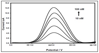
FIG. 1: DPV CURVES OF NIFEDIPINE (10 -100 nM) AT PENCIL GRAPHITE BLEND PASTE ELECTRODE (PGBPE) WITH BLANK BUFFER SAMPLE (DOTTED LINE)
RESULTS AND DISCUSSION:
Fabrication of Pencil Graphite Blend Paste Electrode (PGBPE): The electrode was fabricated by taking pencil graphite lead of HB grade and pulverising it in mortar. The HB pencil graphite powder (85% w/w) was then mixed with 8B pencil graphite for its good electrical conductivity in the 15% w/w proportion.
A uniform paste was then formed with pencil graphite blend and mineral oil (75/25 % w/w) and packed in teflon tubing of 3 mm diameter and copper wire connection provided for taking measurements. Smooth surface of the electrode was obtained by gently touching against soft tissue paper.
The electrode was washed with double distilled water and nitrogen gas dried for 10 minutes. The pencil graphite paste electrode so fabricated was directly used for the characterisation of nifedipine by cyclic voltammetry (CV) and differential pulse voltammetry (DPV). Similarly pencil graphite paste electrodes using only HB and 8B grades were fabricated for comparative studies.
Cyclic Voltammetry Studies: The compound was studied using cyclic voltammetry at different pencil graphite electrodes in the potential range of 1.0 to -0.8 V vs. Ag/AgCl (KCl 3M).
Nifedipine gave one irreversible cathodic peak at -0.40 V owing to the reduction of nitro group to hydroxylamine Fig. 2 and the recordings were taken for different electrodes with 5 - 30% 8B pencil.
The peak current (iP) increased with the scan rate (50 - 500 mV/s), indicating the reduction reaction under study was adsorption-controlled. No corresponding anodic peak was observed indicating the irreversibility of the reaction. The anodic and cathodic peaks obtained were due to oxidation of hydroxylamine to nitroso group and reduction of nitroso group to hydroxyl amine on reverse scan 25. CV curves were obtained for different pencil graphite electrodes Fig. 2.
High peak currents were obtained for pencil graphite blend electrode (a) owing to its increased electrical conductivity compared to other pencil electrodes (b & c), as indicated in Fig. 2.
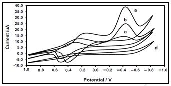
FIG. 2: CV CURVES OF NIFEDIPINE (1 × 10⁻⁴ M) AT PGPE (85% HB AND 15% 8B) (A), PGPE-8B (B), PGPE-HB(C) ALONG WITH BLANK BUFFER SAMPLE (D)
DPV Optimisation: The instrumental method used for the determination of nifedipine was differential pulse voltammetry (DPV), selected for its high sensitivity and good peak profile recordings. The parameters involved such as electrode composition, pH, accumulation potential and accumulation time along with pulse amplitude, pulse width were optimised by measuring the drug peak currents using pulse voltammetry.
Effect of Electrode Composition (8B Graphite): The electrode composition was optimised by studying the effect of 8B graphite on peak currents of nifedipine using differential pulse voltammetry. The peak currents increased from 5 - 15% and then slightly decreased beyond the 15 % value Fig. 3. Hence 15% 8B graphite was used in making the electrode composition for voltammetric studies of nifedipine.
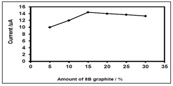
FIG. 3: EFFECT OF AMOUNT OF GRAPHITE (8B) ON PEAK CURRENT OF NIFEDIPINE (100 nM)
Effect of pH, Accumulation Potential, Accumulation Time: Variation of pH was studied in the range 2-10 and the peak current was maximum at 2.0 and then started showing decrease trend, indicating 2.0 as the best pH value Fig. 4. Even the peak potential moved to negative potentials on increasing the pH beyond 2.
This indicated the role of hydrogen ion in nifedipine reduction. An accumulation potential of -0.3 V was applied where the peak current was maximum in the studied range from -0.2 to -1.0 V vs. Ag/AgCl (KCl 3M). Accumulation time of 180 s was found to be best value in the studied range form 0 - 300 s. The accumulation time beyond 180 s had little effect on the peak current of nifedipine Fig. 5A & 5B.
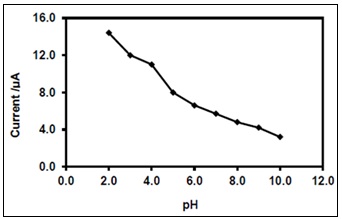
FIG. 4: EFFECT OF PH ON PEAK CURRENT OF NIFEDIPINE (100 NM)
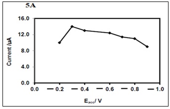
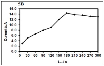
FIG. 5: EFFECT OF ACCUMULATION POTENTIAL, Eacc. (5A) AND ACCUMULATION TIME, tacc. (5B) ON PEAK CURRENT OF NIFEDIPINE (100 nM)
Pulse amplitude (10 - 50 mV) and pulse width (2 - 20 ms) were used for optimising the peak currents and the working values were 20 mV, 10 ms respectively. Optimum values of the DPV procedure were given in Table 1.
TABLE 1: OPTIMISATION OF DPV PARAMETERS FOR NIFEDIPINE
| S. no. | Parameter | Value |
| 1 | pH | B.R : 2.0 |
| 2 | Accumulation potential (Eacc.) | -0.3 V |
| 3 | Accumulation time (tacc.) | 180 s |
| 4 | Pulse amplitude | 20 mV |
| 5 | Pulse width | 10 ms |
| 6 | Step potential | 50 mV |
| 7 | Quiet time | 2 s |
| 8 | Scan rate | 100 mV/s |
Effect of Concentration: The differential pulse voltammetric peak currents of nifedipine increase over the concentration range, 10 – 100 nM using standard addition method. A calibration line with equation Ip(μA) = 0.098C(nM) + 4.309 with R2 = 0.999 was obtained. The detection and quantitation limits were 6 nM and 20 nM respectively based on 3Sb/m and 10 Sb/m, where Sb is the standard deviation of 10 blank measurements and m is the slope of the calibration line.
Stability and Precision: The electrode was dried with nitrogen gas and stored at room temperature after each measurement. Stability of the nifedipine peak current signal (100 nM) was studied for about 30 days and it decreased to 85% over 30 days. Intra-day and inter-day precision was measured by carrying out nifedipine determination for 10 replicates on the same day and on 10 consecutive days with RSD values of 3.4 and 4.9 %.This indicated good stability and reproducibility of the electrode.
Ruggedness and Robustness: The voltammetric method was carried out by three different analysts on three different days and peak signals for nifedipine (100 nM) were determined. Five replicate determinations were performed for each voltammetric measurement. RSD values of < 2% obtained, indicating the ruggedness of the method. Further incremental changes in optimised parameters such as electrode composition, pH, accumulation time and accumulation potential had very little effect on the peak current signals (<2%) for nifedipine (100 nM) indicating the robustness of the method.
Application to Tablet Formulations: The tablets of nifedipine (Nicardia & Calcigard) of 10 and 20 mg were pulverised and dissolved in methanol solution to obtain stock solution. Dilutions were done as described in experimental part. Filtration was carried out using 0.45μm Whatman filter paper. Voltammetric analysis was performed to obtain the excellent recoveries of 97 – 101 % for nifedipine tablets and the corresponding RSD values of 2.5 – 3.8 %. The mean values along with SD were also provided Table 2.
The unknown amount of nifedipine was obtained from calibration plot, obtained by standard addition method.
Interference Studies: The peak current signal for nifedipine (100 nM) was little affected (< 2% decrease) by the presence of common excipients such as magnesium stearate, starch, cellulose, lactose and talc at 100 fold concentration while the peak potential of nifedipine (100 nM) was not affected at all. This suggested that the method has good selectivity because the excipients are inactive at the reduction potential of nifedipine.
TABLE 2: RECOVERY STUDIES FOR NIFEDIPINE TABLETS
| S. no. | Tablet | Taken (mg) | Measured (mg)* | Recovery (%) | Mean ± SD | RSD (%) |
| 1 | Nicardia | 10 | 9.7 | 97.00 | 9.7 ± 0.242 | 2.5 |
| 2 | 20 | 19.6 | 98.00 | 19.6 ± 0.549 | 2.8 | |
| 3 | Calcigard | 10 | 9.8 | 98.00 | 9.8 ± 0.372 | 3.8 |
| 4 | 20 | 20.2 | 101.00 | 20.2 ± 0.727 | 3.6 |
*Average of 5 determinations.
CONCLUSION: A novel analytical method was developed using differential pulse voltammetric technique for the accurate determination of nifedipine in tablet formulations with a little sample preparation and time-consuming steps. The method is quite simple, rapid with good sensitivity and selectivity. The developed electrode possesses good stability, reproducibility of the peak signals and it can be readily applied for nifedipine in different tablet formulations. The voltammetric method so developed using pencil graphite blend paste electrode has considerable sensitivity, selectivity and quite competent to be used for nifedipine analysis in tablet formulations.
ACKNOWLEDGEMENTS: Nil
CONFLICTS OF INTEREST: Nil
REFERENCES:
- Veronico M, Ragno G & Vetuschi C: Simultaneous Assay of Atenolol and Nifedipine by UV Derivative Spectrophotometry and Gas Chromatography Spectroscopy Letters 1995; 28(3): 407-415.
- Jalilia R and Amjadi M: Surface molecular imprinting on silane-functionalized carbon dots for selective recognition of nifedipine. RSC Advances 2015; 5: 74084-74090.
- Liu H, Sun X, Dai Z, Wang Y, Li L, Fan J & Ding Y: A new three-dimensional (3D) molecularly imprinted polymer fluoroprobe based on green–red dual-emission signals of carbon quantum dots and self-polymerization of dopamine (CDs@PDA-MIPs) for sensitive detection of nifedipine. Microchim Acta 2024; 191: 332.
- Guellec CL, Bun H, Giocanti M and Durand A: Determination of Nifedipine in plasma by a rapid capillary gas chromatographic method. Biomedical Chromatography 1992; 6: 20-23.
- Patravale VB, Nair VB and Gore SP: High performance thin layer chromatographic determination of nifedipine from bulk drug and from pharmaceuticals. Journal of Pharmaceutical and Biomedical Analysis 2000; 23: 623-627.
- Gil-Agustí MT, Carda-Broch S, Monferrer-Pons L and Esteve-Romero JS: Photostability studies for micellar liquid chromatographic determination of nifedipine in serum and urine samples. Biomedical Chromatography 2006; 20: 154-160.
- Ahmed S, Alqurshi A and Mohamed AMI: Development of a chromatographic method with multi-criteria decision making design for simultaneous determination of nifedipine and atenolol in content uniformity testing. Talanta 2018; 184: 296 – 306.
- Niopas I and Daftsios AC: Determination of nifedipine in human plasma by solid-phase extraction and high-performance liquid chromatography: validation and application to pharmacokinetic studies. Journal of Pharmaceutical and Biomedical Analysis 2003; 32(6): 1213-1218.
- Patel DP, Sharma P, Sanyal M, Singhal P and Shrivastav PS: Highly sensitive and rapid ultra-performance liquid chromatography–tandem mass spectrometry method for the determination of nifedipine in human plasma and its application to a bioequivalence study. Biomedical Chromatography 2012; 26(12): 1509-1518.
- Palermiti A, Billi M, De Nicolò A, Cusato J, Manca A Antonucci M, Menegatti G, Pappaccogli M, Ponsa L, Fasano C, De Rosa FG, Cantù M, Rabbia F, Veglio F, D’Avolio A: UPLC-MS/MS method for the simultaneous quantification of five calcium channel antagonists’ drugs in human plasma. Biomedicine & Pharmacotherapeutics 2025; 184: 117873
- Shapovalov VA: Determination of Nifedipine by Differential Pulse Adsorptive Stripping Polarography. Journal of Analytical chemistry 2002; 57(2): 157–158.
- Şentürk Z, Özkan SA, Özkan Y: Electroanalytical study of nifedipine using activated glassy carbon electrode. J of Pharma and Biomedical Analysis 1998; 16(5): 801-807.
- Özaltin N, Yardimci C, Süslü I: Determination of nifedipine in human plasma by square wave adsorptive stripping voltammetry. Journal of Pharmaceutical and Biomedical Analysis 2002; 30(3): 573-582.
- Yang X, Sun D, Xie X & Zhang H: Sensitive and rapid determination of nifedipine using polyvinylpyrrolidone-modified carbon paste electrode. Russian Journal of Electrochemistry 2014; 50(5): 453-457.
- Balamurugan M, Alagumalai K, Chen TW, Chen SM, Liu X & Selvaganapathy M: Simultaneous electrochemical determination of nitrofurantoin and nifedipine with assistance of needle-shaped perovskite structure: barium stannate fabricated glassy carbon electrode. Microchimica Acta 2021; 19: 188.
- Wirzal MDH, Rahim A, Yusoff M and Barek J: Voltammetric Determination of Nifedipine at a Hanging Mercury Drop Electrode and a Mercury Meniscus Modified Silver Amalgam Electrode. International Journal of Electrochemical Science 2015; 10(6): 4571- 4584.
- Mokhtari B, Nematollahi D and Salehzadeh H: Electrochemical simultaneous determination of nifedipine and its main metabolite dehydronifedipine using MWCNT modified glassy carbon electrode. Journal of Molecular Liquids 2018; 264: 543-549.
- Gaichore RR and Srivastava AK: Voltammetric determination of nifedipine using a β-cyclodextrin modified multi-walled carbon nanotube paste electrode. Sensors and Actuators B: Chemical 2013; 188: 1328-1337.
- Baghayeri M, Namadchian M and Beitollahi H: Determination of nifedipine using nanostructured electrochemical sensor based on simple synthesis of Ag nanoparticles at the surface of glassy carbon electrode: Application to the analysis of some real samples. Journal of Electroanalytical Chemistry 2013; 697: 53-59.
- Khairy M, Khorshed AA and Banks CE: Simultaneous voltammetric determination of antihypertensive drugs nifedipine and atenolol utilizing MgO nanoplatelet modified screen-printed electrodes in pharmaceuticals and human fluids. Sensors and Actuators B: Chemical 2017; 252: 1045-1054.
- Agarwal N, Savalia R and Chatterjee S: Nanostructured zinc oxide film amalgamated with functionalized carbon nanotubes for facile electrochemical determination of nifedipine. Colloids and Surfaces B: Biointerfaces 2021; 201: 111635.
- Arumugam B, Muthukutty B and Ramaraj SY: Electrochemical reduction of Procardia drug with aid of silver phosphate/strontium phosphate nanoparticles (AgP/SrP NPs) modified glassy carbon electrode. Microchemical Journal 2020; 159: 105565. https://doi.org/10.1016/j.microc.2020.105565
- David IG, Popa DE and Bulendra M: Pencil graphite electrode: A Versatile Tool for electroanalysis. Journal of Analytical Methods in Chemistry 2017; 1905968. https://doi.org/10.1155/2017/1905968
- Annu, Swati Sharma, Rajeev Jain and Antony Nitin Raja: Pencil graphite electrode is an highly sensitive sensor for different inorganic and organic analytes. Journal of The Electrochemical Society 2020; 167(3): 037501. Doi: 10.1149/2.0012003JES
- Squella, JA, Bollo S and Nunez-Vergara LJ: Recent Developments in the Electrochemistry of Some Nitro Compounds of Biological Significance. Current Organic Chemistry 2005; 9(6); 565-581.
How to cite this article:
Kiran YM, Kumar MS and Sreedhar NY: Efficient voltammetric determination of nifedipine at pencil graphite blend paste electrode. Int J Pharm Sci & Res 2026; 17(9): 2715-20. doi: 10.13040/IJPSR.0975-8232.17(9).2715-20.
All © 2026 are reserved by International Journal of Pharmaceutical Sciences and Research. This Journal licensed under a Creative Commons Attribution-NonCommercial-ShareAlike 3.0 Unported License.
Article Information
15
2715-2720
1538 KB
5
English
IJPSR
Y. Madhu Kiran, M. Sunil Kumar and N. Y. Sreedhar *
Electroanalytical Lab, Department of Chemistry, Sri Venkateswara University, Tirupati, Andhra Pradesh, India.
nyschem01@gmail.com
23 April 2026
23 June 2026
25 June 2026
10.13040/IJPSR.0975-8232.17(9).2715-20
01 September 2026
 Download