




GREEN SYNTHESIS OF SILVER NANOPARTICLES USING PTERIS VITTATA L. EXTRACT AND STUDIES ON ITS ANTIBACTERIAL ACTIVITY AGAINST ESCHERICHIA COLI
HTML Full TextGREEN SYNTHESIS OF SILVER NANOPARTICLES USING PTERIS VITTATA L. EXTRACT AND STUDIES ON ITS ANTIBACTERIAL ACTIVITY AGAINST ESCHERICHIA COLI
P. N. Mysore, D. Boruah, V. V. Antad, D. Bodas and A. S. Limaye *
Department of Botany, Nowrosjee Wadia College, Pune, Maharashtra, India.
ABSTRACT: The aim of the present investigation is to synthesize and characterize silver nanoparticles (AgNP) for their antibacterial activities. Aqueous frond extract of Pteris vittata L. was used for synthesis of silver nanoparticles. The phytosynthesized silver nanoparticles were characterized using UV-Vis spectrophotometer, SEM, TEM, FE-SEM, EDS, FT-IR. Morphologically, they are spherical in shape where average size was found to be 28 nm. Phytosynthesized silver nanoparticles were further studied for their antibacterial potential. Various concentrations of silver nanoparticles were used to check antibacterial activity against E. coli (ATCC 25922) strain. Stock solution of silver nanoparticles was prepared in DMSO at 1mg/ml. Among various concentrations studied, 350 µg/ml was effective and showed optimum zone of inhibition (13 mm diameter). Hence, the present study is beneficial to focus on effective use of Pteris vittata L. for the green synthesis of AgNP’s with minimum amount of plant material as well as independent of specific pH or temperature requirements.
Keywords: Green synthesis, Silver nanoparticles, Pteris vittata L., E. coli, antibacterial activity
INTRODUCTION: Nanotechnology, at present is one of the most active and rapidly evolving areas in science and technology. Discovering new applications of different types of nanoparticles and standardizing their synthesis are core to nanotechnology 1. Nanoparticles are molecular structures which range in size from 1 to 100 nm 2.
Nanoparticles have shown widespread applications ranging from energy storage devices like batteries, fuel and solar cells, electronic and optical devices, scratch-free paints, surface coatings, sports equipment, environmental remediation to antimicrobial agents, cancer therapy, biolabeling, drug delivery, etc. 3–5.
These nanomaterials differ in their physical properties compared to their bulk counterparts. This difference can be attributed to difference in surface to volume ratio 6. Among the range of nanoparticles, silver nanoparticles have gained enormous attention of researchers because of distinctive properties, such as good conductivity, chemical stability, catalytic and antibacterial activity 7. Due to such properties, they have been extensively used in water purification, disinfectants, pharmaceuticals, wound dressing, various medical implants, or bone cement, etc. 8.
Nanoparticles can be synthesized by physical, chemical, or biological methods. Physical and chemical methods are expensive, time consuming and require hazardous chemicals and are therefore harmful to the environment 9. Since, biological methods are based on the use of plant extracts or certain non-harmful bacteria, these are eco-friendly 10. Additionally biosynthesized NPs have biocompatibility and therefore are safer for their use in biomedical applications 11.
Various plants ranging from algae to angiosperms have been investigated for their potential in the phytosynthesis of nanoparticles. Among these plant species ferns have not been explored to their true potential 12. Synthesis of silver nanoparticles has been reported using the extracts of a few Pteris species. Use of Pteris argyreae, Pteris confuse and Pteris biaurita for synthesis of silver nanoparticles 13. Till date very few reports are available on the green synthesis of silver nanoparticles using Pteris vittata with their biological applications.
Elemental silver as well as salts of silver and their derivatives are well known to possess antimicrobial activities and are non-toxic to humans when used in low concentrations 14. AgNP have also proved their potential to be used as an antibiotic and have been found to be effective even against multidrug resistant microorganisms 15. Such antimicrobial activity is fetching the AgNPs much attention in various industries such as textile, food packaging, cosmetics, pharmaceuticals, etc. 16.
Escherichia coli is a gram-negative, rod-shaped bacteria. It is a common microflora found in animals and human beings and is a part of normal intestinal flora. Many pathotypes of E. coli are available and are known to cause various diseases including diarrhea, dysentery, meningitis, extra intestinal infections, urinary tract infections, etc. 17–19.
MATERIALS AND METHODS:
Preparation of Plant Extract: Healthy, disease free fronds of P. vittata were collected from Pune and nearby localities and their identification was confirmed from Botanical Survey of India, Western Regional Circle (BSIWRC) (voucher Number: BSI/WRC/PM/Tech.Pl.Id/2023/01). Leaflets were removed and washed thoroughly under running tap water and dried with the help of absorbent paper. 1 g leaves were homogenised using 10 ml distilled water. Homogenate was filtered. Filtrate was centrifuged at 5000 rpm for 2 min. Supernatant was collected and stored at 4℃ until further use.
Synthesis of AgNP’s: Aqueous solution of silver nitrate (1mM) was prepared and used for synthesis of AgNP’s. Three different ratios of silver salt to plant extract were evaluated: 99:1 (v/v), 97:3 (v/v) and 95:5 (v/v). Five different pH ranges were analysed from pH 5 to pH 9. The pH of the reaction mixture was systematically adjusted to each pH. Additionally, the effect of incubation temperature on the synthesis of nanoparticles was investigated across a range of 18°C to 45°C. The reaction mixture was incubated for 24 hrs at room temperature. The change in colour from green to dark brown was checked periodically.
Characterisation of AgNP’s:
UV-Vis Spectrum Analysis: UV-Vis spectrum for synthesized silver nanoparticles was analysed between 350 – 650 nm.
SEM Analysis: The morphological features of synthesized silver nanoparticles was studied by Scanning Electron Microscope (JSM-6480 LV). The SEM slides were prepared by making a smear of the solution on slide. A thin layer of platinum was coated to make the samples conductive. Then the samples were characterized in the SEM at an accelerating voltage of 20 KV.
TEM Analysis: The morphology and size of silver nanoparticles was analysed by transmission electron microscopy (TEM). A drop of solution containing sample was deposited on a carbon coated copper grid and studied directly using TEM with a 100 kV accelerated voltage.
FE-SEM & EDS Analysis: The structural morphology and chemical composition of synthesized nanoparticles were studied using Field Emission Scanning Electron Microscope (FESEM) model FEI Nova NanoSEM 450. The FESEM micrographs reveal the nature of nanoparticles clearly.
FT-IR Analysis: FT-IR analysis is used to find out the chemical groups which are responsible for the reduction of silver nitrate and synthesis of silver nanoparticles. The sample was dried at 75℃ and the dried powder was characterized in the range of 4000–400 cm-1.
Determination of Antibacterial Activity of Phytosynthesized AgNP’s: The disk diffusion method was used to understand the antimicrobial ability of the phytosynthesized silver nanoparticles.
Bacterial Strains used: E. coli (ATCC 25922)
Disc Preparation: Whatman filter paper number 42 (GE healthcare, ash less, diameter 110 mm) was used for disc preparation. Disks were autoclaved before use. Silver nanoparticles were dissolved in 0.1% DMSO. Then disks were prepared by pouring 100 µL of test concentration, and were incubated for 2 hrs at 37°C in an incubator. After 2 hrs of incubation disks were used for the assay.
Antibacterial Assay: Commercially available discs were used as a standard for the assay. Gentamycin and Ampicillin with concentration of 10µg/ ml were used for present assay. Additionally, commercially available McFarland standard was used to prepare the bacterial suspension. The suspension was prepared by adjusting the turbidity according to the 0.5 McFarland standard (1 X 10⁸ CFU/ml to 2 X 10⁸ CFU/ml). Then the bacterial suspension was incubated overnight at 37°C in an incubator in a nutrient broth and 0.5 ml of bacterial suspension was used for the assay. 0.5 ml of bacterial inoculum from overnight culture was spread on MH agar plates. Commercially available Gentamycin and ampicillin were used as positive control against the bacteria Escherichia coli (ATCC 25923). 0.1% DMSO disc was placed as vehicle control. Plates then were incubated with prepared discs with different concentration Viz., 50µg/mL; 150µg/mL; 250µg/mL; 350µg/mL, in an incubator for 18-24 hrs.
RESULTS:
Synthesis of Silver Nanoparticles: after addition of plant extract to the silver nitrate solution, it started to change colour from pale green to brown. This colour change indicated the formation of silver nanoparticles. Silver salt to plant extract ratio of 99:1 yielded maximum uniform size. Similarly, pH 7 and temperature range of 25°C ± 2 corresponding to room temperature was found to be optimum to yield maximum uniform size.
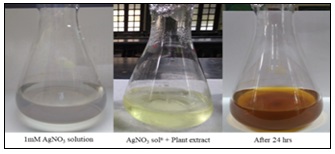
FIG. 1: CHANGE IN REACTION COLOR AFTER AgNP SYNTHESIS
Characterisation of Silver Nanoparticles: UV-Vis Spectrum in the present study UV-Vis spectrum of green synthesized silver nanoparticles was taken from 350 to 650 nm. AgNP’s showed the absorption peak at 442 nm.
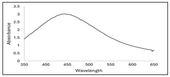
FIG. 2: UV-VIS. SPECTRUM OF PHYTOSYNTHESIZED AgNP
SEM and TEM Analysis: EM analysis of the phytosynthesized nanoparticles revealed the shape of these silver nanoparticles to be spherical where average size was found to be 28 nm.
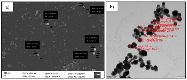
FIG. 3: (A) SEM AND (B) TEM OF PHYTOSYNTHESIZED AgNP
FE-SEM & EDS Analysis: Spherical shape of the synthesized nanoparticles was confirmed by FE-SEM. EDS spectrum showed the characteristic peak at around 3 keV corresponding to Ag. Another peak was observed corresponding to C. EDS analysis revealed percentage relative composition as Ag (50.59) and C (49.41). No other element was observed in the sample. Presence of C can be attributed to the capping elements associated with phytosynthesized AgNP.
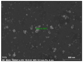
FIG. 4: FE-SEM ANALYSIS OF PHYTOSYNTHESIZED AgNP
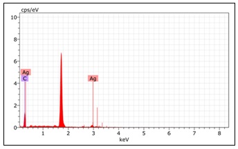
FIG. 5: EDS SPECTRUM OF PHYTOSYNTHESIZED AgNP
FT-IR Analysis: FTIR spectroscopic analysis showed two prominent peaks at wave numbers 3275.47 cm-1 and 1635.63 cm-1. These peaks correspond to polymeric O-H stretch of polyphenols, tannins and/or flavonoids and amide respectively.
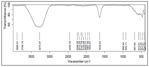
FIG. 6: FTIR ANALYSIS OF PHYTOSYNTHESIZED AgNP
Antibacterial Activity of Phytosynthesized AgNP’s: Ag NPs showed remarkable preliminary antibacterial activity based on zone of inhibition against E. coli (ATCC 25922) which is human pathogenic bacteria. As it was the aqueous extracts, and the stock concentration was 1mg/ml, getting the zone of inhibition at 350 µg/ml was very encouraging.
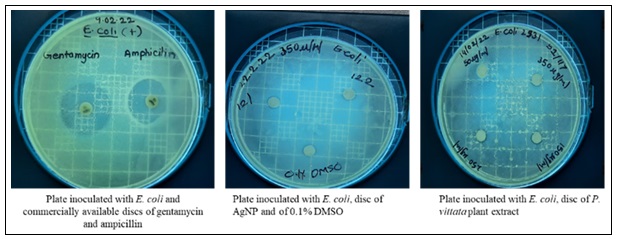
FIG. 7: ANTIBACTIRIAL ACTIVITY OF PHYTOSYNTHESIZED AgNP
TABLE 1: ANTIBACTERIAL ACTIVITY OF PHYTOSYNTHESIZED AGNP AGAINST E. COLI
| Sr. no. | Concentration | Zone of inhibition of AgNP | Zone of inhibition of plant extract |
| 1 | 50 µg/ml | 8 mm | 6 mm |
| 2 | 150 µg/ml | 10 mm | 7 mm |
| 3 | 250 µg/ml | 11mm | 8 mm |
| 4 | 350 µg/ml | 13 mm | 8 mm |
DISCUSSIONS: Most of the research groups have reported the use of 1 mM AgNO3 as silver salt for the synthesis of silver nanoparticles using plants 7, 20, 21. Generally, plant extract to silver salt ratio is maintained as 1:9 for production of nanoparticles. However, in the present study we got good results with significantly lower ratio of 1:100. This suggests that the plant extract used in the present investigation showed maximum reducing as well as capping activity even at a minimum concentration which might be attributed to higher activity and/or concentration of the bioactive compounds in the extract comprising of various secondary metabolites such as polyphenols, tannins, flavonoids, amide etc.
The formation of AgNPs was confirmed through UV–Visible spectroscopic analysis, which revealed an absorption peak within the characteristic surface plasmon resonance (SPR) region reported for silver nanoparticles. Antonysamy Johnson et. al. 19 have reported the presence of AgNP with absorption spectrum around 400-420 nm while Nadzir et. al. 22 confirmed the absorption bands at 468-471 nm. Das G. et. al. 23, reported the absorption maxima at 488 nm. Whereas Sant et. al. 9, reported the peak at 420 nm. The peak we got in the present study Fig. 2 is in accordance with the reported range. Differences in the size and shape of nanoparticles and their degree of aggregation may lead to variation in the absorption maxima. Generally, peaks at lower wavelengths indicate smaller and comparatively monodispersed nanoparticles while the red shift towards the higher wavelength suggests increased size as well as the aggregation 19. Therefore, the SPR peak observed in the present study positively suggest the formation of stable nanoparticles.
Varied shapes of the phytosynthesized nanoparticles have been reported by different research groups. Ibrahim H. et.al. 24, reported spherical shape which is consistent with the shape observed in the present study while Karunakaran G. et.al. 25, reported the presence of both ellipsoidal AgNP as well as spherical AgNP. This variation in shape might be attributed to the composition of the plant extract as different bioactive compounds influence the process of nucleation and stabilization differently during the formation of nanoparticles. Spherical nanoparticles are generally considered more stable due to uniform surface energy distribution. Hence, the present study indicates phytosynthesis of AgNP by controlled nucleation and stabilization mediated by the phytochemicals present in P. vittata extract.
Field Emission Scanning Electron Microscopy (FE-SEM) analysis provided detailed insight into the surface morphology and structural characteristics of the phytosynthesized silver nanoparticles. The FE-SEM micrographs Fig. 4 clearly revealed that the nanoparticles were predominantly spherical in shape with a fairly uniform distribution. The particles appeared well-dispersed with minimal aggregation, indicating effective stabilization by phytochemicals present in the P. vittata extract. The surface of the nanoparticles was observed to be smooth and homogeneous, suggesting controlled nucleation and growth during the phytosynthesis process. The nanoscale size range observed in FE-SEM analysis was consistent with SEM and TEM findings and further confirming the formation of nano sized particles. Slight agglomeration was noticed in some regions which may be attributed to the natural tendency of nanoparticles to minimize surface energy or during drying process of sample preparation. However, the overall morphology indicated successful synthesis of stable and discrete silver nanoparticles. The FE-SEM observations strongly support that plant derived biomolecules acted as reducing as well as capping agents contributing to the stability and uniform morphology of the synthesized AgNPs.
Most of the reported FTIR peaks for AgNP phytosynthesis correspond to phenol, alkene, amide or amine group depending on the plant species or extract used for phytosynthesis. The present study suggests the involvement of amide, polyphenols, tannins and/or flavonoids in the phytosynthesis of AgNP Fig. 6. These biomolecules act as reducing agent for the conversion of silver ions (Ag+) to metallic silver (Ag0) nanoparticles. They might also play a role as capping agent, enhancing the stability of the synthesized nanoparticles.
Antibacterial properties of phytosynthesized silver nanoparticles have widely been reported. Antibacterial activity of phytosynthesized AgNPs against E. coli was previously reported by Maiti S. et. al. 26. De Britto et. al. 27 have confirmed the bactericidal activity of AgNPs synthesized using fern extracts. They showed bactericidal activities against Shigella sp., Staphylococcus aureus and Salmonella Typhi. Bhor et. al. 28 also have reported presence of antibacterial activity of AgNP phytosynthesized using fern extracts against Xanthomonas axonopodis, Klebsiella pneumonia, E. coli. Staphylococcus sp., S. aureus and Proteus morgani Loo et. al. 21 have confirmed the antibacterial activity of phytosynthesized AgNP against E. coli, Klebsiella pneumoniae and Salmonella sp. The metal NPs synthesized using pteridophytes, previously have shown enhanced antibacterial activity which can be attributed to the antioxidant and antibacterial activity of the plant themselves 11. Such synergistic effect may enhance the overall antibacterial efficacy of the synthesized nanoparticles.
CONCLUSION: The present study shows effective use of Pteris vittata for the green synthesis of AgNP’s. It provides a method for the phytosynthesis of AgNPs which is independent of specific pH and/or temperature requirements and requires only minimum amount of plant material. Hence, it gives an easy and effective method for phytosynthesis of AgNPs. This study demonstrates antibacterial activity of AgNPs against the bacteria E. coli. However, further studies including broader microbial screening and toxicity evaluation are required to explore its antibacterial potential and pharmaceutical applications.
ACKNOWLEDGEMENT: Authors are grateful to Principal, Fergusson College and Principal as well as Head, Department of Botany, Nowrosjee Wadia College for providing necessary laboratory and library facilities to carry out this research.
CONFLICT OF INTEREST: Authors have NO conflict of interest.
REFERENCES:
- Gardea-Torresdey JL, Parsons JG, Gomez E, Peralta-Videa J, Troiani HE, Santiago P and Yakaman MJ: Formation and Growth of Au Nanoparticles inside Live Alfalfa Plants. Nano Letters 2002; 2(4): 397–401. DOI: 10.1021/nl015673+.
- Willard MA, Kurihara LK, Carpenter EE, Calvin S and Harris VG: Chemically prepared magnetic nanoparticles. International Materials Reviews 2004; 49(3–4): 125–70. DOI: 10.1179/095066004225021882.
- Nair GM, Sajini T and Mathew B: Advanced green approaches for metal and metal oxide nanoparticles synthesis and their environmental applications. Talanta Open 2022; 5. DOI:10.1016/j.talo.2021.100080.
- Hasan S: A Review on Nanoparticles: Their Synthesis and Types. Research Journal of Recent Sciences 2015; 4: 2502.
- Baig N, Kammakakam I, Falath W and Kammakakam I: Nanomaterials: A review of synthesis methods, properties, recent progress, and challenges. Mater Adv 2021; 2(6): 1821–71. DOI: 10.1039/D0MA00807A.
- Srikar SK, Giri DD, Pal DB, Mishra PK and Upadhyay SN: Green Synthesis of Silver Nanoparticles: A Review. Green and Sustainable Chemistry 2016; 06(01): 34–56. DOI: 10.4236/gsc.2016.61004.
- Sharma VK, Yngard RA and Lin Y: Silver nanoparticles: Green synthesis and their antimicrobial activities. Adv Colloid Interface Sci 2009; 145(1–2): 83–96. DOI: 10.1016/j.cis.2008.09.002
- Aboyewa JA, Sibuyi NRS, Meyer M and Oguntibeju OO: Green synthesis of metallic nanoparticles using some selected medicinal plants from southern Africa and their biological applications. Plants 2021; 10(9): 1929. DOI: 10.3390/plants10091929.
- Sant DG, Gujarathi TR, Harne SR, Ghosh S, Kitture R and Kale S: Adiantum philippense Frond Assisted Rapid Green Synthesis of Gold and Silver Nanoparticles. Journal of Nanoparticles 2013; 2013(1): 182320. DOI: 10.1155/2013/182320.
- Aboyewa JA, Sibuyi NRS, Meyer M and Oguntibeju OO: Gold nanoparticles synthesized using extracts of Cyclopia intermedia, commonly known as honeybush, amplify the cytotoxic effects of doxorubicin. Nanomaterials 2021; 11(1): 132. DOI: 10.3390/nano11010132.
- Das RK, Pachapur VL, Lonappan L, Naghdi M, Pulicharla R and Maiti S: Biological synthesis of metallic nanoparticles: plants, animals and microbial aspects. Nanotechnology for Environmental Engineering 2017; 2(1): 18. DOI: 10.1007/s41204-017-0029-4.
- Fierascu I, Fierascu RC, Ungureanu C, Draghiceanu OA and Soare LC: Application of polypodiopsida class in nanotechnology–potential towards development of more effective bioactive solutions. Antioxidants 2021; 10(5): 748. DOI: 10.3390/antiox10050748.
- Ullah R, Siraj M, Zarshan F, Abbasi BA, Yaseen T, Waris A and Iqbal J: A comprehensive overview of fabrication of biogenic multifunctional metal/metal oxide nanoparticles and applications. Reviews in Inorganic Chemistry 2024; 45(2): 411–436. DOI: 10.1515/revic-2024-0031.
- Bruna T, Maldonado-Bravo F, Jara P and Caro N: Silver nanoparticles and their antibacterial applications. Int J Mol Sci 2021; 22(13): 7202. DOI: 10.3390/ijms22137202
- Losasso C, Belluco S, Cibin V, Zavagnin P, Mičetić I and Gallocchio F: Antibacterial activity of silver nanoparticles: Sensitivity of different Salmonella Front Microbiol 2014; 5: 227. DOI: 10.3389/fmicb.2014.00227
- Savithramma N, Rao ML, Rukmini K and Devi PS: Antimicrobial activity of silver nanoparticles synthesized by using medicinal plants. Int J Chemtech Res 2011; 3(3): 1394–1402.
- Cooke EM: Escherichia coli – an overview. Journal of Hygiene 1985; 95(3): 523–30. DOI: 10.1017/S022217240006065X.
- Vila J, Sáez-López E, Johnson JR, Römling U, Dobrindt U and Cantón R: Escherichia coli: An old friend with new tidings. FEMS Microbiol Rev 2016; 40(4): 437–463. DOI: 10.1093/femsre/fuw005.
- Johnson JR and Russo TA: Extraintestinal pathogenic Escherichia coli: “The other bad E coli.” Journal of Laboratory and Clinical Medicine 2002; 139(3): 155–162. Available from: https://doi.org/10.1067/mlc.2002.121550
- Antonysamy Johnson MA, Shibila T, Amutha S, Menezes IRA, Da Costa JGM and Sampaio NFL: Synthesis of silver nanoparticles using Odontosoria chinensis (L.) J. sm. and evaluation of their biological potentials. Pharmaceuticals 2020; 13(4): 66. DOI: 10.3390/ph13040066.
- Loo YY, Rukayadi Y, Nor-Khaizura MAR, Kuan CH, Chieng BW and Nishibuchi M: In-vitro antimicrobial activity of green synthesized silver nanoparticles against selected Gram-negative foodborne pathogens. Front Microbiol 2018; 9: 1555. DOI: 10.3389/fmicb.2018.01555.
- Nadzir MM, Idris FN and Hat K: Green synthesis of silver nanoparticle using Gynura procumbens aqueous extracts. AIP Conference Proceedings 2019; 2124(1): 030018. DOI: 10.1063/1.5117140.
- Das G, Patra JK and Shin HS: Biosynthesis, and potential effect of fern mediated biocompatible silver nanoparticles by cytotoxicity, antidiabetic, antioxidant and antibacterial, studies. Materials Science and Engineering: C 2020; 114: 111011. DOI: 10.1016/j.msec.2020.111011.
- Ibrahim HMM: Green synthesis and characterization of silver nanoparticles using banana peel extract and their antimicrobial activity against representative microorganisms. Journal of Radiation Research and Applied Sciences 2015; 8(3): 265–275. DOI: 10.1016/j.jrras.2015.01.007.
- Karunakaran G, Jagathambal M, Venkatesh M, Suresh Kumar G, Kolesnikov E and Dmitry A: Hydrangea paniculata flower extract-mediated green synthesis of MgNPs and AgNPs for health care applications. Powder Technology 2017; 305: 488–494. DOI: 10.1016/j.powtec.2016.10.034.
- Maiti S, Krishnan D, Barman G, Ghosh SK and Laha JK: Antimicrobial activities of silver nanoparticles synthesized from Lycopersicon esculentum Journal of Analytical Science and Technology 2014; 5(1): 40. DOI: 10.1186/s40543-014-0040-3.
- De Britto JA, Sheeba Gracelin HD and Jeya Rathna Kumar BP: Antibacterial activity of silver nanoparticles synthesized from a few medicinal ferns. Int J of Pharma Research and Development 2014; 3(3): 224–227.
- Bhor G, Maskare S, Hinge S, Singh L and Nalwade A: Synthesis of silver nanoparticles by using leaflet extract of Nephrolepis exaltata L. and evaluation of antibacterial activity against human and plant pathogenic bacteria. Asian J of Pharma Technology & Innov 2014; 2(7): 23–31.
How to cite this article:
Mysore PN, Boruah D, Antad VV, Bodas D and Limaye AS: Green synthesis of silver nanoparticles using Pteris vittata L. extract and studies on its antibacterial activity against Escherichia coli. Int J Pharm Sci & Res 2026; 17(8): 2427-34. doi: 10.13040/IJPSR.0975-8232.17(8).2427-34.
All © 2026 are reserved by International Journal of Pharmaceutical Sciences and Research. This Journal licensed under a Creative Commons Attribution-NonCommercial-ShareAlike 3.0 Unported License.
Article Information
16
2427-2434
1082 KB
290
English
IJPSR
P. N. Mysore, D. Boruah, V. V. Antad, D. Bodas and A. S. Limaye *
Department of Botany, Nowrosjee Wadia College, Pune, Maharashtra, India.
aslimaye@nowrosjeewadiacollege.edu.in
15 March 2026
22 May 2026
19 June 2026
10.13040/IJPSR.0975-8232.17(8).2427-34
01 August 2026
 Download