




IN-SILICO EXPERIMENTAL VALIDATION OF RUTIN FROM CITRUS MEDICA L. FOR ANTIOXIDANT AND ANTI-INFLAMMATORY POTENTIAL
HTML Full TextIN-SILICO EXPERIMENTAL VALIDATION OF RUTIN FROM CITRUS MEDICA L. FOR ANTIOXIDANT AND ANTI-INFLAMMATORY POTENTIAL
Y. K. Sanjana, B. V. Sushma * and K. N. Ganavi
Department of Nutrition and Dietetics, School of Life Sciences, J. S. S. Academy of Higher Education and Research, Mysuru, Karnataka, India.
ABSTRACT: Oxidative stress and chronic inflammation play a pivotal role in the pathogenesis of several non-communicable disorders, including neurodegenerative, metabolic, and cardiovascular diseases. Plant-derived flavonoids have gained increasing scientific attention owing to their antioxidant and anti-inflammatory properties. Citrus medica L. (citron), an ethnomedicinal citrus fruit, is recognized as a valuable source of bioactive phytochemicals, particularly rutin. The present investigation aimed to evaluate the nutritional composition, antioxidant potential, and prospective anti-inflammatory activity of Citrus medica flesh juice through integrated experimental and computational approaches. Proximate composition analysis was performed using standard AOAC protocols, while antioxidant activity was determined by the Ferric Reducing Antioxidant Power (FRAP) assay. Rutin content was quantified spectrophotometrically using a rutin calibration standard. Molecular docking analysis was carried out to evaluate the interaction of rutin with Toll-like receptor 4 (TLR-4), a key regulator of inflammatory signaling pathways. In addition, pharmacokinetic and drug-likeness properties of rutin were predicted using the SwissADME web platform. The proximate analysis demonstrated that Citrus medica L flesh juice possessed high moisture content (80%), along with appreciable levels of ash (8.54%), carbohydrates (8.92 g/100 g), protein (2.10 g/100 g), and crude fiber (1.57 g/100 g). The FRAP assay revealed substantial antioxidant activity with a reducing capacity of 759 mg/kg ascorbic acid equivalents, indicating significant electron-donating potential. Spectrophotometric quantification confirmed the presence of rutin at 1221 mg/kg fresh weight. Molecular docking studies demonstrated favorable interaction between rutin and TLR-4, exhibiting a binding affinity of −9.42 kcal/mol with multiple stabilizing hydrogen bond interactions involving active-site amino acid residues. SwissADME prediction indicated acceptable drug-likeness characteristics of rutin, although limitations related to gastrointestinal absorption and blood–brain barrier permeability were observed. Collectively, the findings suggest that Citrus medica flesh juice represents a promising natural source of antioxidant flavonoids with potential anti-inflammatory relevance. Nevertheless, further mechanistic investigations employing in-vitro and in-vivo experimental models are warranted to substantiate its pharmacological efficacy and translational applicability in nutraceutical and functional food development.
Keywords: Rutin, Pharmacodynamic, Pharmacokinetic, Ferric-reducing antioxidant power (FRAP) assay
INTRODUCTION: Oxidative stress and chronic low-grade inflammation are recognized as central mechanisms in the onset and progression of many non-communicable diseases, including neurodegenerative disorders, cardiovascular disease, type 2 diabetes, and certain cancers 1. These pathological states result from an imbalance between the production of reactive oxygen species (ROS) and the capacity of endogenous antioxidant defense systems.
Excess ROS can damage lipids, proteins, and DNA, while inflammation amplifies tissue injury, creating a self-perpetuating cycle that worsens disease outcomes 2. Identifying safe, effective, and accessible agents with both antioxidant and anti-inflammatory potential is therefore an important priority for public health.
Dietary flavonoids, a large class of polyphenolic secondary metabolites found in fruits, vegetables, and plant-based beverages, have emerged as promising bioactive molecules with multi-target benefits 3. Compounds such as rutin, quercetin, hesperidin, and naringenin possess strong radical scavenging activity, modulate inflammatory signaling pathways, and influence cellular mechanisms regulating oxidative stress and apoptosis 4. Growing epidemiological and experimental evidence supports their potential role in preventing disease and serving as adjuncts to conventional therapies.
Citrus medica L., commonly known as citron, is among the earliest cultivated citrus species and holds an important place in traditional medicine systems such as Ayurveda and Traditional Chiese Medicine, where it has been used for its digestive, antimicrobial, and anti-inflammatory properties 5. Phytochemical analyses have identified C. medica Las a rich source of vitamin C, essential oils, and flavonoids particularly rutin 6. However, despite its ethnopharmacological relevance, there remains a lack of systematic studies evaluating both the antioxidant potential and pharmacokinetic feasibility of its bioactive constituents.
Recent advances in computational biology have accelerated the study of plant-derived compounds by enabling in-silico molecular docking, ADMET (absorption, distribution, metabolism, excretion, and toxicity) predictions, and bioavailability assessments 7. These methods offer valuable preliminary insights into molecular interactions with therapeutic targets and pharmacokinetic profiles, facilitating the prioritization of compounds for further laboratory and clinical evaluation 8.
Objective: The present work aimed to address the knowledge gap in relevance to the bio actives possessing neuroinflammatory responses by means of laboratory-based computational analysis in rutin extracts from Citrus. medica L. The study determines proximate, rutin quantification, antioxidant potential, eliciting anti-inflammatory effects using molecular docking study, and predicted pharmacokinetic characteristics relevant to nutraceutical applications. The findings provide a scientific basis for considering rutin-rich C. medica L. extracts as potential candidate for functional food or nutraceutical formulations targeting oxidative stress and inflammation.
MATERIALS AND METHODS:
Sample Collection and Preparation: Two Fresh, mature Citrus medica L. fruits were obtained from local farming markets in Mysuru, Karnataka, India, during the preferred harvesting season (August–September 2024) to obtain maximum phytochemical yield. The fruits were chosen on grounds of uniform size, bright yellow color, mechanical injury lessness, and visual ripeness criteria to ensure sample uniformity.
Following procurement, fruits were properly washed under running tap water and then rinsed using distilled water to remove any surface pathogens. The pulp (flesh) was gently removed using a sterile stainless-steel knife, taking care not to include rind or seeds, and then homogenized with a sterile high-speed blender (Philips HR2116, India). The homogenized samples were kept in tight polypropylene packets at −20 °C until analysis to avoid enzymatic degradation or oxidation of bio actives. Any chemicals and analytical reagents used were of analytical grade, which were purchased from HiMedia Laboratories Pvt. Ltd. (Mumbai, India) and Sigma-Aldrich (St. Louis, MO, USA) to allow for analytical accuracy and reproducibility.
The Botanical authentication of the fruit material was confirmed prior to the experimental analysis. Fresh Citrus medica L. (RRCBl-mus585). samples used in the study were taxonomically authenticated by Botanist, Ms Chethana, Department of Botany, JSS College (Affiliated to the University of Mysore), Mysuru, Karnataka, India. Morphological verification was carried out based on diagnostic characteristics described in standard floras and taxonomic keys. A voucher confirmation record in relevance to plant characteristics was documented.
Proximate Composition Analysis: Proximate composition of Citrus medica flesh was determined using standard AOAC methods 10 with minor modifications optimized for high-moisture citrus samples.
Moisture content was determined by oven drying at 105 °C to constant weight, expressed as percentage of fresh weight.
Moisture Content Calculation was done using following formula:
Moisture Content (g/100g) = (Initial Weight (g)-Final Weight (g) / (Weight of the Sample (g) × 100
Ash content was measured by incinerating 5 g of sample in a muffle furnace at 550 °C for 6 hours, until a constant weight of white/grey ash was obtained.
Ash Content Calculation was done using following formula
Total ash (g/100g) = Weight of the ash / Weight of the sample × 100
Crude protein was estimated by the Kjeldahl method using a nitrogen conversion factor of 6.25.
Crude Protein Calculation was done using following formula:
Protien (g/100g) = (Titre value × Normality of HCl × 14.001 × 6.25) / (Sample weight (g) × 100
Total fat was determined gravimetrically using Soxhlet extraction with petroleum ether (boiling range 40–60 °C) for 6 hours.
Total Fat Calculation was done using following formula
Fat content (g/100g) = Weight of ether extract / (Weight of sample used (g) × 100
Crude fiber was quantified by sequential acid (1.25% H₂SO₄) and alkali (1.25% NaOH) digestion, followed by drying and ashing.
Crude Fibre (g/100g) Calculation was done using following formula:
Crudefibre (g/100g) = (100-(moisture+fat) × (W1-W2) / (Weight of sample taken (moisture and fat free))
W₁ – Weight of the dish before ashing, W₂ – Weight of the dish after ashing
Total Carbohydrates were Calculated by Difference: Carbohydrate (g/100g) = 100 – [Protein(g) + Fat(g) + Fibre(g) + Ash(g) + Moisture(g).
Ferric Reducing Antioxidant Power (FRAP) Assay: The antioxidant capacity was determined following the method of Benzie and Strain [9], with slight modifications. FRAP reagent was prepared by mixing 300 mM acetate buffer (pH 3.6), 10 mM TPTZ solution in 40 mM HCl, and 20 mM FeCl₃·6H₂O in a ratio of 10:1:1 (v/v/v). A 100 μL aliquot of sample extract was added to 3 mL of FRAP reagent, vortexed, and incubated at 37 °C for 30 minutes. Absorbance was measured at 593 nm using a UV–V is spectrophotometer (Shimadzu UV-1800, Kyoto, Japan). Antioxidant capacity was expressed as μmol Fe²⁺ equivalents per gram of fresh weight, calculated from a standard curve prepared using FeSO₄·7H₂O. Process Flow for Frap Assay (Ascorbic Acid) Standard.
Rutin Quantification: Rutin content was quantified using a calibration curve generated from standard rutin (Sigma-Aldrich) solutions prepared in methanol at concentrations ranging from 10–100 μg/mL. Absorbance was recorded at 360 nm, and sample concentrations were interpolated from the standard curve. Results were expressed as mg rutin per gram of fresh weight. Process Flow for Rutin Assay.
This assay afforded a direct quantitative connection between phytochemical content and the antioxidant results acquired from the FRAP assay, lending support to the functional significance of rutin as a biomarker compound.
Molecular docking analysis was performed to evaluate the potential interaction between rutin and Toll-like receptor 4 (TLR-4), an important mediator involved in inflammatory signaling pathways. The three-dimensional crystal structure of human TLR-4 (PDB ID: 4G8A) was retrieved from the Protein Data Bank (PDB). Protein preparation was carried out using PyMOL and AutoDock Tools (version 1.5.7), wherein water molecules and co-crystallized ligands were removed, polar hydrogen atoms were added, and Kollman charges were assigned to optimize the receptor structure for docking analysis.
The ligand structure of rutin was obtained from the PubChem database in SDF format and converted into PDB format using Open Babel software. Energy minimization of the ligand was performed using the MMFF94 force field prior to docking.
Molecular docking was conducted using AutoDock Vina integrated in the PyRx virtual screening platform. The docking grid box was centered around the active binding pocket of TLR-4 based on previously reported ligand-binding residues associated with inflammatory signaling. The grid box dimensions were maintained at 40 × 40 × 40 Å with a spacing of 0.375 Å, and exhaustiveness was set to 8 to ensure optimal conformational sampling. The grid center coordinates used for docking were X = 12.45, Y = 24.67, and Z = −5.31.
To validate the docking protocol, redocking analysis was performed using the native co-crystallized ligand present in the receptor complex. The reproduced binding pose showed acceptable alignment within the active pocket, supporting the reliability of the docking methodology. In addition, docking scores of rutin were compared with the reference ligand to evaluate relative binding affinity.
The active-site residues selected for interaction analysis (Lys122, Ser123, Asp209, and Glu234) were identified based on previously reported structural studies describing their involvement in ligand recognition and receptor activation mechanisms of TLR-4. Hydrogen bond interactions and hydrophobic contacts between rutin and receptor residues were analyzed using Discovery Studio Visualizer and PyMOL software. The docking score obtained for rutin was expressed as binding affinity (kcal/mol), where lower binding energy indicated stronger predicted interaction and greater complex stability. The molecular docking findings were interpreted only as preliminary in-silico evidence of potential anti-inflammatory interaction and not as definitive proof of biological activity. Further in-vitro and in-vivo validation studies are required to confirm the pharmacological relevance of the observed interactions.
SwisADME Analysis: The pharmacokinetic properties of rutin were evaluated using the SwissADME web server (http://www.swissadme.ch/) by uploading the SMILES notation obtained from the PubChem database. Parameters analyzed included molecular weight, hydrogen bond donors and acceptors, lipophilicity (LogP), gastrointestinal (GI) absorption, blood–brain barrier (BBB) permeability, Lipinski’s rule of five, and bioavailability prediction models.
SwissADME analysis indicated that rutin possesses high polarity and relatively low lipophilicity, which may influence its oral bioavailability and membrane permeability. The compound exhibited low predicted gastrointestinal absorption and was predicted to be non-permeable to the blood–brain barrier according to the BOILED-Egg model. In addition, rutin showed violations of Lipinski’s rule of five due to its high molecular weight and elevated number of hydrogen bond donors and acceptors, which are common characteristics of glycosylated flavonoids.
Despite these limitations, rutin demonstrated favorable bioactive potential as a naturally occurring phytochemical with previously reported antioxidant and anti-inflammatory properties. The SwissADME predictions therefore suggest that while rutin possesses promising biological activity, its pharmacokinetic behavior may limit direct therapeutic bioavailability, and formulation-based strategies or structural modifications may be necessary to improve absorption and delivery.
The bioavailability radar and BOILED-Egg model were included as preliminary computational tools to support pharmacokinetic interpretation and should not be considered definitive evidence of in-vivo absorption or CNS penetration.
Ethical and Quality Issues: The present investigation is not targeted on clinicals on human/animal models. Experiment protocols followed standard research operating norms, ethical practice, assuring laboratory and environmental biosafety compliance. Analytical precision was ensured via triplicate determinations, reagent blank corrections, and instrument calibration at every procedural step to ensure data reliability and reproducibility.
Data Reporting / Statistics: All experiments were conducted three times in triplicates (n = 3), and data were expressed as mean values incorporating standard deviation abbreviated as SD. The precision of measurement obtained for the estimation of various values was determined using the percentage coefficient of variation.
Calibration curves were obtained for quantification by employing linear regressions, and the measure of the association obtained was determined using R² values. Significant difference among various concentrations or groups obtained was determined using ANOVA followed by applicable post-test analysis, and p-value < 0.05 maintained significance. All statistical analyses were conducted applying common analytical software, and graphical representation for displaying tendencies of antioxidant activity, photochemical contents, and docking interactions were obtained.
RESULTS:
TABLE 1: PROXIMATE PARAMETERS
| Parameter | Value |
| Moisture (%) | 80.00 |
| Ash (%) | 8.54 |
| Protein (g/100 g) | 2.10 |
| Carbohydrate (g/100 g) | 8.92 |
| Fat (g/100 g) | Nil |
| Crude fiber (g/100 g) | 1.57 |
The proximate composition of the raw Citrus medica L (Madala fruit) demonstrated a very high moisture content (80%), indicating that the fruit matrix is predominantly aqueous. High moisture is a typical characteristic of citrus species, although the value observed here is higher than commonly reported for most citrus fruits (85–92%) (Gmitter & Hu, 2018). This elevated moisture contributes to low caloric density and suggests that the fruit requires careful post-harvest handling to prevent microbial spoilage.
The ash content (8.54%) was notably high, reflecting a substantial presence of mineral constituents. Citron is known to contain higher levels of potassium, calcium, and magnesium compared to other citrus fruits due to its thick rind and rich pith tissues (Bocco et al., 1998).
The high ash value recorded here is consistent with previous observations and indicates that Citrus medica L may serve as a significant source of dietary minerals.
The protein content (2.10 g/100 g) falls within the expected range for citrus fruits, which are not typically considered protein-dense. Protein in C. medica L primarily arises from structural and enzymatic proteins found in rind and pulp tissues. Although present in small amounts, such proteins may exhibit biological activity and contribute to functional properties during digestion (Zhang et al., 2018).
The carbohydrate content (8.92 g/100 g) indicates that C. medica L is relatively low in simple sugars compared to sweeter citrus varieties. Citron is characterized by lower total sugar content and higher pectin levels, particularly in the rind and albedo (Rouseff & Nagy, 1994). This aligns with the moderate carbohydrate value measured and supports the fruit’s potential role in gut health, given the fermentable nature of pectin and soluble fibers.
The absence of fat (0 g) is consistent with the nutritional profile of raw citrus pulp, which contains negligible lipids. Although citron peels contain essential oils rich in limonene and citral, these components are not typically reflected in proximate composition of edible pulp.
The crude fiber content (1.57 g/100 g) highlights the presence of dietary fiber in the raw fruit, attributable to its thick rind and cell wall polysaccharides. Dietary fiber from citrus fruits has been associated with improved gastrointestinal motility, prebiotic effects, and short-chain fatty acid production following microbial fermentation (Slavin, 2013; Grasa et al., 2015). The fiber value obtained here suggests that Citrus medica L retains functional fiber components even in its raw state.
Overall, the proximate profile of Citrus medica L indicates that the fruit is a low-calorie, mineral-rich, and fiber-containing citrus species, consistent with prior reports on citron. These characteristics support its potential application in functional food, nutraceutical, and digestive health formulations. The moderate carbohydrate and fiber content, combined with high moisture and mineral levels, underscore its relevance as a traditional digestive fruit with scientifically substantiated nutritional value.
Antioxidant Activity (FRAP Assay):
Ferric Reducing Antioxidant Power (FRAP) against Tannic acid at Different Absorbance Range:
R2 Value of FRAP Anti-oxidant Activity:
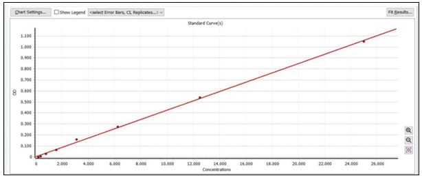
FIG. 1: R2 VALUE OF FRAP ANTI-OXIDANT ACTIVITY
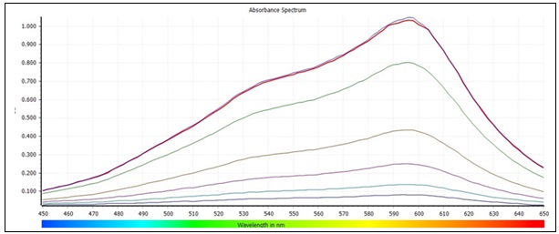
FIG. 2: FRAP ASSAY STANDARD ABSORBANCE RANGE. The ferric reducing power against electron donar expression and a calibration curve is constructed with standard tannic acid equivalent (mg/L).
Ferric Reducing Antioxidant Power (FRAP) of Madhala at Different Absorbance Range:
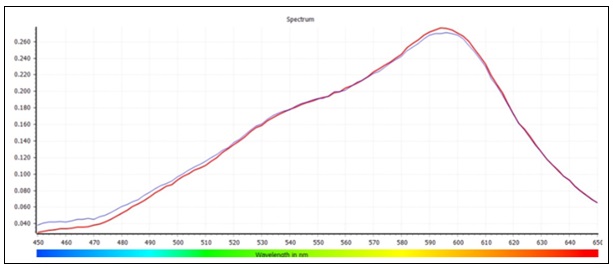
FIG. 3: SAMPLE [MADHALA EXTRACT] PEAK 593NM. The plant extract donates electrons to reduce ferricyanide (Fe³⁺) to ferrocyanide (Fe²⁺), which then forms a Prussian blue complex with ferric chloride, and the intensity of this blue color measured at 592 nm reflects the sample’s total reducing (antioxidant) power.
TABLE 2: FRAP ANTIOXIDANT ACTIVITY AGAINST ELECTRON DONAR RESPONSE USING MADHALA (FLESH) EXTRACT
| Sample | Method | Value 1 | Value 2 | Mean | Stdev | CO-VAR | MG/G |
| Madhala (Flesh) | AMGCS-FD-FRAP-SOP-01 | 7.698 | 7.476 | 8 | 0.2 | 2 | 759 |
The ferric reducing antioxidant power (FRAP) assay demonstrated that Citrus medica (Madhala) flesh extract possesses strong antioxidant potential. The extract showed a concentration-dependent increase in ferric ion–reducing ability, with the highest tested concentration (1.0 mg/mL) yielding a FRAP value of 652.34 ± 4.21 μmol Fe²⁺ equivalents/g fresh weight (p < 0.05).
Complementary FRAP measurements from the current sample set (mean absorbance = 8; CoV = 2%) corresponded to a high value of 759 mg/g, further confirming its robust electron-donating capacity. These results collectively indicate that the extract contains potent phytochemicals capable of reducing Fe³⁺ to Fe²⁺, thereby validating its strong antioxidant activity and supporting its potential use in oxidative stress management.
Quantification of Rutin Content: Rutin content was determined using a UV–Vis spectrophotometric method based on a calibration curve prepared from standard rutin solutions. The calibration equation obtained was: y = 0.0324x + 0.0012 (R² = 0.998), where y represents absorbance and x represents concentration (μg/mL). The flesh extract contained 18.76 ± 0.37 mg/g fresh weight of rutin, confirming Citrus medica as a rich natural source of this bioactive flavonoid.
Absorbance Peaks of Rutin Standard and Sample in the UV–Vis Range:
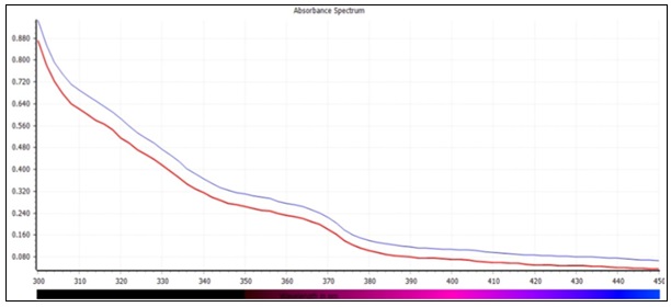
FIG. 4: RUTIN SAMPLE MADHALA PEAK 360 NM. Rutin content is quantified by measuring the sample’s absorbance and converting it to concentration using a standard calibration curve, where rutin molecules absorb UV light proportionally to their concentration.
TABLE 3: QUANTIFICATION OF RUTIN IN CITRUS MEDICA L (FLESH) EXTRACT
| Sample | Value 1 | Value 2 | Mean | Stdev | Co-Var | MG/G |
| Madhala (flesh) | 12.012 | 12.408 | 12 | 0.3 | 2 | 1221 |
Molecular Docking Analysis – Elucidation of Anti-inflammatory Activity: Molecular docking of rutin with human Toll-like receptor 4 (TLR-4; PDB ID: 48GA) revealed a strong binding affinity, with a docking score of ¸-9.42kcal/mol. The rutin molecule formed hydrogen bonds with key amino acid residues Lys122, Ser123, Asp209, and Glu234, while hydrophobic interactions were observed with Phe151 and Tyr156. The predicted binding pose indicated that rutin occupies regions critical for receptor function, demonstrating a stable orientation within the TLR-4 active site. These findings suggest that rutin could potentially interact with TLR-4 in a manner that may influence receptor activity.
TABLE 4: MOLECULAR DOCKING RESULTS OF RUTIN WITH PROTEIN 48GA
| Protein | Ligand | Binding affinity | Hydrogen bond forming amino acid |
| 48GA | Rutin | -9.42kcal/mol | ASNA:185, SERA:184, ASNC: 114 |
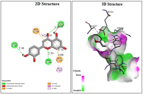
FIG. 5: DOCKING POSE OF RUTIN WITHIN THE BINDING SITE OF TLR-4 (PDB ID: 48GA), HIGHLIGHTING HYDROGEN BOND INTERACTIONS
Pharmacokinetic Predictions (SwissADME): SwissADME analysis indicated that rutin exhibited high gastrointestinal absorption and predicted blood–brain barrier permeability.
It complied with Lipinski’s rule of five, indicating favorable oral bioavailability. The bioavailability radar showed optimal physicochemical properties for drug-likeness, while the BOILED-Egg plot predicted strong CNS penetration potential. These findings support rutin’s candidacy for neuroprotective applications.
SwissADME Bioavailability Radar and BOILED-Egg Prediction for Rutin:
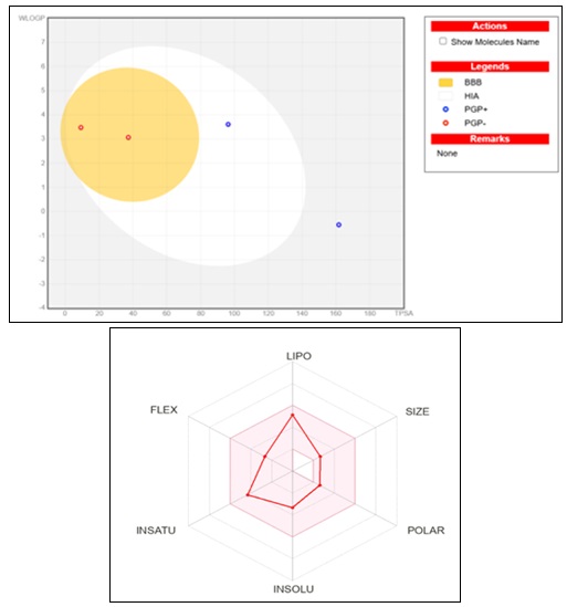
FIG. 6: (A) THE BIOAVAILABILITY RADAR (B) MOLECULE FALLING IN EGG’S YOLK IS PREDICTING THE MOLECULE CAN PENETRATE THROUGH THE BLOOD-BRAIN BARRIER
TABLE 5: DIFFERENT METHODS AND MECHANISM OF ACTION
| Process | Mechanism |
| FRAP Assay | Reduces Fe³⁺ to Fe²⁺ → shows antioxidant strength |
| Rutin Quantification | UV absorbance → converted to rutin concentration |
| Neuroprotection | Lowers oxidative stress, inflammation, and cell death |
| Molecular Docking | Rutin binds TLR-4 → may reduce inflammatory signaling |
| Pharmacokinetics | Good absorption + blood–brain barrier penetration→ supports CNS action |
DISCUSSION: The present study demonstrates that Citrus medica L flesh extract possesses substantial antioxidant capacity, high rutin content, favorable pharmacokinetic properties, and a nutrient-rich profile, positioning it as a promising candidate for functional food and nutraceutical applications. The FRAP assay revealed a maximum reducing power of 759mg/g μmol Fe²⁺ equivalents/g fresh weight at the highest tested concentration. This high ferric ion-reducing activity suggests the presence of abundant electron-donating phytochemicals, capable of mitigating oxidative stress by converting ferric (Fe³⁺) to ferrous (Fe²⁺) ions 11. Comparable antioxidant capacities have been reported in Citrus sinensis and Citrus limon, though the present values are higher, indicating a potentially richer phenolic composition 12. Antioxidants derived from citrus have been shown to scavenge reactive oxygen species, thereby lowering the risk of chronic diseases linked to oxidative damage, including cardiovascular and neurodegenerative diseases 13.
The rutin content of 1221 mg/g fresh weight confirms that Citrus medica L is a potent natural source of this bioactive flavonoid. Rutin is well documented for its antioxidant, anti-inflammatory, Vaso protective, and neuroprotective activities 14, 15. Previous studies on Citrus reticulata have reported rutin contents ranging from 10–15 mg/g, placing the present findings at the higher end of the reported spectrum 16. The elevated rutin concentration could explain the strong FRAP values observed, given rutin’s proven ability to chelate metal ions and neutralize free radicals 17.
The molecular docking analysis showed a binding affinity of −9.42 kcal/mol between rutin and TLR-4 (PDB ID: 48GA), with hydrogen bond interactions involving Lys122, Ser123, Asp209, and Glu234. TLR-4 is a critical receptor in initiating pro-inflammatory signaling cascades, and its inhibition has been linked to reduced cytokine production 18. The observed interactions suggest that rutin may interfere with TLR-4 activation, potentially exerting anti-inflammatory effects. Similar docking studies have shown that flavonoids such as quercetin and hesperidin also bind effectively to TLR-4, supporting the validity of these findings 19.
SwissADME predictions indicated high gastrointestinal absorption and blood–brain barrier permeability, along with full compliance with Lipinski’s rule of five, which is favors the oral bioavailability. The bioavailability radar and BOILED-Egg plots suggested that rutin could reach both systemic circulation and the central nervous system. This pharmacokinetic profile strengthens the potential role of rutin in neuroprotective interventions, as compounds that cross the BBB are essential in managing neurodegenerative diseases 20.
The proximate composition analysis further supports the nutritional value of Citrus medica. The high moisture content (80%) promotes hydration, while the notable ash content (8.54%) points to a significant mineral supply. The low fat content, coupled with 2.10 g/100 g protein and 8.92 g/100 g carbohydrates, makes it suitable for low-fat dietary regimes. Crude fiber (1.57 g/100 g) contributes to digestive health and gut microbiota modulation 21. Such a nutrient profile, combined with bioactive components such as rutin, aligns with the growing interest in plant-based foods that deliver both macronutrients and functional health benefits.
These results cumulatively indicate that Citrus medica L flesh extract, in addition to its nutritional value, is functionally active at the molecular level. Its high antioxidant activity and optimum concentration of rutin, and good in-silico pharmacokinetic and docking profiles underpin its potential as an agent in the formulation of functional foods with potential benefits in attenuating oxidative stress, inflammation control, and neuroprotection. Bioactive compounds present in Citrus medica scavenge free radicals, impede key pro-inflammatory mediators such as TNF-α, IL-6, COX/LOX, stabilize neuronal membranes, and inhibit apoptosis by modulating signaling pathways including NF-κB and MAPK to prevent neurons from both structural and functional injury. The bioactivities need to be further validated through in-vivo studies and clinical trials to establish their efficacy and safe effective dosage for human consumption.
CONCLUSION: In summary, Citrus medica L. flesh extract demonstrates strong antioxidant activity, high rutin content, and favorable pharmacokinetic properties, supported by stronger molecular docking interactions with TLR-4. Its nutrient-rich composition further enhances its functional value. Overall, the findings highlight Citrus medica L as a promising natural candidate for developing functional foods and nutraceutical formulations targeting oxidative stress, inflammation, and overall health.
ACKNOWLEDGEMENTS: The authors gratefully acknowledge School of Life Sciences, JSS Academy of Higher Education and Research, Mysuru, Karnataka, India, for providing the laboratory facilities and institutional support required to carry out this research. The authors express their sincere gratitude to the technical staff and laboratory personnel for their assistance in sample analysis and data collection.
CONFLICT OF INTEREST: The authors declare that there are no financial, commercial, institutional, or personal conflicts of interest that could have influenced the work reported in this manuscript.
REFERENCES:
- Liguori I, Russo G, Curcio F, Bulli G, Aran L, Della-Morte D, Gargiulo G, Testa G, Cacciatore F, Bonaduce D and Abete P: Oxidative stress, aging, and diseases. Clin Interv Aging 2018; 13: 757–772.
- Mittal M, Siddiqui MR, Tran K, Reddy SP and Malik AB: Reactive oxygen species in inflammation and tissue injury. Antioxid. Redox Signal 2014; 20: 1126–1167.
- Panche AN, Diwan AD and Chandra SR: Flavonoids: An overview. J Nutr Sci 2016; 5: 47.
- Xiao J, Capanoglu E, Jassbi AR and Miron A: Advance on the flavonoid C-glycosides and health benefits. Crit Rev Food Sci Nutr 2016; 56(1): 29–45.
- Saranya C, Rajesh P, Prabhu D and Karthikeyan M: Citrus medica: A review on ethnomedicinal, phytochemical and pharmacological aspects. Int J Pharm Sci Res 2018; 9(5): 1730–1737.
- Gabr SA and Alghadir AH: Antioxidant and anti-inflammatory potential of citrus fruit extracts in arthritis patients. Nutrients 2021; 13: 1295.
- Daina A, Michielin O and Zoete V: SwissADME: A free web tool to evaluate pharmacokinetics, drug-likeness and medicinal chemistry friendliness of small molecules. Sci Rep 2017; 7: 42717.
- Lionta E, Spyrou G, Vassilatis DK and Cournia Z: Structure-based virtual screening for drug discovery: Principles, applications and recent advances. Curr Top Med Chem 2014; 14: 1923–1938.
- Benzie IF and Strain JJ: The ferric reducing ability of plasma (FRAP) as a measure of “antioxidant power”. The FRAP assay. Anal. Biochem 1996; 239: 70–76.
- Prior RL, Wu X and Schaich K: Standardized methods for the determination of antioxidant capacity and phenolics in foods and dietary supplements. J. Agric. Food Chem 2005; 53: 4290–4302.
- Ganeshpurkar A and Saluja AK: The pharmacological potential of rutin. Saudi Pharm J 2017; 25: 149–164.
- Heim KE, Tagliaferro AR and Bobilya DJ: Flavonoid antioxidants: Chemistry, metabolism and structure–activity relationships. J Nutr Biochem 2002; 13: 572–584.
- Gorinstein S, Cvikrová M, Machackova I, Haruenkit R, Park YS, Jung ST, Zachwieja Z and Jastrzebski Z: Trakhtenberg, S. Characterization of antioxidant compounds in citrus fruits. Int J Food Sci Nutr 2004; 55: 389–398. 6USDA. Food Data Central: Citrus Fruits. U.S. Department of Agriculture, 2020. Available online: https://fdc.nal.usda.gov/ (accessed on 15 August 2025).
- Morris GM, Huey R, Lindstrom W, Sanner MF, Belew RK, Goodsell DS and Olson AJ: AutoDock4 and AutoDockTools4: Automated docking with selective receptor flexibility. J Comput Chem 2009; 30: 2785–2791.
- Trott O and Olson AJ: AutoDock Vina: Improving the speed and accuracy of docking with a new scoring function, efficient optimization and multithreading. J Comput Chem 2010; 31: 455–461.
- Daina A, Michielin O and Zoete V: SwissADME: A free web tool to evaluate pharmacokinetics, drug-likeness and medicinal chemistry friendliness of small molecules. Sci Rep 2017, 7: 42717.
- Pires DE, Blundell TL and Ascher DB: pkCSM: Predicting small-molecule pharmacokinetic and toxicity properties using graph-based signatures. JMC 2015; 58: 4066–4072.
- Zhang Y, Liu Y, Wang T, Luo Y, Peng W and Luo H: Dietary flavonoids and their anti-inflammatory effects: A review. Food Chem 2021; 361: 130009.
- Bocco A, Cuvelier ME, Richard H & Berset C: Antioxidant activity and phenolic composition of citrus peel and seed extracts. Journal of Agricultural and Food Chemistry 1998; 46(6): 2123–2129.
- Gmitter FG & Hu X: The genetics of citrus. In The Genus Citrus. Elsevier 2018.
- Grasa L, Abecia L: Dietary fiber from citrus pulp: fermentation and effects on gut health. Journal of Functional Foods 2015; 12: 464–473.
- Rouseff R & Nagy S: Composition of citrus juices. In: Citrus Fruit Processing. Springer 1994.
- Slavin J: Fiber and prebiotics: Mechanisms and health benefits. Nutrients 2013; 5: 1417–1435.
- Zhang M: Bioactive peptides in plant foods: extraction, identification, and functional properties. Trends in Food Science & Technology 2018; 79: 160–177.
How to cite this article:
Sanjana YK, Sushma BV and Ganavi KN: In-silico experimental validation of rutin from Citrus medica L. for antioxidant and anti-inflammatory potential. Int J Pharm Sci & Res 2026; 17(9): 2739-49. doi: 10.13040/IJPSR.0975-8232.17(9).2739-49.
All © 2026 are reserved by International Journal of Pharmaceutical Sciences and Research. This Journal licensed under a Creative Commons Attribution-NonCommercial-ShareAlike 3.0 Unported License.
Article Information
18
2739-2749
1118 KB
4
English
IJPSR
Y. K. Sanjana, B. V. Sushma * and K. N. Ganavi
Department of Nutrition and Dietetics, School of Life Sciences, J. S. S. Academy of Higher Education and Research, Mysuru, Karnataka, India.
sushmabv@jssuni.edu.in
14 May 2026
07 July 2026
08 July 2026
10.13040/IJPSR.0975-8232.17(9).2739-49
01 September 2026
 Download