




MICROBIOLOGICAL SPECTRUM AND DIAGNOSTIC APPROACH IN NEONATAL SEPSIS
HTML Full TextMICROBIOLOGICAL SPECTRUM AND DIAGNOSTIC APPROACH IN NEONATAL SEPSIS
Nidhi Pal *, Anju Rani, Jagriti Bansal, Kushal Singh, Ajay Kumar Sahni and Harmesh Manocha
College of Allied and Healthcare Science, GIMS, Greater Noida, Uttar Pradesh, India.
ABSTRACT: Background: Neonatal sepsis is a life-threatening systemic infection caused by bacteria, fungi, or viruses, characterized by hemodynamic instability and variable clinical presentations. Early and accurate diagnosis remains challenging and typically relies on a combination of clinical assessment, laboratory parameters, and microbiological confirmation, most commonly through blood culture. Materials and Methods: A hospital-based prospective cross-sectional study was conducted in the Government Institute of Medical Sciences (GIMS), Greater Noida, from June 1, 2024, to May 31, 2025. A total of 511 blood samples from neonates with suspected sepsis were processed using the automated BACTEC blood culture system. Isolates were identified and antimicrobial susceptibility testing was performed using the VITEK 2 Compact system. Results: Of the 511 suspected cases, 144 (28.2%) were culture-positive. Fungal isolates (budding yeast cells) were the most frequent, accounting for 63 (43.8%) cases, followed by Gram-negative bacilli in 54 (37.5%) cases and Gram-positive cocci in 27 (18.8%) cases. Among fungal pathogens, Candida albicans was predominant (46 isolates; 31.9%), with a higher proportion observed in early-onset sepsis (43.7%) compared to late-onset sepsis (22.8%). Candida pelliculosa was the second most common fungal isolate. Among Gram-negative organisms, Klebsiella pneumoniae (14.6%) was the most frequently isolated, followed by Pseudomonas aeruginosa (6.3%), Escherichia coli (4.9%), Serratia marcescens (4.2%), and Acinetobacter spp. (3.5%). Among Gram-positive cocci, coagulase-negative staphylococci (CONS) were predominant (10.4%). Most bacterial isolates, both Gram-negative and Gram-positive, were associated with late-onset sepsis. Gram-positive organisms demonstrated high susceptibility to linezolid and vancomycin, while Gram-negative isolates showed variable susceptibility patterns, indicating the need for ongoing local antimicrobial surveillance. Conclusion: Timely identification of causative pathogens is critical for the effective management of neonatal sepsis. The use of automated diagnostic systems enhances detection efficiency and reduces turnaround time. However, reliance primarily on culture-based methods may underestimate the true burden of infection due to prior antibiotic exposure, low-level bacteremia, or fastidious organisms. Integration of rapid molecular diagnostics alongside conventional culture methods is recommended to improve diagnostic yield and support targeted antimicrobial stewardship.
Keywords: Neonatal sepsis; Bloodstream infection; Candida albicans; Klebsiella pneumoniae; Antimicrobial resistance
INTRODUCTION: Neonatal sepsis is a life-threatening systemic infection of bacterial, viral, or
fungal origin, characterized by hemodynamic instability and a wide range of clinical manifestations, including temperature instability (fever or hypothermia), respiratory distress, lethargy or irritability, poor feeding, convulsions, jaundice, abdominal distension, and bleeding tendencies 1. Despite advances in neonatal care, it remains a major contributor to neonatal morbidity and mortality worldwide.
Clinically, neonatal sepsis is classified into early-onset sepsis (EOS) and late-onset sepsis (LOS) based on the timing of presentation. EOS occurs within the first 72 hours of life and is commonly associated with vertical transmission of pathogens from the maternal genital tract. Key risk factors include premature rupture of membranes (PROM), chorioamnionitis, maternal fever, prolonged labor, and perinatal infections. In contrast, LOS presents after 72 hours of life and is typically acquired from nosocomial or community sources, with risk factors such as prematurity, prolonged hospitalization, invasive procedures (e.g., mechanical ventilation and intravascular catheterization), and inadequate early breastfeeding 2. The microbiological profile of neonatal sepsis varies geographically. In developing countries, Gram-negative bacilli are more commonly implicated, whereas Gram-positive organisms predominate in developed settings. Fungal infections, particularly those caused by Candida species, are increasingly recognized, especially in neonatal intensive care units. Globally, neonatal sepsis accounts for a substantial proportion of neonatal deaths, with the highest burden observed in South Asia 3. In India, neonatal infections remain one of the leading causes of neonatal mortality, alongside prematurity and congenital anomalies 4.
Despite its clinical importance, region-specific data on the epidemiology and antimicrobial susceptibility patterns of neonatal sepsis in North India remain limited. Continuous surveillance of causative pathogens and their resistance patterns is essential to guide empirical therapy and improve clinical outcomes. Therefore, the present study was undertaken to determine the incidence, microbiological profile, and antimicrobial susceptibility patterns of neonatal sepsis in a tertiary care setting in Greater Noida, North India.
MATERIALS AND METHODS:
Study Design and Setting: This study was designed as a prospective hospital-based observational surveillance study conducted in the Neonatal Unit of the Government Institute of Medical Sciences (GIMS), Greater Noida, over a period of one year from June 1, 2024, to May 31, 2025. A total of 511 neonates with clinically suspected sepsis were included, and their blood samples were analyzed microbiologically.
Study Population: All neonates admitted to the neonatal intensive care unit (NICU) or neonatal wards with clinical suspicion of sepsis were enrolled. Clinical suspicion was based on standard signs and symptoms, including temperature instability, respiratory distress, poor feeding, lethargy, irritability, seizures, and other features suggestive of systemic infection.
Sample Collection and Blood Culture Processing: Blood samples were collected under strict aseptic precautions using sterile techniques. A volume of 1–2 mL of blood was obtained (as per neonatal safety recommendations) and inoculated directly into aerobic BACTEC blood culture bottles at the bedside. The inoculated bottles were incubated in the automated BACTEC blood culture system and monitored continuously for up to 5 days. Bottles flagged positive by the system were subcultured onto Blood agar and MacConkey agar plates and incubated at 37°C for 18–24 hours. If no growth was observed, plates were further incubated for an additional 24 hours before being reported as negative.
This standardized approach resolves the earlier discrepancy and ensures consistency in blood volume and culture methodology.
Microbiological Identification and Antimicrobial Susceptibility Testing: Isolates obtained from positive cultures were subjected to Gram staining and identified using the VITEK 2 Compact automated system (bioMérieux, France). Antimicrobial susceptibility testing (AST) for bacterial isolates was performed using the same system, and results were interpreted according to the Clinical and Laboratory Standards Institute (CLSI) guidelines.
For fungal isolates (yeasts), preliminary identification was based on colony morphology, Gram staining (budding yeast cells), and growth characteristics. Species-level identification was performed using the VITEK 2 Compact system with appropriate yeast identification cards. Antifungal susceptibility testing (AFST) was performed where feasible using VITEK 2 AST cards, and results were interpreted according to CLSI guidelines 5.
Criteria for Differentiating Contamination and True Infection:
To distinguish true candidemia or bacteremia from contamination, the following criteria were applied:
- Isolation of the same organism from repeat cultures (where available).
- Presence of clinical signs consistent with sepsis.
- Laboratory markers supporting infection (e.g., elevated CRP, abnormal leukocyte counts).
- Clinical response to targeted antimicrobial/antifungal therapy.
For coagulase-negative staphylococci (CONS) and Candida species, isolates were considered clinically significant only when supported by compatible clinical and laboratory findings.
Diagnostic Criteria: A diagnosis of culture-confirmed neonatal sepsis was established when a pathogenic organism was isolated from blood culture in a neonate with clinical features of sepsis. While blood culture was considered the reference standard, the limitations of culture-based diagnosis (e.g., low sensitivity due to prior antibiotic exposure or low-level bacteremia) were acknowledged.
Data Collection: Microbiological data were obtained from the Department of Microbiology. All data were recorded in structured formats, verified for accuracy, and securely stored with restricted access.
Statistical Analysis: Descriptive statistics were used to summarize risk factors and clinical characteristics. Comparative analysis between culture-positive and culture-negative sepsis cases was conducted.
RESULTS: Blood culture was sent for 511 suspected neonates with clinical sepsis out of which 144 (28.2%) neonates culture positive organisms were isolated. 55% and 44% of cases were LOS and EOS respectively Fig. 1. Gram-Negative organisms and budding yeast cell were isolated in 37.5% and 43.8% of cases of clinical sepsis respectively and Gram-Positive cocci were isolated from 18.8% of cases. Distribution of microorganisms in early and late onset of sepsis has mentioned in Table 1.
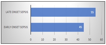
FIG. 1: DISTRIBUTION OF EOS AND LOS (PERCENTAGE)
TABLE 1: DISTRIBUTION OF MICROORGANISM ASSOCIATED WITH NEONATAL SEPSIS
| Organism | No. | % | EOS | EOS % | LOS | LOS% |
| C. gullermondi | 6 | 4.2 | 3 | 4.69 | 3 | 3.80 |
| C. albicancs | 46 | 31.9 | 28 | 43.75 | 18 | 22.78 |
| C. pellucilolus | 11 | 7.6 | 0 | 0.00 | 11 | 13.92 |
| Total fungi | 63 | 43.8 | 34 | 53.13 | 29 | 36.71 |
| Chromobacter | 3 | 2.1 | 3 | 4.69 | 0 | 0.00 |
| E. coli | 7 | 4.9 | 0 | 0.00 | 7 | 8.86 |
| K. pneumonia | 21 | 14.6 | 9 | 14.06 | 12 | 15.19 |
| K. oxytoca | 3 | 2.1 | 3 | 4.69 | 0 | 0.00 |
| P. auruginosa | 9 | 6.3 | 4 | 6.25 | 5 | 6.33 |
| S. marcescence | 6 | 4.2 | 0 | 0.00 | 6 | 7.59 |
| Acinetobacter | 5 | 3.5 | 0 | 0.00 | 5 | 6.33 |
| Total GNB | 54 | 37.5 | 19 | 29.69 | 35 | 44.30 |
| S. aureus | 4 | 2.8 | 3 | 4.69 | 1 | 1.27 |
| S. saprophyticus | 8 | 5.6 | 3 | 4.69 | 5 | 6.33 |
| CONS | 15 | 10.4 | 6 | 9.38 | 9 | 11.39 |
| Total GPC | 27 | 18.8 | 12 | 18.75 | 15 | 18.99 |
DISCUSSION: Neonatal sepsis remains a major cause of morbidity and mortality, particularly in low- and middle-income countries, where healthcare-associated infections and antimicrobial resistance pose significant challenges. The present study provides insight into the microbiological profile of neonatal sepsis in a tertiary care setting in North India, with emphasis on pathogen distribution, onset patterns, and diagnostic approaches. The overall blood culture positivity rate of 28.2% in this study is comparable to previously reported Indian data (20–40%) 3. Variations in culture yield may be influenced by factors such as prior antibiotic exposure, adequacy of blood volume collected, and diagnostic methodology. The use of automated systems such as BACTEC likely contributed to improved detection rates and faster turnaround time. Late-onset sepsis (LOS) accounted for a higher proportion of cases (55%) compared to early-onset sepsis (EOS), consistent with reports from tertiary care NICUs 4, 6. This pattern reflects the increasing role of hospital-acquired infections associated with prolonged hospitalization, invasive procedures, and intensive supportive care. Although detailed patient-level risk factor analysis was not comprehensively presented, the observed predominance of LOS suggests a strong association with NICU-related exposures such as mechanical ventilation, intravascular catheterization, and parenteral nutrition.
A particularly striking finding of this study is the high proportion of fungal isolates (43.8%), with Candida albicans as the predominant organism. This distribution is notably higher than that reported in most Indian studies, where fungal sepsis typically accounts for 5–20% of cases 7, 8. Several factors may explain this observation. First, the study population likely included a high proportion of preterm and low birth weight neonates, who are inherently at greater risk of invasive candidiasis. Second, widespread use of broad-spectrum antibiotics in NICU settings may suppress bacterial flora and predispose to fungal overgrowth. Third, invasive interventions such as central venous catheterization, mechanical ventilation, and prolonged parenteral nutrition are well-established risk factors for candidemia. In addition, the possibility of unit-specific colonization or a localized outbreak cannot be excluded. Environmental persistence of Candida species and transmission via healthcare workers have been documented in NICU settings. The presence of non-albicans Candida species, such as Candida pelliculosa and Candida guilliermondii, which are often associated with nosocomial transmission, further supports this possibility. These findings highlight the need for strict infection control practices, including hand hygiene, catheter care protocols, and periodic environmental surveillance. The distinction between true infection and contamination is particularly important in interpreting microbiological data. Coagulase-negative staphylococci (CONS) and Candida species are known to be potential contaminants, especially in single blood cultures. In the present study, efforts were made to correlate microbiological findings with clinical features; however, the lack of repeat cultures and limited availability of adjunct biomarkers may have affected the ability to definitively classify all isolates as true pathogens. Therefore, some isolates particularly CONS may represent contamination or colonization rather than true bloodstream infection. This limitation underscores the importance of integrating clinical, laboratory, and microbiological data for accurate diagnosis.
Among bacterial pathogens, Gram-negative bacilli, particularly Klebsiella pneumoniae, predominated, followed by Pseudomonas aeruginosa, Escherichia coli, and Serratia marcescens. This is consistent with several Indian studies that report Gram-negative organisms as the leading cause of neonatal sepsis, especially in LOS 9, 10. These organisms are well known for their ability to survive in hospital environments and develop multidrug resistance, making them a major therapeutic challenge. Gram-positive cocci accounted for a smaller proportion of isolates (18.8%), with CONS being the most common. While CONS are increasingly recognized as significant pathogens in NICU settings, especially in association with indwelling devices, their role remains controversial due to the possibility of contamination. In contrast, Staphylococcus aureus remains an important pathogen due to its potential for severe invasive disease. Although the abstract refers to antimicrobial susceptibility patterns, detailed organism-wise resistance data were not comprehensively presented in this manuscript, which limits the clinical applicability of the findings. Available data suggest that Gram-positive organisms retained high susceptibility to vancomycin and linezolid, consistent with other regional studies 11. However, increasing resistance among Gram-negative organisms remains a concern and highlights the need for continuous surveillance and institution-specific antibiograms to guide empirical therapy. The use of automated diagnostic platforms such as BACTEC and VITEK 2 improved the efficiency of pathogen detection and identification in this study. However, reliance on culture-based methods alone may underestimate the true burden of neonatal sepsis due to prior antibiotic exposure, low-level bacteremia, or fastidious organisms. Integration of molecular diagnostic methods could enhance detection rates and provide more comprehensive pathogen profiling. This study has several limitations. The absence of detailed demographic and clinical risk factor analysis limits the ability to establish strong clinicomicrobiological correlations. Antifungal susceptibility testing was not uniformly performed, restricting interpretation of resistance patterns among Candida species. The lack of repeat cultures in all cases makes differentiation between contamination and true infection challenging. Additionally, being a single-center study, the findings may not be generalizable to other settings.
CONCLUSION: Neonatal sepsis continues to pose a significant clinical challenge, particularly in resource-limited settings. This study highlights an unusual predominance of fungal pathogens, particularly Candida albicans, alongside Gram-negative bacteria in a tertiary care NICU in North India. The findings underscore the importance of considering invasive candidiasis in high-risk neonates, especially those with prolonged hospitalization, invasive device use, and prior antibiotic exposure. Early and accurate microbiological diagnosis using automated systems can facilitate timely and targeted therapy. However, improved clinicomicrobiological correlation, inclusion of detailed risk factor analysis, and incorporation of advanced diagnostic techniques are essential to enhance diagnostic accuracy. Continuous surveillance, strict infection control practices, and antimicrobial stewardship programs are critical to improving neonatal outcomes.
ACKNOWLEDGEMENTS: The authors sincerely acknowledge the support and cooperation of the Microbiology Department technicians for their technical assistance and help in laboratory procedures. We also thank the students who contributed to the study in various capacities. Special thanks are extended to the staff of the Department of Pediatrics for their valuable assistance, coordination, and support during the course of this work
CONFLICTS OF INTEREST: There is no conflict of interest among authors.
REFERENCES:
- Jatsho J: Clinical and bacteriological profile of neonatal sepsis: a prospective hospital-based study. Int J Pediatr 2020; 2020: 1–9.
- Nikita, Payal N, Bedi N, Sharma M and Khandait M: Epidemiology, risk factors and prevention strategies for neonatal sepsis: a review. ACBR 2025; 9: 28–34.
- Upadhyay S: Incidence, bacteriological profile and risk factor analysis of neonatal sepsis in a peri-urban set up of North India. Int J Contemp Pediatr 2020; 7(6): 1349–1354.
- Raturi A and Chandran S: Neonatal sepsis: aetiology, pathophysiology, diagnostic advances and management strategies. Clin Med Insights Pediatr 2024; 18: 11795565241281337.
- Clinical and Laboratory Standards Institute (CLSI): Performance standards for antimicrobial susceptibility testing. CLSI supplement M100, 33rd ed. Wayne, PA: CLSI; 2023.
- Li Z, Xiao Z, Li Z, Zhong Q, Zhang Y and Xu F: 116 cases of neonatal early-onset or late-onset sepsis: A single center retrospective analysis on pathogenic bacteria species distribution and antimicrobial susceptibility. Int J Clin Exp Med 2013; 6(8): 693-9.
- Benjamin DK: Neonatal candidiasis: epidemiology, risk factors, and clinical judgment. Pediatrics 2010; 126(4): 865-873.
- Jain A: Candidaemia in neonatal intensive care units: experience from a tertiary care hospital in India. Indian J Med Microbiol 2013; 31(4): 337–341
- Tripathi S and Malik GK: Neonatal sepsis: past, present and future; a review article. IJMU 2010; 5(2): 45–54.
- Roy I, Jain A, Kumar M and Agarwal SK: Bacteriology of neonatal septicaemia in a tertiary care hospital of northern India. Indian J Med Microbiol 2002; 20(3): 156–159.
- National Neonatal Perinatal Database. Report 2019–2020. New Delhi: NNF India.
How to cite this article:
Pal N, Rani A, Bansal J, Singh K, Sahni AK and Manocha H: Microbiological spectrum and diagnostic approach in neonatal sepsis. Int J Pharm Sci& Res 2026; 17(8): 2469-73. doi: 10.13040/IJPSR.0975-8232.17(8).2469-73.
All © 2026 are reserved by International Journal of Pharmaceutical Sciences and Research. This Journal licensed under a Creative Commons Attribution-NonCommercial-ShareAlike 3.0 Unported License.
Article Information
21
2469-2473
515 KB
309
English
IJPSR
Nidhi Pal *, Anju Rani, Jagriti Bansal, Kushal Singh, Ajay Kumar Sahni and Harmesh Manocha
College of Allied and Healthcare Science, GIMS, Greater Noida, Uttar Pradesh, India.
nidhipal44@gmail.com
15 March 2026
22 May 2026
19 June 2026
10.13040/IJPSR.0975-8232.17(8).2469-73
01 August 2026
 Download