




MINIMUM INHIBITORY CONCENTRATION OF LINEZOLID IN CLINICAL ISOLATES OF METHICILLIN-RESISTANT STAPHYLOCOCCUS AUREUS AT A TERTIARY CARE HOSPITAL
HTML Full TextMINIMUM INHIBITORY CONCENTRATION OF LINEZOLID IN CLINICAL ISOLATES OF METHICILLIN-RESISTANT STAPHYLOCOCCUS AUREUS AT A TERTIARY CARE HOSPITAL
Reetu Gogoi *, Vishal Sharma and Shilpa Arora
Department of Microbiology, Guru Gobind Singh Medical College & Hospital, Faridkot, Punjab, India.
ABSTRACT: Background: Methicillin-resistant Staphylococcus aureus (MRSA) is a major cause of hospital and community-acquired infections, with substantial impact on infection rates, morbidity, mortality, and healthcare costs. Vancomycin, remained a longstanding cornerstone in MRSA treatment, butthe emergence of vancomycin resistant MRSA (VRSA), necessitates alternative antimicrobial solutions. Linezolid, stands out as a promising candidate. Linezolid antibiotic is available as an oral formulation for resistant staphylococcal infections. It has unique advantages such as an absence of renal toxicity and improved lung parenchymal diffusion compared. However, emerging resistance to linezolid has become a concern worldwide. There is clonal dissemination of Linezolid Resistant Staphylococcus aureus (LRSA) within or across healthcare settings which demands continuous surveillance to determine the emergent risk of resistant strains and to establish guidelines for appropriate use. Objectives: To determine the minimum inhibitory concentration (MIC) of linezolid among MRSA isolates and to compare Kirby-Bauer disk diffusion method with MIC testing by agar dilution method for linezolid resistance detection. Methods: A hospital-based descriptive study was conducted over one year in the Department of Microbiology, Guru Gobind Singh Medical College & Hospital, Faridkot. Two hundred clinically significant MRSA isolates obtained from various clinical specimens were included. Identification and antimicrobial susceptibility testing were performed according to CLSI guidelines. Linezolid susceptibility was assessed using Kirby–Bauer disk diffusion and agar dilution MIC methods. Results: Of the 200 MRSA isolates, maximum MRSA were isolated from pus (67.5%) followed by blood (30.5%). All MRSA isolates were susceptible to linezolid by both disk diffusion and MIC methods. MIC distribution showed that 85.5% isolates had MIC ≤0.25 µg/mL, 8% had MIC 0.5 µg/mL, 3.5% had MIC 1 µg/mL, and 3% had MIC 2 µg/mL. No isolate demonstrated resistance (MIC≥8 µg/mL). MIC50, MIC90, and geometric mean MIC were 0.25 µg/mL, 0.5 µg/mL, and 0.2952 µg/mL respectively. Perfect agreement was observed between disk diffusion and agar dilution method (Kappa = 1). Conclusion: Linezolid demonstrated excellent in vitro activity against MRSA isolates in the present study. Routine surveillance of MIC trends remains essential for early detection of emerging resistance and prevention of linezolid MIC creep.
Keywords: MRSA, Linezolid, Minimum inhibitory concentration, MIC50, MIC90, Antimicrobial resistance
INTRODUCTION: Methicillin-resistant Staphylococcus aureus (MRSA) is a major multidrug-resistant pathogen responsible for a wide range of infections in both community and hospital settings 1.
MRSA has become a significant global public health problem 2. MRSA infections are associated with increased morbidity, mortality, prolonged hospital stay, and greater healthcare costs 3.
MRSA strains are broadly classified into healthcare-associated MRSA (HA-MRSA), community-associated MRSA (CA-MRSA), and livestock-associated MRSA (LA-MRSA) 4. HA-MRSA commonly spreads in hospitals through direct contact with contaminated hands, wounds, or medical devices, while CA-MRSA is increasingly seen among healthy individuals and is often associated with skin and soft tissue infections 3. Several risk factors contribute to MRSA infection, including prolonged hospitalization, ICU admission, recent antibiotic use, invasive procedures, hemodialysis, HIV infection, open wounds, indwelling catheters, and residence in nursing homes 5. Skin and soft tissue infections are the most common manifestations, but MRSA can also cause severe invasive diseases such as osteomyelitis, septic arthritis, infective endocarditis, bloodstream infections, ventilator-associated pneumonia, and necrotizing pneumonia. Community-associated MRSA is especially associated with cellulitis, diabetic foot ulcers, and necrotizing fasciitis 6, 7.
Vancomycin has traditionally been considered the drug of choice for serious MRSA infections because most strains remain susceptible to it 3. However, extensive use of glycopeptide antibiotics has led to the emergence of strains with reduced susceptibility to vancomycin, including vancomycin-intermediate S. aureus (VISA) and vancomycin-resistant S. aureus (VRSA) 8. Linezolid, the first member of the oxazolidinone class of antibiotics, has emerged as an important therapeutic option for MRSA infections, especially VISA and VRSA strains 9. It acts by inhibiting bacterial protein synthesis through binding to the 50S ribosomal subunit and blocking initiation of translation 10. Unlike many other protein synthesis inhibitors, linezolid acts at the initial stage of protein synthesis, giving it a unique mechanism of action 11. It has excellent activity against multidrug-resistant Gram-positive bacteria and does not show cross-resistance with other antibiotic classes 12.
One of the major advantages of linezolid is its excellent oral bioavailability, allowing easy transition from intravenous to oral therapy 10, 13. It is widely used for skin and soft tissue infections, diabetic foot infections, pneumonia, infective endocarditis, meningitis, and eradication of MRSA colonization 10. Despite its effectiveness, resistance to linezolid has started emerging worldwide. The first case of linezolid-resistant Staphylococcus aureus (LRSA) was reported within a year of its clinical introduction 14. Several laboratory methods are available for detection of linezolid resistance, including Kirby-Bauer disk diffusion, E-test, broth dilution, agar dilution, VITEK 2, PCR, and pulsed-field gel electrophoresis 15. In routine laboratories, disk diffusion is commonly used because it is simple and economical. However, disk diffusion results can sometimes be variable. Therefore, resistant isolates should be confirmed using MIC determination methods, which provide a more precise and quantitative assessment of antibiotic susceptibility 16. Considering the increasing emergence of linezolid-resistant MRSA strains and the limitations of routine susceptibility testing methods, accurate detection of resistance has become crucial for effective patient management and infection control. Hence, the present study was undertaken to compare the disk diffusion method with MIC determination method for the detection of linezolid resistance in MRSA isolates.
MATERIALS AND METHODS: A hospital-based descriptive study was conducted over a period of one year from September 2024 to August 2025, in the Department of Microbiology, Guru Gobind Singh Medical College & Hospital, Faridkot, Punjab.Convenient sampling method was used. According to the hospital statistics, availability and feasibility, the first consecutive 200 MRSA isolates from patients of all age groups and both sexes were included. Repeat samples from the same patient were excluded for this study. The study was conducted after taking ethical approval from the institution’s ethical committee (No.BFUHS/2K26p-TH/1792).
All the clinical specimens received in the Microbiology department were inoculated on culture plates [blood agar and MacConkey’s agar; cystine lactose electrolyte deficient (CLED) agar for urine specimens] and incubated at 37 °C for 24- 48 hours and direct microscopy was performed 16.
Control Strains used in the Study: S. aureus ATCC 25923 and MRSA 43300 Methicillin resistance was detected using cefoxitin disc (30µg), which is a surrogate marker and a potent inducer of MECA gene. The strains of S. aureus with a zone diameter of ≤ 21mm were considered as MRSA.
S. aureus Isolation and Identification 17: Identification was done by colony morphology and gram staining. Bacterial colonies showing Gram positive cocci arranged in clusters were further subjected to catalase test, slide and tube coagulase test. The bacteria that were catalase positive and both slide and tube coagulase test positive were identified as S. aureus.
Antibiotic Susceptibility Testing (Antibiogram) 17: In accordance with CLSI standards, Kirby-Bauer's disc diffusion method was used to evaluate the antibiotic sensitivity pattern of all S. aureus isolates to different antibiotics. The antibiotics (Hi-Media Ltd., Mumbai, India) used were Ampicillin, 10μg; Erythromycin, 15μg; Cefoxitin, 30μg; Ciprofloxacin, 5μg; Amikacin, 10μg; Clindamycin, 2μg; Cotrimoxazole, 1.25/23.75μg; Linezolid, 30μg; Nitrofurantoin, 300μg; Norfloxacin, 10μg; and Vancomycin was tested by MIC agar dilution method.
Inoculum Preparation 17: 0.5 McFarland turbidity standard inoculum of the isolated strain was applied to Mueller-Hinton agar (MHA) plates and the plates were incubated at 37C for 18-24 hours. The zone of inhibition diameters were then measured with the help of caliper and interpreted as per CLSI criteria. In case of cefoxitin, the plates were incubated for 24 hours 26. Linezolid zone diameter≥ 21 mm with a 30μg disc was considered as sensitive and ≤20 mm as resistant.
Maintenance and Preservation of Culture Strains 16: Organisms grown in appropriate media for 18 hours were preserved in brain heart infusion (BHI) broth with 20% glycerol and stored without significant loss of viability at 20 C until MIC was done.
Minimum Inhibitory Concentration of Linezolid by Agar Dilution Method 16, 17: The minimum inhibitory concentration (MIC) of linezolid for S. aureus isolates was determined by agar dilution method. Mueller-Hinton agar (MHA) was supplemented with different concentration of linezolid according to CLSI guidelines.
Stock Solution Preparation: The following formula was used to create a stock solution of linezolid:
W = V × C × 1000 / P
Where, W = Weight of antibiotic to be dissolved, V= Volume, C = final Concentration of solution, P = Potency.
Using the following formula, C1 V1 = C2 V2, where:
C1 = Concentration of linezolid stock solution, V1 = Unknown Volume of stock solution needed to make the dilution concentration, C2 = Required concentration of linezolid for dilution, V2 = Required volume of concentration of linezolid for dilution, serial two-fold dilutions of linezolid were prepared:(64, 32, 16, 8, 4, 2, 1, 0.5, 0.25) μg /mL above known concentrations of linezolid were incorporated in plates of mueller-hinton agar and each plate (with different concentration of linezolid) thus obtained was spotinoculated (using 10μL bacterial culture) to find out MIC of all the isolates. The plates were incubated for 24 hours at 37°C. The lowest concentration of linezolid agar plate that entirely prevented the growth of isolate was the minimum inhibitory concentration (MIC) of linezolid for that particular isolate. MIC ≥ 8 μg /mL were considered resistant; MIC of ≤ 4 μg/mL as susceptible to linezolid for MRSA isolates as per CLSI 17.
RESULTS: A total of 13816 samples were received in the department of Microbiology over one year and processed. Of all the specimens received, 33.4% were of blood, 32.4% of urine, 17.17% of pus, 11.01% specimens were of sterile body fluids, 4% specimens were of sputum, 1% were of catheter tip (1%), 0.6% of bronchoalveolar lavage, 0.4% of stool and 0.02% of biopsy tissue (0.02%). Maximum number of 61.13% Staphylococcus aureus (S. aureus) isolates were obtained from pus samples, then followed by 35.37% from blood, 2.62% from sterile body fluids and 0.43% from catheter tip and biopsy tissue.
Methicillin resistance using cefoxitin disc (30μg) method showed 90.83% S. aureus were Methicillin resistant Staphylococcus aureus (MRSA) and 9.17% methicillin sensitive Staphylococcus aureus (MSSA). Of all the MRSA isolated, first 200 consecutive MRSA strains were considered for this study.
More number of MRSA isolates were obtained from patients with age group 21-40 years (47%) followed by 27% from age group 41-60 years. Of all the MRSA strains isolated, 70.5% isolates were from males and 29.5% from females. Male to female ratio was 2.39:1.
Maximum MRSA were isolated from pus (67.5%), as shown in Table 1. Fig. 1 shows majority of MRSA isolates were obtained from specimens received from medicine department (23.5%), followed by plastic surgery (17%).
TABLE 1: DISTRIBUTION OF MRSA ISOLATES AMONG VARIOUS CLINICAL SPECIMENS (N=200) AND SPECIMEN-WISE LINEZOLID MIC ANALYSIS (µg/mL)
| Type of Clinical specimen | Number of MRSA Isolates | Percentage | MIC 50
(µg/mL) |
MIC 90
(µg/mL) |
Geometric Mean
(µg/mL) |
| PUS | 135 | 67.5% | 0.25 | 0.50 | 0.318 |
| Blood | 61 | 30.5% | 0.25 | 0.25 | 0.25 |
| Pleural Fluid | 2 | 1% | 0.25 | 0.25 | 0.25 |
| Cerebrospinal Fluid | 2 | 1% | 0.25 | 0.25 | 0.25 |
| Total | 200 | 100% | 0.25 | 0.50 | 0.2952 |
Pus isolates demonstrated significantly higher MIC90 and geometric mean MIC values of linezolid compared to other specimens (p <0.001) Table 1.
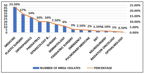
FIG. 1: DISTRIBUTION OF MRSA (n=200) IN VARIOUS CLINICAL DEPARTMENTS
The Antimicrobial Susceptibility Testing (AST) of 200 MRSA isolates showed hundred percent susceptibility to linezolid. The maximum resistance among MRSA isolates was observed against ampicillin (100%), followed by 92% resistance to ciprofloxacin Fig. 2 and vancomycin was found to be susceptible by using MIC agar dilution method (MIC<2µg/ml). Inducible clindamycin resistance detected by D test was found to be positive for 25.2% of MRSA isolates Table 2.
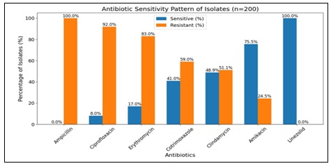
FIG. 2: ANTIMICROBIAL SUSCEPTIBILITY PROFILE OF VARIOUS ANTIBIOTICS OF MRSA ISOLATES BY KIRBY BAUER DISK DIFFUSION (n=200)
TABLE 2: RESISTANCE PATTERN OF CLINDAMYCIN AMONG MRSA ISOLATES FROM PUS SAMPLES (n=135)
| Resistance pattern of clindamycin | Number of MRSA isolates | Percentage |
| E-R; CD-R (MLSBc) Phenotype | 69 | 51.11% |
| E-R; CD-R MLSBi Phenotype (D-Test Positive) | 34 | 25.2% |
| E-R, CD-S MS Phenotype (D-Test Negative) | 43 | 31.85% |
| E-S, CD-S | 23 | 17.03% |
E-R – Erythromycin resistant, CD-R – Clindamycin resistant, CD-S – Clindamycin sensitive. MLSBc – Macrolide-lincosamide-streptogramin resistant, (Constitutive resistance). MLSBi – Macrolide-lincosamide-streptogramin resistant, (Inducible resistance; D-test positive). MS phenotype – Macrolide, (Erythromycin) resistant; lincosamide (clindamycin) sensitive (D-test negative).
Linezolid minimum inhibitory concentration (MIC) determinedusing agar dilution method is shown in Table 2. No MRSA isolates showed MIC ≥ 4 μg/ml. Both MIC and disk diffusion method, showed 100% susceptibility to Linezolid in MRSA isolates Table 3.
MIC50, MIC90 and Geometric mean MIC of linezolid for all MRSA isolates were calculated to be 0.25 μg/ml, 0.5 μg/ml and 0.2952 μg/ml respectively Fig. 3. MIC90 and geometric mean MIC of linezolid for pus isolates were significantly higher than other specimens Table 1.
Although statistically not significant, linezolid geometric mean MIC reduced from 0.3 μg/ml in the first six months to 0.29 μg/ml in the next six months Fig. 4.
TABLE 3: MINIMUM INHIBITORY CONCENTRATION (MIC) OF LINEZOLID BY AGAR DILUTION METHOD (in µg/mL)
| MIC value (in µg/mL) | NO. of MRSA Isolates | Percentage of MRSA Isolates |
| ≤ 0.25 | 171 | 85.5% |
| 0.5 | 16 | 8% |
| 1 | 7 | 3.5% |
| 2 | 6 | 3% |
| 4 | 0 | 0% |
| Total | 200 | 100% |
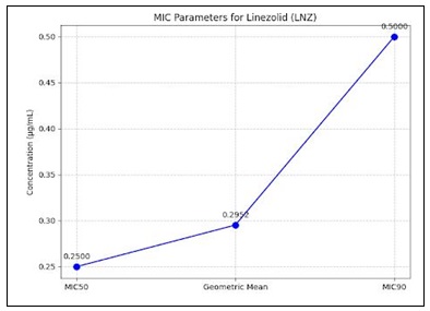
FIG. 3: MIC50, MIC90 AND GEOMETRIC MEAN MIC OF MRSA ISOLATES (n=200)
TABLE 3: COMPARISON OF DISC DIFFUSION AND MIC METHOD (AGAR DILUTION) FOR DETECTION OF LINEZOLID RESISTANCE IN MRSA ISOLATES
| Disc Diffusion | MIC | |
| Sensitive | 200 | 200 |
| Resistant | 0 | 0 |
| Total | 200 | 200 |
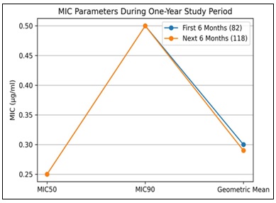
FIG. 4: MINIMUM INHIBITORY CONCENTRATION (MIC) (µg/mL) STATISTICS FOR LINEZOLID FROM 2024 TO 2025
DISCUSSION: Due to the emergence of VISA, hVISA, and VRSA strains, linezolid has become an important alternative for treating severe MRSA infections including hospital-acquired pneumonia, community-acquired pneumonia, and skin and soft tissue infections 18. However, reports of reduced susceptibility to linezolid among gram-positive cocci have gradually emerged 19. Resistance mechanisms of linezolid include mutations in the domain V region of the 23S rRNA gene, acquisition of the plasmid-mediated cfr gene, and alterations in ribosomal proteins L3 and L4. The most common mechanism is the G2576T point mutation in the 23S rRNA gene 20.
Minimum Inhibitory Concentration (MIC) is considered the best indicator for selecting appropriate antimicrobial therapy in serious infections 21.
In the present study of the total 13,816 clinical specimens processed for culture and sensitivity testing, blood samples constituted the majority, followed by urine. Similar distributions have been reported in studies by Kansal R et al. and Regmi S et al. 22, 23. Of all the samples processed, 229 isolates (1.65%) were identified as S. aureus. The majority of these isolates were recovered from pus samples (61.13%) as similar to studies conducted by Sharlee R et al., and Moses VK et al. where pus samples yielded the highest number of S. aureus isolates 9, 24. In the present study, 90.83% of isolates were identified as MRSA, while only 9.17% were methicillin-sensitive Staphylococcus aureus (MSSA). The prevalence of MRSA in this study was significantly higher than that reported in many previous studies, although similar high prevalence was observed by Kansal R et al. 23. Variations in MRSA prevalence across studies may be attributed to environmental factors, irrational antibiotic use, differences in infection control practices, and varying inclusion criteria. First 200 consecutive MRSA isolates were included in this study.
Most MRSA isolates were obtained from patients aged 21–40 years, followed by those aged 41–60 years, which correlated with the findings of Kansal R et al. 23. Male patients accounted for 70.5% of MRSA isolates, consistent with studies conducted by Arumugam A et al. and Sharlee R et al. 8, 9. Among clinical specimens, pus samples contributed the majority of MRSA isolates followed by blood, pleural fluid, and cerebrospinal fluid. This is in accordance with studies conducted by Kansal R et al., Moses VK et al., and Husain A et al. 23, 24, 25. Surgical departments such as plastic surgery, general surgery, pediatric surgery, and neurosurgery accounted for the highest proportion of MRSA isolates, followed by medicine and orthopedics departments. Similar trends were observed in studies conducted by Kansal R et al., Moses VK et al., and Srinivasan S et al. 23, 24, 26.
Antimicrobial susceptibility testing using the Kirby–Bauer disk diffusion method demonstrated extensive multidrug resistance among MRSA isolates. All isolates were resistant to ampicillin, while high resistance rates were observed for ciprofloxacin (92%), erythromycin (83%), cotrimoxazole (59%) and clindamycin (51.11%). For amikacin resistance rate was 24.5%. Similar resistance patterns have been reported by Chitnis S et al. and Bawankar NS et al. 27, 28. Importantly, no resistance to linezolid was detected in the present study. Studies conducted by Niveditha N et al., Xu Yanlei et al., Arumugam A et al., Husain A et al., and Chitnis S et al., also reported 100% susceptibility of MRSA isolates to linezolid 2, 5, 8, 25, 27. In the current investigation, 25.2% of MRSA isolates showed inducible clindamycin resistance, which is higher than 14% inducible resistance observed by Kansal R et al., from the same institution in 2024 indicating the declining susceptibility to clindamycin, and erythromycin 23. While Sharma V et al., Sharma NK et al., and Kumar R et al. reported similar percentages of 33.7%, 21.38%, and 27% 29, 30, 31.
All MRSA isolates were evaluated for vancomycin susceptibility using the agar dilution MIC method. Every isolate showed vancomycin MIC values below 2 µg/mL, indicating 100% susceptibility. Similar findings have been documented by Anitha TK et al., Husain A et al., Xu Yanlei et al., Niveditha N et al., and Hortiwakul R et al. 21, 25, 32, 33, 34.
The major focus of the study was the evaluation of linezolid susceptibility using both Kirby–Bauer disk diffusion and agar dilution MIC methods. All MRSA isolates showed inhibition zone diameters greater than 21 mm with linezolid discs, confirming susceptibility by disk diffusion testing. MIC values ranged from ≤0.25 µg/mL to 2 µg/mL. The majority of isolates (85.5%) demonstrated MIC values ≤0.25 µg/mL, while smaller proportions showed MICs of 0.5 µg/mL, 1 µg/mL, and 2 µg/mL. No isolate demonstrated MIC values greater than 4 µg/mL. These findings were comparable to those reported by Arumugam A et al. 8, although Bawankar NS et al. reported higher MIC values and even linezolid-resistant isolates 28.
A perfect concordance was observed between disk diffusion and MIC methods with100% percentage agreement. Similar findings were reported in several Indian and international studies 8, 9, 10, 25, 27, 32, 33, 34, 35. Although disk diffusion is more practical for routine laboratory use, MIC determination remains important for detecting early shifts in susceptibility and possible emergence of resistance 28. While no linezolid-resistant MRSA isolates were identified in this study, some isolates had MIC values at the upper end of the susceptible range, which is concerning because previous studies by Bawankar NS et al., Husain A et al., and Wali M et al. have documented reduced susceptibility and frank resistance to linezolid 25, 28, 36.
The study calculated linezolid MIC50, MIC90, and geometric mean MIC values as 0.25 µg/mL, 0.5 µg/mL, and 0.2952 µg/mL respectively. Pus isolates showed significantly higher MIC90 and geometric mean MIC values compared to blood and fluid isolates, similar to observations made by Xu Y et al. 32. Over the study period, MIC50 and MIC90 remained stable while geometric mean MIC showed a slight decrease, unlike the “MIC creep” reported by Husain A et al., and Miyazaki M et al. 25, 37.
CONCLUSION: The study concludes that linezolid remain highly effective against MRSA isolates in this institution. However, MIC values on higher side of the susceptible range highlight the need for continuous surveillance, antibiotic stewardship, and strict infection control practices. Linezolid should be reserved for severe MRSA infections. The study also acknowledges limitations including its single-center design, predominance of pus samples, lack of molecular typing methods, and use of agar dilution instead of more precise gradient methods like E-test. Further multicentric studies with molecular analysis are recommended to better understand resistance patterns and clonal dissemination of MRSA strains.
ACKNOWLEDGEMENT: None
Ethical Approval: The study was performed after taking approval from the institution’s Ethical committee.
Funding Source: NIL
A.I used: None
CONFLICTS OF INTEREST: None
REFERENCES:
- Oliveira D, Borges A and Simões M: Staphylococcus aureus toxins and their molecular activity in infectious diseases. Toxins (Basel) 2018; 10(6): 252.doi: 10.3390/toxins10060252.
- Centre for Disease Control and Prevention. Antibiotic resistance threats in the United States, 2019. Atlanta, GA: US Department of Health and Human Services, CDC 2019.
- Siddiqui AH and Koirala J: Methicillin resistant Staphylococcus aureus. In: Stat Pearls [internet]. Treasure Island (FL): Stat Pearls Publishing 2025.
- Alghamdi BA, Al-Johani I, Al-Shamrani JM, Alshamrani HM, Al-Otaibi BG and Master KA: Anti-microbial resistance in methicillin-resistant Staphylococcus aureus. Saudi J Biol Sci 2023; 30(5): 1-12.doi: 10.1016/j.sjbs.2023.103604
- National Nosocomial Infections Surveillance System. National Nosocomial Infections Surveillance (NNIS) System Report, data summary from January 1992 through June 2004, issued October 2004. Am J Infect Control. 2004 Dec;32(8):470-85.doi: 10.1016/S0196655304005425
- Khan TM, Kok YL, Bukhsh A, Lee LH, Chan KG and Goh BH: Incidence of methicillin resistant Staphylococcus aureus (MRSA) in burn intensive care unit: a systematic review. Germs 2018; 8(3): 113-125.doi: 10.18683/germs.2018.1138.
- Clebak KT and Malone MA: Skin Infections. Prim Care 2018; 45(3): 433-454.doi: 10.1016/j.pop.2018.05.004.
- Arumugam A, Bhat S and Kanungo R: Correlation between oxacillin MIC values with those of linezolid against clinical isolates of MRSA. Indian J Microbiol Res 2023; 10(4): 216-21.https://doi.org/10.18231/j.ijmr.2023.038
- Sharlee R and Sumangala B: A Study to Comparison of MIC of Linezolid on MRSA by Micro Broth Dilution and E Strip Method in Teaching Hospital, Karnataka, India. Int J Curr Microbiol App Sci 2020; 9(03): 2848-56.https://doi.org/10.20546/ijcmas.2020.903.328
- Gandham P: Linezolid resistant Staphylococcus aureus. Int J of Res Med Sci 2014; 2(4): 1253–1256.DOI:10.5455/2320-6012.ijrms20141103
- Swaney SM, Aoki H, Ganoza MC and Shinabarger DL: The oxazolidinone linezolid initiation of protein synthesis in bacteria. Antimicrob Agents Chemother 1998; 42(12): 3251-5.DOI: 10.1128/AAC.42.12.3251
- Chien JW, Kucia ML and Salata RA: Use of linezolid, an oxazolidinone, in the treatment of multidrug-resistant gram-positive bacterial infections. Clin Infect Dis 2000; 30(9): 1246-51. DOI: 10.1086/313597
- Grau S and Rubio-Terres C: Pharmacoeconomics of linezolid. Expert Opin Pharmacother 2008; 9(6): 987-1000.DOI: 10.1517/14656566.9.6.987
- Barber M: Methicillin-resistant staphylococci. J Clin Pathol 1961; 14(4): 385–93.DOI: 10.1136/jcp.14.4.385
- Tenovar FC, Williams PP and Stocker S: Accuracy of six antimicrobial susceptibility methods for testing linezolid against staphylococci and enterococci. J Clin Microbiol 2007; 45(9): 2917-22. doi: 10.1128/JCM.00913-07
- Miles SR and Amyes BGS: Laboratory control of antimicrobial therapy. In: Mackie and McCartney practical medical microbiology. Collee JG, Marmion BP, Fraser AG, Simmons A, editors. 14th edition. Churchill Livingstone 2015; 155-64.
- Clinical and Laboratory Standards Institute. Performance standards for antimicrobial susceptibility testing (34th edition). CLSI supplement M100. Pennsylvania: CLSI 2025.
- Purrello SM, Garau J, Giamarellos E, Mazzei T, Pea F and Soriano A: Methicillin-resistant Staphylococcus aureus infections: a review of the currently available treatment options. J Global Antimicrob Resist 2016;7:178–186.DOI: 10.1016/j.jgar.2016.07.010
- AbdAlhafiz AI, Elleboudy NS, Aboshanab KM, Aboulwafa MM and Hassouna NA: Phenotypic and genotypic characterization of linezolid resistance and the effect of antibiotic combinations on methicillin-resistant Staphylococcus aureus clinical isolates. Ann Clin Microbiol Antimicrob 2023; 22(23): 1-16.doi: 10.1186/s12941-023-00574-2
- Besier S, Ludwig A and Zander J: Linezolid resistance in Staphylococcus aureus: gene dosage effect, stability, fitness costs and cross-resistances. Antimicrob Agents Chemother 2008; 52(4): 1570-2. DOI: 10.1128/AAC.01098-07
- Anitha TK., Rao MR, Shankaregowda R, Mahale RP and Chitharagi VB: Evaluation of Vancomycin Minimum Inhibitory Concentration in the clinical isolates of Methicillin Resistant Staphylococcus aureus (MRSA), J Pure Appl Microbiol 2019; 13(3): 1797-1801.https://doi.org/10.22207/JPAM.13.3.56
- Regmi S, Amatya J and Labh SN: Antimicrobial Resistance Pattern of Methicillin-Resistant Staphylococcus aureus (MRSA) Strains Isolated from Clinical Specimens in Kathmandu, Nepal. Arch Clin Microbiol 2020; 11(4): 116.DOI:10.36648/1989-8436.11.4.116
- Kansal R, Sharma V and Arora S: A study of vancomycin intermediate resistance and hetero-resistance in clinical isolates of methicillin resistant Staphylococcus aureus at a tertiary care hospital. IJPSR 2024; 15(2): 577-583.DOI: 10.13040/IJPSR.0975-8232.15(2).577-83
- Moses VK, Kandi V and Rao SKD: Minimum Inhibitory Concentrations of Vancomycin and Daptomycin Against Methicillin-resistant Staphylococcus Aureus Isolated from Various Clinical Specimens: A Study from South India Cureus 2020; 12(1): 1-8.DOI: 10.7759/cureus.6749
- Husain A, Rawat V, Umesh, Kumar M and Verma PK: Vancomycin, linezolid and daptomycin susceptibility pattern among clinical isolates of methicillin-resistant Staphylococcus aureus (MRSA) from Sub-Himalyan Center. J Lab Physicians 2018; 10(2): 145-148. DOI: 10.4103/JLP.JLP_92_17
- Srinivasan S, Sheela D, Mathew R, Bazroy J and Kanungo R: Risk factors and associated problems in the management of infections with methicillin resistant Staphylococcus aureus. Indian J Med Microbiol 2006; 24(3): 182-5.
- Chitnis S, Katara G, Hemvani N, Pareek S and Chitnis DS: In-vitro activity of daptomycin & linezolid against methicillin resistant Staphylococcus aureus & vancomycin resistant enterococci isolated from hospitalized cases in Central India. Indian J Med Res 2013; 137: 191-96.
- Bawankar NS, Agrawal GN and Zodpey SS: Linezolid susceptibility in MRSA isolates: insights into resistance and concordance in phenotypic detection methods. Int J Res Med Sci 2024; 12(2): 507-11. DOI: https://doi.org/10.18203/2320-6012.ijrms20240220
- Sharma V and Sharma A: Inducible clindamycin resistance in clinical isolates of Staphylococcus aureus in a teaching hospital. JAMDSR 2016; 4(5): 86.
- Sharma NK, Garg R, Baliga S and Bhat G: Nosocomial infections and drug susceptibility patterns in methicillin sensitive and methicillin resistant Staphylococcus aureus. J Clin Diagn Res 2013; 7(10): 2178.DOI: 10.7860/JCDR/2013/6750.3463
- Kumar R, Sarma M and Shalini G: Detection of inducible clindamycin resistance in Staphylococcus aureus from various samples in a tertiary care hospital. Int J Res Med Sci 7(12): 4696–4702. DOI: https://doi.org/10.18203/2320-6012.ijrms20195541
- Xu Y, Wang B, Zhao H, Wang X, Rao L and Ai W: In-vitro activity of vancomycin, teicoplanin, linezolid and daptomycin against methicillin-resistant Staphylococcus aureus Isolates Collected from Chinese Hospitals in 2018-2020. Infect Drug Resist 2021; 14: 5449-56.doi: 10.2147/IDR.S340623
- Niveditha N and Sujatha S: Worrisome trends in rising minimum inhibitory concentration values of antibiotics against methicillin resistant Staphylococcus aureus -Insights from a tertiary care centre, South India. Braz J Infect Dis 2015; 19(6): 585-89.
- Hortiwakul R, Chayakul P and Ingviya N: In-vitro Activities of linezolid, vancomycin, fosfomycin and fusidic acid against methicillin-resistant Staphylococcus aureus (MRSA). J Infect Dis Antimicrob Agents 2004; 21(1): 7-10.
- Genç S, Duymaz FZ and Dündar D: Temporal changes in linezolid minimum inhibitory concentration values in vancomycin-resistant Enterococci and Methicillin-resistant Staphylococcus aureus Strains. Mediterr J Infect Microb Antimicrob 2019; 8: 6.DOI: 10.4274/mjima.galenos.2019.2019.6
- Wali M, Shah MS, Rehman TU, Wali H, Hussain M and Zaman L: Detection of linezolid resistance cfr gene among MRSA isolates. J Infect Public Health 2022; 15(10): 1142-46. DOI: 10.1016/j.jiph.2022.09.002
- Miyazaki M, Nagata N, Miyazaki H, Matsuo K, Takata T and Tanihara S: Linezolid Minimum Inhibitory Concentration (MIC) Creep in Methicillin-Resistant Staphylococcus aureus (MRSA) Clinical Isolates at a Single Japanese Centre. Biol Pharm Bull 2014; 37(4): 679-82. DOI: 10.1248/bpb.b13-00670
How to cite this article:
Gogoi R, Sharma V and Arora S: Minimum inhibitory concentration of linezolid in clinical isolates of methicillin-resistant Staphylococcus aureus at a tertiary care hospital. Int J Pharm Sci & Res 2026; 17(11): 3596-04. doi: 10.13040/IJPSR.0975-8232.17(11).3596-04.
All © 2026 are reserved by International Journal of Pharmaceutical Sciences and Research. This Journal licensed under a Creative Commons Attribution-NonCommercial-ShareAlike 3.0 Unported License.
Article Information
26
3596-3604
691 KB
9
English
IJPSR
Reetu Gogoi *, Vishal Sharma and Shilpa Arora
Department of Microbiology, Guru Gobind Singh Medical College & Hospital, Faridkot, Punjab, India.
reetz004@gmail.com
17 July 2026
29 August 2026
23 October 2026
10.13040/IJPSR.0975-8232.17(11).3596-04
01 November 2026
 Download