




MOLECULAR DOCKING ANALYSIS OF HETEROCYCLIC LIGANDS TARGETING NUDT5 PROTEIN IN BREAST CANCER
HTML Full TextMOLECULAR DOCKING ANALYSIS OF HETEROCYCLIC LIGANDS TARGETING NUDT5 PROTEIN IN BREAST CANCER
R. Govinda Rajan, S. Udhaya Vani, A. P. S. M. Krishna *, Ch. Dhanalakshmi Manimadhuri, G. Harika and S. K. Nujhat Aara
Department of Pharmaceutical Chemistry, Hindu College of Pharmacy, Guntur, Andhra Pradesh, India.
ABSTRACT: Breast cancer is the most common malignancy affecting women worldwide. Molecular docking is an important computational technique in drug discovery interaction between ligands and target proteins. In present study docking analysis binding affinity of 51 heterocyclic ligands against the breast cancer target protein NUDT5 (PDB ID:5NWH) are nucleotide- metabolizing enzymes (NUDIX) hydrolases. It plays a major role in hormone- dependent gene regulation and proliferation in breast cancer cells. The heterocyclic ligands docking analysis were analyzed by Autodock 4.0.1. The docking was validated by re docking co-crystallized inhibitor 9CH into active binding site of NUDT5 protein and evaluated binding energy, RMSD values and molecular interactions. The results were found ligand “GR39 exhibited the lowest binding energy” (-14.12kcal/mol), when compared with reference inhibitor 9CH [-10.59kcal/mol] and standard drugs like STD1 Tamoxifen (-9.65), STD2 Cyclophosphamide (-5.83), STD3 Doxorubicin (-11.74). However the present study is limited to molecular docking analyis. Further studies include in-vitro, in-vivo, ADME and cytotoxicity studies.
Keywords: Breast cancer, Molecular docking and NUDT5 protein
INTRODUCTION: Breast cancer is a malignant tumor that develops in the breast tissues, primarily in the ducts or lobules. It is the most frequently diagnosed cancer among women globally and accounts for a significant proportion of cancer-related deaths 1. It is a heterogenous disease characterized by the molecular subtypes and multiple histological include Ductal carcinoma in situ and Invasive ductal carcinoma most common (80%) 2.
At the molecular level, breast cancer is classified based on receptor status into Hormone receptor- positive and Triple-negative breast cancer. Breast cancer progression involves complex biological process such as Metastasis is primary cause of mortality, occurring years after initial treatment due to residual cancer stem cells and micro-environmental interactions 3.
Molecular docking is widely used in structure-based drug design to screen compounds and optimize drug candidates before experimental testing. The protein NUDT5 (NUDIX hydrolase 5) PDB ID:5NWH is a member of Nudix family of nucleotide-metabolizing enzymes. It functions primarily as an ADP-ribose pyrophosphatase, catalyzing the conversion of ADP-ribose into AMP OR ATP depending on cellular conditions. ATP plays Chromatin remodeling, Gene transcription and Hormone-dependent signaling pathways Fig. 1. This ATP production is particularly important in progesterone and estrogen signaling in breast cancer cells, linking metabolism directly to gene regulation 4. To overcome the problem and side effects of cancer, the present marketed drugs showing various adverse reactions, during treatments. In view of that, we want to develop new entities for the treatment of breast cancer cells 5.
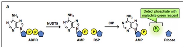
FIG. 1: DEVELOPMENT OF POTENT NUDT5 INHIBITORS FROM A HIGH-THROUGHPUT SCREENING CAMPAIGN. A REPRESENTATION OF THE ENZYME-COUPLED MALACHITE GREEN ASSAY (MG ASSAY). ADPR IS HYDROLYZED TO AMP AND R5P BY NUDT5, THEN R5P IS CONVERTED TO FREE INORGANIC PHOSPHATE DETECTED BY THE MALACHITE GREEN REAGENT 5
MATERIALS AND METHODS:
Softwares: Chemdraw 8.0, Chemspider, Protein Data Bank, Pub Meb, Autodock 4.0.1, Discovery Studio V24, Crystal Protein-PDB CODE:5NWH Fig. 2 6.
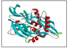
FIG. 2: CRYSTAL PROTEIN STRUCTURE OF 5NWH
Ligands:
Standard Ligand: Inhibitor (9CH), STDI (Tamoxifen), STD2 (cyclophosphamide), STD3 (Doxorubicin) 7.
Test Ligand: 51 selected ligands of various heterocyclic derivatives were collected from thesis 8.
The 51 ligands were collected from various literatures, based on antioxidant activty, the selected ligands pharmacophores nucleus were presence in selected ligands such as chromen 2-one, Tetrahydropyrimide,-1H-pyrrole, 1,5 benzodiazepene, 1,4 benzodiazepene, pyrazinyl, pyrrolidine-1-yl, biphenyl, 1,3,5 triazine, 2H-chromen-3-yl, quinoline, phenoxyphenyl, 1-2—hydroxyphenyl, pyrimidin, tiazine, and imadazo [2,1-b] [1,3,4]thiadiazole 9.
Protein Data Bank Information of 5NWH:
Classification: Hydrolase Organism(s): Homo sapiens Expression System: Escherichia coli 10.
Mutation(s): NO
Study of Molecular Docking:
Protein Preparation: Macromolecule was downloaded from protein data bank website (RCSB PDB) 10 Download the macromolecule in PDB formate 11 open discovery studio for protein cleaning, ligand molecules, water molecules and other ions were removed using discovery studio. Open autodock 4.0.1 set preference and make default. Edit option and delete water, under edit select hydrogen and add polar hydrogen only and add kollman charges then check total residues and save molecule in PDBQT format.
Ligand Preparation: Structures of 51 selected ligand molecules, standard and reference inhibitor were drawn by using chemdraw 8.0 software 12. Save the structures in mol format. Covert the mol format structures to pdb format by using BIOVIA 13. Retrieve the standard ligand molecules from chemspider website (http://www.chemspider.com/) and download the 3d structure of inhibitor from the protein data bank 14.
Molecular Docking: The ligand prepared and docked against protein using autodock 4.0.1. Output -lamarckian GA -save the file in DPF format. Launch the auto dock. Open the DLG file in the folder and check the lowest binding energy, runs, RMSD and estimated inhibition constant 15.
Visualization: Open the PDBQT saved file in discovery studioand draw the 2D and 3D diagram 16.
Docking Validation: Docking validation was performed by re-docking the co-crystallized inhibitor 9CH into the active site of the NUDT5 protein (PDB ID: 5NWH) using AutoDock 4.0.1. The reference inhibitor showed a binding energy of -10.59 kcal/mol with an RMSD value of 1.557 Å and inhibition constant of 17.30 nM, indicating reliable docking accuracy. The ligand exhibited significant interactions with key active-site residues including TRP A28, TRP B46, ARG A51, GLU A166, and GLY B135. Since the RMSD value was below 2 Å, the docking protocol was considered validated and suitable for further docking studies 15, 16.
RESULTS AND DISCUSSION: It is of interest to design inhibitors for the breast cancer target NUDT5 using molecular docking based virtual screening followed by molecular docking 17.
STD1 (Tamoxifen) was shown more binding energy (-9.65), when compared with selected ligands of GR01, GR02, GR03, GR04, GR05, GR06, GR07, GR08, GR09, GR10, GR11, GR12, GR13, and GR1. Reference inhibitor (9CH) was shown more binding energy (-10.59), when compared with selected ligands of GR03, GR04, GR05, GR06, GR08, GR12, GR16, GR21, GR25, GR26, GR28, GR37, GR38, GR39, GR46, and GR47 respectively.
For docking results, amino acid residues linked with Reference inhibitor (9CH) binding position in the receptor at TRP A 28 (Pi-Pi Stacked Bond), TRP B 46 (Pi-Pi stacked bond), THR B 45 (Pi-Pi Stacked Bond), GLU B 47 (Carbon Hydrogen Bond), ARG A 51 (Conventional Hydrogen bond), GLU A 166 (Carbon Hydrogen Bond), GLY B 135 (Carbon Hydrogen Bond), ALA A 96 (Pi Alkyl Bond), ILE A 141 (Alkyl Bond). The Pose Run, RMSD value and the Inhibition Constant for Reference inhibitor (9CH) was found 41, 1.557 and 17.30 nM respectively.
GR16, GR17, GR18, GR19, GR20, GR21, GR22, GR23, GR25, GR26, GR28, GR30, GR37, GR38, GR39, GR41, GR44, GR46, GR47, GR49 Respectively: For docking results, amino acid residues with STD1 (Tamoxifen) binding position in the receptor at TRP A 28 (Pi-Pi T shaped bond), TRP B 46(Pi-Pi Stacked Bond), ARG A 51(Pi Alkyl bond), LEU A 98(Carbon Hydrogen Bond), GLU A 166 (Carbon Hydrogen Bond). The Pose Run, RMSD value and the Inhibition Constant for STD1 were found 6, 20.390 and 85.01nM respectively 18.
STD2 (cyclophosphamide) was shown more binding energy (-5.83), when compared with All the selected ligands. For docking results, amino acid residues with STD2 (cyclophosphamide) binding position in the receptor at TRP B 46 (Pi cation bond), ARG A 51 (Conventional Hydrogen Bond), GLY B 135 (Pi Cation Bond), TRP A 28 (Pi Alkyl bond). The Pose Run, RMSD value and the Inhibition Constant for STD2 were found 34, 19.875 and 53,670 NM respectively 19.
STD3 (Doxorubicin) was shown more binding energy (-11.74), when compared with selected ligands of GR25, GR37, GR38, GR39 respectively. For docking results, amino acid residues with STD3 (doxorubicin) binding position in the receptor at ASP B 164 (Conventional Hydrogen Bond), GLU B 166(Carbon Hydrogen Bond), GLY B 165 (Conventional Hydrogen Bond), TYR A 36 (Conventional Hydrogen Bond), TRP A 46(Pi-Pi T shaped bond), PHE B 165 (Carbon Hydrogen Bond), VAL B 168 (Alkyl bond), ARG B 51(Conventional Hydrogen Bond). The Pose Run, RMSD value and the Inhibition Constant for STD3 (doxorubicin) were found 47, 20.366 and 2.50nM respectively 20.
Grid values of co-ordinates axis X, Y, Z are 2.086, -9.948, -17.613
Spacing of atoms 0.375
Angle of Dimensions X, Y, Z – 126,126,126
Docking Studies: The selected ligands, standards and inhibitors interaction with protein were performed by using Autodock 4.0.1 and the output of docking results was calculated from dlg file Table 2. In comparison binding energy with Reference Inhibitor (-10.59), STD1 (-9.65), STD2 (-5.83), STD3 (-11.74); the selected ligand GR39 was showed lowest energy (-14.12). For docking results, amino acid residues with selected ligand GR39 binding position in the receptor at TRP B 46 (Napthyl ring, Pi-Pi stacked bond), ARG A 51(phenyl ethyl ring, Pi Cation bond), ARG A 84(Pi Cation bond), GLN B 15(Amino phenyl ring, Conventional Hydrogen Bond ), TYR B 36(Phenyl ring, Pi Donar Hydrogen Bond). The Pose Run, RMSD value and the Inhibition Constant for selected ligand GR39 were found 47, 30.726 and 44.59nM respectively. Fig. 3 showing the 2D and 3D structures of GR39 selected ligand interaction with the crystal protein 5NWH 21-22.
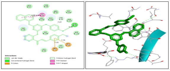
FIG. 3: 3D AND 2D STRUCTURES OF GR39 (E) (-1-(4-(4-(4-AMINOPHENYLAMINO)-6-(NAPHTHALEN-1-YLOXY)-1,3,5-TRIAZINE-2-YLAMINO) PHENYL)-3-(4-ETHYLPHENYL) PROP-2-EN-1-ONE)
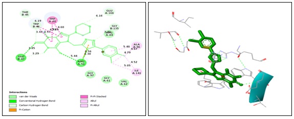
FIG. 4: 3D AND 2D STRUCTURE OF INHIBITOR (9CH)
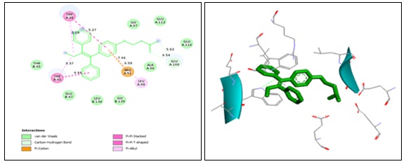
FIG. 5: 3D AND 2D STRUCTURE OF STD1 (TAMOXIFEN)
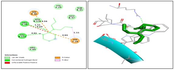
FIG. 6: 3D AND 2D STRUCTURE OF STD2 (CYCLOPHOSPHAMIDE)
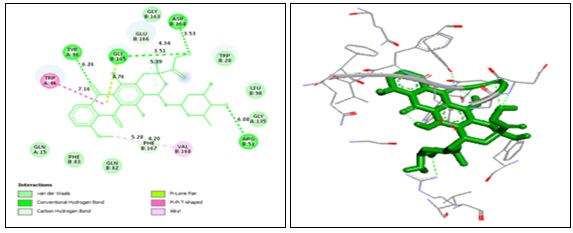
FIG. 7: 3D AND 2D STRUCTURE OF STD3 (DOXORUBICIN)
TABLE 1: CHEMICAL PROPERTIES OF SELECTED LIGANDS, STANDARD DRUGS AND INHIBITOR’S
| S. no. | Code | Structurere | IUPAC Name | Molecular formula | Mol. wt |
| 1 | GR01 | 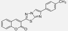 |
3-[6-(4-methylphenyl)imidazole[2,1-6][1,3,4]thiadiazol-2yl]-2H-Chromen-2-one | C20H13N3O2S | 359.4
|
| 2 | GR02 | 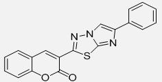 |
3-(6-phenyl)imidazo(2,1-6)(1,3,4)thiadiazol-2-yl)2H-chromen-2-one | C19H11N3O2S
|
345.37 |
| 3 | GR03 | 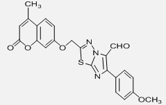 |
6-(4methoxyphenyl)-2-{[(4-methyl-2-oxo-2H-chromen-7yl)oxy]methyl}imidazo(2,1-6)(1,3,4)thiadazole-5carbaldehyde | C23H17N3O5S
|
447.46 |
| 4 | GR04 | 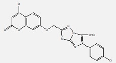 |
2-((4-chloro-2-oxo-2H-chromen-7-yloxy)methyl)-6-(4-chlorophenyl)imidazo[2,1-b][1,3,4]thiadiazole-5-carbaldehyde | C21H11Cl2N3O4S
|
472.3 |
| 5 | GR05 | 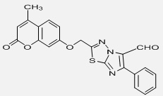 |
2-{[(4-methyl-2-oxo-2H-chromen-7-yl)oxy]methyl}-6-phenylimidazo(2,1-6)(1,3,4)thiadazole-5-carbaldehyde | C22H15N3O4S
|
417.44 |
| 6 | GR06 | 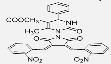 |
Methyl 1-[(19z,20z)-3,4-bis(2-nitrobenzylidene)-2,5-dioxopyrolidin-1-yl-)-1,2,3,4-tetrahydro-6-methyl-4-phenyl pyrimide-5-carboxylate | C31H23N5O9
|
609.54 |
| 7 | GRO7 | 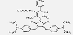 |
Methyl 1-[(19z,20z)-3,4-bis(4-(dimethylamino)benzylidene)-2,5-dioxopyrolidin-1-yl-)-1,2,3,4-tetrahydro-6-methyl-4-phenyl pyrimide-5-carboxylated | C35H35N5O5
|
605.68 |
| 8 | GR08 | 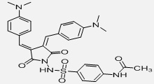 |
N-1-(4-[(3-(E)-1-[4-(dimethylamino)phenyl]methylidene-4(z)-1[4(dimethylamino)phenyl]methylidene-2,5-dioxotetrahydro-1H-1-pyrrolyl)amino]sulphonyl-phenyl)acetamide | C35H35N5O5
|
605.68 |
| 9 | GR09 | 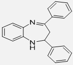 |
2,3-dihydro-2-4-diphenyl-1H-1,5benzodiazepine | C21H18N2
|
298.38 |
| 10 | GR10 | 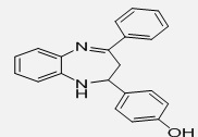 |
4-[2-3-dihydro-4-phenyl-1H-1,5benzodiazepine-2yl)phenol | C21H18N2O
|
314.38 |
| 11 | GR11 | 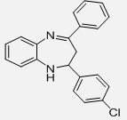 |
2-(4-chlorophenyl)-2,3-dihydro-4-phenyl-H-1,5benzodiazepine | C21H17ClN2
|
332.83 |
| 12 | GR12 | 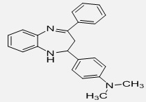 |
4-((E)-2,3-dihydro-4-phenyl-1H-1,5benzodiazepine-2-yl)-N,Ndimethyl benzene amine | C23H23N3
|
341.45 |
| 13 | GR13 | 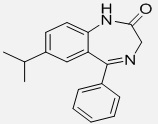 |
7-isopropyl-5-phenyl-1H-1,4benzodiazepine-2(3H)-one | C18H18N2O
|
278.35 |
| 14 | GR14 | 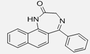 |
5-phenyl-1H-naptho[1,2][1,4]diazepine-2(3H)one | C19H14N2O
|
286.33 |
| 15 | GR15 | 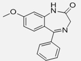 |
8-methoxy-5-phenyl-1H-1,4benzodiazepine-2(3H)-one | C16H14N2O2
|
266.29 |
| 16 | GR16 | 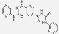 |
(N1-(2-pyrazinyl)-(E)-3-[4-({[(2-pyridalamino)carbonyl]amino}sulfonyl)phenyl]-2-propenamide] | C19H16N6O4S
|
424.43 |
| 17 | GR17 | 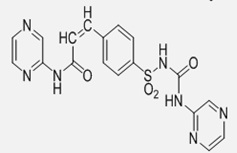 |
(N1-(2-pyrazinyl)-(E)-3-[4-({[(2-pyrazinylamino)carbonyl]amino}phenyl]-2-propenamide] | C18H15N7O4S
|
425.42 |
| 18 | GR18 | 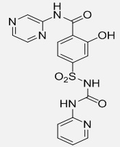 |
(N1-(2-pyrazinyl)-2-hydroxy-[4-({[(2-pyrazinylamino)carbonyl]amino}sulfonyl)benzamide | C17H14N6O5S
|
414.4 |
| 19 | GR19 | 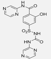 |
(N1-(2-pyrazinyl)-2-hydroxy-[4-({[(2-pyrazinylamino)carbonyl]amino}sulfonyl)benzamide | C16H13N7O5S
|
415.38 |
| 20 | GR20 | 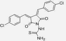 |
1-(3z,4E)-3,4bis(4-chloro benylidene)-2,5-dioxopyrrolidin-1-yl) Thio urea | C19H13Cl2N3O2S
|
418.3 |
| 21 | GR21 | 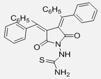 |
1-2,5-dioxo-3,4-bis(diphenylmethylene)pyrrolidine-1-yl)thiourea | C31H23N3O2S
|
501.6 |
| 22 | GR22 | 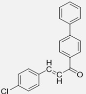 |
(E)-1-([1,1,-biphenyl]-4-yl)-3-(4-cholor phenyl)prop-2-en-1-one | C21H15ClO
|
318.8 |
| 23 | GR23 | 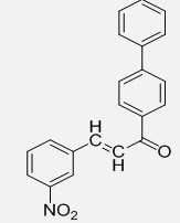 |
(E)-1-([1,1,-biphenyl]-4-yl)-3-(3-nitrophenyl)prop-2-en-1-one | C21H15NO3
|
329.35 |
| 24 | GR24 | 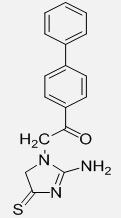 |
1-([1,1-biphenyl]-4-yl)-2-(2-amino-4,5-dihydro-1H-imidazol-1-yl)ethan-1-on | C17H15N3OS
|
309.39 |
| 25 | GR25 | 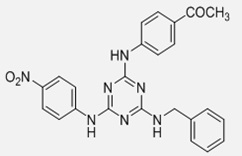 |
1-(4-[4-(4-nitrophenylamino)-6-(benzylamino)-1,3,5-triazin-2-ylamino)phenyl]ethanone | C24H21N7O3
|
455.47 |
| 26 | GR26 | 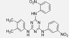 |
N2-(3,4-dimethylphenyl)-N6-(2-nitrophenyl)-N4-(3-nitrophenyl)-1,3,5-triazine-2,4,6-triamine | C24H22N8O4
|
486.48 |
| 27 | GR27 | 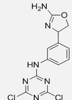 |
N-(3-(2-amino-4,5-dihydro oxazol-4-yl)-phenyl)-4,6-dichloro-1,3,5-triazin-2-amine | C12H10Cl2N6O
|
325.15 |
| 28 | GR28 | 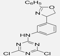 |
4,6-dichloro-N-(3-(4,5-dihydro-2-phenyloxazol-4-yl)phenyl)-1,3,5-triazin-2-amine | C18H13Cl2N5O
|
386.23 |
| 29 | GR29 | 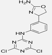 |
N-(4-(2-amino-4,5-dihydro oxazol-4-yl)-phenyl)-4,6-dichloro-1,3,5-triazin-2-amine | C12H10Cl2N6O
|
325.15 |
| 30 | GR30 | 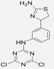 |
N-(4-(2-amino-4,5-dihydro thiazol-4-yl)-phenyl)-4,6-dichloro-1,3,5-triazin-2-amine | C12H10Cl2N6S
|
341.22 |
| 31 | GR31 | 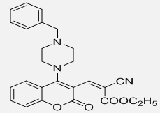 |
(2z)-ethyl3-(4-(4-benzylpiperazin-1-yl)-2-oxo-2H chromen-3-yl)-2-cyanoacrylate | C26H25N3O4
|
443.49 |
| 32 | GR32 | 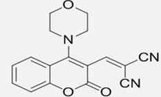 |
2-((4-morpholino-2-oxo-2H-chromen-3-yl)methylene)malononitrile | C17H13N3O3
|
307.3 |
| 33 | GR33 | 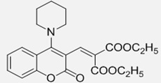 |
Diethyl2-((2-oxo-4-(piperidine-1-yl)-2H-chromen-3-yl)methylene)malonate | C22H25NO6
|
399.44 |
| 34 | GR34 | 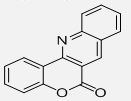 |
6H-chromeno[4,3-b]quinolin-6-one | C16H9NO2
|
247.25 |
| 35 | GR35 | 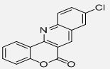 |
9-chloro-6H-chromeno[4,3-b]quinolin-6-one | C16H8ClNO2
|
281.69 |
| 36 | GR36 | 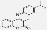 |
6H-chromen[4,3-b]quinoline-6-one | C19H15NO2
|
289.33 |
| 37 | GR37 | 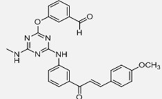 |
(E)-1-(4-(methylamino)-6-(benzaldehyde-1-yloxy)-1,3,5-triazine-2-ylamino)phenyl)-3-(methoxyphenyl)prop-2-en-1-one | C27H23N5O4
|
481.5 |
| 38 | GR38 | 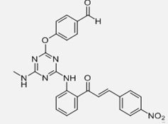 |
(E)-1-(4-(methylamino)-6-(benzaldehyde-1-yloxy)-1,3,5-triazine-2-ylamino)phenyl)-3-(nitrophenyl)prop-2-en-1-one | C26H20N6O5
|
496.47 |
| 39 | GR39 | 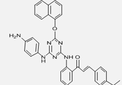 |
(E)-1-(4-(4-(4-aminophenylamino)-6-(naphthalen-1-yloxy)-1,3,5-triazine-2-ylamino)phenyl)-3-(4-ethylphenyl)prop-2-en-1-one | C36H30N6O2
|
578.66 |
| 40 | GR40 | 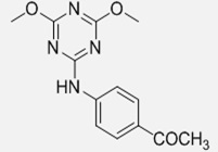 |
1-(4-(4,6-dimethoxy-1,3,5-triazine-2-ylamino)phenyl)ethanone | C13H14N4O3
|
274.28 |
| 41 | GR41 | 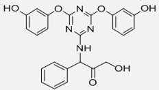 |
1-(4,6-bis(3-hydroxyphenoxy)-1,3,5-triazin-2-ylamino)-3-hydroxy-1-phenylpropan-2-one | C19H18N4O6S
|
430.43 |
| 42 | GR42 | 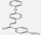 |
1-(4-nitrophenyl)-3-(4-phenoxyphenyl)prop-2-en-1-one | C21H15NO4
|
345.35 |
| 43 | GR43 | 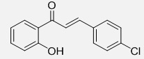 |
3-(4-chlorophenyl)-1-(2-hydroxyphenyl)prop-2-en-1-one | C15H11ClO2
|
258.7 |
| 44 | GR44 | 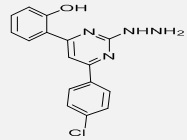 |
2-(2-hydrazino-6-(4-chlorophenyl)pyrimidin-4-yl)phenol | C16H13ClN4O
|
312.75 |
| 45 | GR45 | 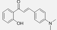 |
3-(4-(dimethylamino)phenyl)-1-(2-hydroxyphenyl)prop-2-en-1-one | C17H17NO2
|
267.32 |
| 46 | GR46 | 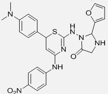 |
3-(4-(4-nitrophenylamine)-6-(4-dimethylamino)phenyl)-6H-1,3-thiazin-2-ylamino)-2-(furan-2-yl))imidazolidine-4-one
|
C25H25N7O4S
|
519.58 |
| 47 | GR47 | 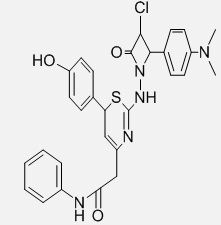 |
1-(4-phenylacetamido)-6-(4-hydroxyphenyl)-6H-1,3-thiazin-2-ylamino)-4-(4-(dimethylamino)phenyl)-3-chloroazetedin-2-one | C29H28ClN5O3S
|
562.08 |
| 48 | GR48 | 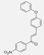 |
1-(4-nitrophenyl)-3-(4-phenoxyphenyl)prop-2-en-1-one | C21H15NO4
|
345.35 |
| 49 | GR49 | 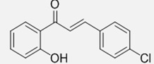 |
3-(4-chlorophenyl)-1-(2-hydroxyphenyl)prop-2-en-1-one | C21H15NO4
|
345.35 |
| 50 | GR50 | 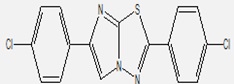 |
2,6-bis(4-chlorophenyl) imidazo[2,1-b] [1,3,4]thiadiazole | C16H9Cl2N3S
|
346.23 |
| 51 | GR51 | 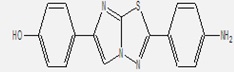 |
4-(2-(4-aminophenyl) imidazo[2,1-b][1,3,4]thiadiazol-6-yl)phenol | C16H15N7O2 | 337.34 |
| 52 | Inhibitor | 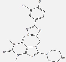 |
7-[[5-(3,4-dichlorophenyl)-1,3,4-oxadiazol-2-yl]methyl]-1,3-dimethyl-8-piperazin-1-yl-purine-2,6-dione | C20H20Cl2N8O3
|
491.33 |
| 53 | STD1 | 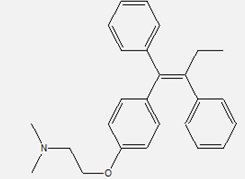 |
2-{4-[(1Z)-1,2-Diphenyl-1-buten-1-yl]phenoxy}-N,N-dimethylethanamin | C26H29NO
|
371.51 |
| 54 | STD2 | 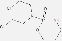 |
2-(bis(2-chloroethyl)amino)tetrahydro-2H-1,3,2-oxazaphosphorine 2-oxide
|
C12H28Cl2N3O2P
|
348.25 |
| 55 | STD3 | 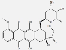 |
(8S,10S)-10-{[(2R,4S,5S,6S)-4-Amino-5-hydroxy-6-methyltetrahydro-2H-pyran-2-yl]oxy}-8-glycoloyl-6,8,11-trihydroxy-1-methoxy-7,8,9,10-tetrahydro-5,12-tetracenedione | C27H29NO11 | 543.52 |
TABLE 2: DOCKING VALIDATION RESULTS OF SELECTED LIGANDS, STANDARD DRUGS AND INHIBITOR
| S. no. | Code | Lowest Binding Energy | Run | RMSD | Inhibition Constant (Nm) |
| 1 | GR01 | -10.11 | 10 | 20.842 | 39.01 |
| 2 | GR02 | -9.73 | 44 | 19.766 | 73.89 |
| 3 | GR03 | -10.96 | 42 | 17.240 | 9.23 |
| 4 | GR04 | -10.67 | 15 | 20.058 | 15.20 |
| 5 | GR05 | -11.14 | 25 | 30.720 | 6.81 |
| 6 | GR06 | -11.27 | 15 | 14.41 | 5.45 |
| 7 | GR07 | -9.88 | 2 | 17.564 | 57.58 |
| 8 | GR08 | -10.69 | 21 | 16.289 | 14.62 |
| 9 | GR09 | -9.73 | 28 | 15.760 | 73.67 |
| 10 | GR10 | -9.76 | 39 | 25.877 | 70.04 |
| 11 | GR11 | -10.13 | 35 | 16.210 | 37.83 |
| 12 | GR12 | -11.30 | 17 | 16.419 | 5.18 |
| 13 | GR13 | -10.03 | 9 | 26.957 | 44.16 |
| 14 | GR14 | -9.87 | 30 | 26.729 | 58.58 |
| 15 | GR15 | -8.44 | 42 | 27.418 | 651.27 |
| 16 | GR16 | -10.84 | 22 | 21.987 | 11.38 |
| 17 | GR17 | -10.37 | 4 | 21.906 | 25.18 |
| 18 | GR18 | -10.10 | 24 | 22.229 | 39.41 |
| 19 | GR19 | -9.84 | 6 | 16.500 | 61.08 |
| 20 | GR20 | -9.74 | 49 | 21.782 | 72.82 |
| 21 | GR21 | -10.63 | 36 | 28.176 | 16.10 |
| 22 | GR22 | -9.80 | 41 | 22.173 | 65.41 |
| 23 | GR23 | -10.59 | 30 | 29.195 | 17.24 |
| 24 | GR24 | -8.84 | 29 | 20.690 | 332.21 |
| 25 | GR25 | -13.14 | 33 | 24.165 | 232.57 |
| 26 | GR26 | -11.70 | 47 | 22.571 | 2.67 |
| 27 | GR27 | -9.56 | 18 | 17.957 | 98.70 |
| 28 | GR28 | -10.61 | 27 | 24.557 | 16.75 |
| 29 | GR29 | -9.56 | 19 | 17.767 | 98.93 |
| 30 | GR30 | -9.86 | 24 | 17.925 | 58.82 |
| 31 | GR31 | -9.28 | 45 | 25.546 | 158.00 |
| 32 | GR32 | -9.62 | 48 | 27.210 | 88.47 |
| 33 | GR33 | -9.21 | 12 | 17.264 | 177.49 |
| 34 | GR34 | -9.19 | 7 | 25.796 | 184.50 |
| 35 | GR35 | -9.28 | 20 | 26.988 | 158.16 |
| 36 | GR36 | -9.59 | 8 | 27.674 | 93.20 |
| 37 | GR37 | -13.33 | 24 | 19.650 | 170.11 |
| 38 | GR38 | -13.08 | 5 | 32.374 | 257.38 |
| 39 | GR39 | -14.12 | 47 | 30.726 | 44.59 |
| 40 | GR40 | -8.51 | 49 | 25.623 | 581.19 |
| 41 | GR41 | -9.75 | 34 | 20.465 | 71.56 |
| 42 | GR42 | -9.46 | 15 | 16.978 | 117.06 |
| 43 | GR43 | -8.24 | 26 | 25.157 | 917.00 |
| 44 | GR44 | -9.74 | 39 | 27.274 | 72.83 |
| 45 | GR45 | -8.12 | 34 | 18.677 | 1,130 |
| 46 | GR46 | -11.46 | 4 | 30.091 | 3.95 |
| 47 | GR47 | -11.35 | 15 | 27.740 | 4.80 |
| 48 | GR48 | -9.63 | 11 | 32.194 | 88.05 |
| 49 | GR49 | -8.76 | 25 | 20.865 | 379.97 |
| 50 | GR50 | -9.56 | 20 | 19.622 | 98.42 |
| 51 | GR51 | -9.17 | 18 | 20.684 | 188.56 |
| 52 | Inhibitor 9CH | -10.59 | 41 | 1.557 | 17.30 |
| 53 | STD1 Tamoxifen | -9.65 | 6 | 20.390 | 85.01 |
| 54 | STD2 Cyclophosphamide | -5.83 | 34 | 19.875 | 53,670 |
| 55 | STD3 Doxorubicin | -11.74 | 47 | 20.366 | 2.50 |
CONCLUSION: Breast cancer is one of the most common types of malignancies in women in world wide. The risk of breast increases with age Between 45 to 50 in females. So we conducted docking studies on the marketed drugs, Reference Inhibitor with selected ligands on breast cancer crystal protein (PDB: 5NWH).
In this study the crystal structure of 5NWH receptor bound to 9CH was replaced with 51 selected ligands as potential target for breast Cancer. So among 51 selected ligands GR39 (-14.12) showed lowest binding energy compared with Reference Inhibitor (-10.59), STD1 (-9.65), STD2 (-5.83), STD3 (-11.74). In future, studies will be performed to establish the in-vitro, in-vivo on breast cancer cell lines.
ACKNOWLEDGEMENT: Nil
CONFLICTS OF INTEREST: Nil
REFERENCES:
- Siegel RL, Miller KD, Fuchs HE and Jemal A: Cancer statistics, 2026. CA Cancer J Clin 2026; 76(1): 17-48.
- Sung H, Ferlay J, Siegel RL, Laversanne M, Soerjomataram I and Jemal A: Global cancer statistics 2020. CA Cancer J Clin 2021; 71(3): 209-49.
- Waks AG and Winer EP: Breast cancer treatment: a review. JAMA 2019; 321(3): 288-300.
- Wright RHG and Beato M: NUDT5: a metabolic oncoprotein in hormone signaling and cancer. Trends Cancer 2018; 4(4): 268-72.
- Page BDG, Valerie NCK, Wright RHG, Wallner O, Isaksson R and Carter M: Targeted NUDT5 inhibitors block hormone signaling in breast cancer cells. Nat Commun 2018; 9: 250.
- Trott O and Olson AJ: AutoDock Vina: improving the speed and accuracy of docking. J Comput Chem 2010; 31(2): 455-61.
- Jordan VC: Tamoxifen: a most unlikely pioneering medicine. Nat Rev Drug Discov 2003; 2(3): 205-13.
- Wishart DS, Burley SK, Berman HM, Christie C, Duarte JM, Feng Z and Westbrook J: RCSB PDB: biological macromolecular structures. Nucleic Acids Res 2019; 47(1): 464-74.
- Patrick GL: An introduction to medicinal chemistry. Ed 7th Oxford: Oxford University Press 2021.
- Burley SK, Berman HM, Christie C, Duarte JM, Feng Z and Westbrook J: RCSB PDB: biological macromolecular structures. Nucleic Acids Res 2019; 47(1): 464-74.
- Berman HM, Westbrook J, Feng Z, Gilliland G, Bhat TN and Weissig H: The Protein Data Bank. Nucleic Acids Res 2000; 28(1): 235-42.
- RCSB Protein Data Bank. Structure of human NUDT5 (PDB ID: 5NWH). Available from: https://www.rcsb.org/
- Morris GM, Huey R, Lindstrom W, Sanner MF, Belew RK and Goodsell DS: AutoDock4 and AutoDockTools4. J Comput Chem 2009; 30(16): 2785-91
- (http://www.chemspider.com/
- https://autodock.scripps.edu/
- Biovia: Discovery Studio Visualizer user guide. San Diego (CA): Dassault Systems 2024.
- Jain AN: Scoring functions for protein-ligand docking. Curr Protein Pept Sci 2006; 7(5): 407-20.
- Jordan VC and Brodie AMH: Development and evolution of therapies targeting estrogen receptor. Steroids 2007; 72(1): 7-25.
- Emadi A, Jones RJ and Brodsky RA: Cyclophosphamide and cancer. Nat Rev Clin Oncol 2009; 6(11): 638-47.
- Minotti G, Menna P, Salvatorelli E, Cairo G and Gianni L: Anthracyclines: doxorubicin. Pharmacol Rev 2004; 56(2): 185-229.
- Morris GM, Huey R and Lindstrom W: Autodock4 and Autodock tools; automated docking with selective receptor flexibility. J Comput Chem 2009; 30(16): 2785-2791
- Biovia DS: Discovery studio visualizer, version 2021.San diego: Dassault systems BIOVIA 2021.
How to cite this article:
Rajan RG, Vani SU, Krishna APSM, Manimadhuri CD, Harika G and Aara SKN: Molecular docking analysis of heterocyclic ligands targeting NUDT5 protein in breast cancer. Int J Pharm Sci & Res 2026; 17(9): 2660-75. doi: 10.13040/IJPSR.0975-8232.17(9).2660-75.
All © 2026 are reserved by International Journal of Pharmaceutical Sciences and Research. This Journal licensed under a Creative Commons Attribution-NonCommercial-ShareAlike 3.0 Unported License.
Article Information
10
2660-2675
11121 KB
6
English
IJPSR
R. Govinda Rajan, S. Udhaya Vani, A. P. S. M. Krishna *, Ch. Dhanalakshmi Manimadhuri, G. Harika and S. K. Nujhat Aara
Department of Pharmaceutical Chemistry, Hindu College of Pharmacy, Guntur, Andhra Pradesh, India.
annavarapukrishna37@gmail.com
16 May 2026
27 May 2026
19 June 2026
10.13040/IJPSR.0975-8232.17(9).2660-75
01 September 2026
 Download