




NEUROINFLAMMATION IN DIABETIC ENCEPHALOPATHY: MECHANISTIC INSIGHTS, BIOMARKERS, AND EMERGING THERAPEUTIC TARGETS
HTML Full TextNEUROINFLAMMATION IN DIABETIC ENCEPHALOPATHY: MECHANISTIC INSIGHTS, BIOMARKERS, AND EMERGING THERAPEUTIC TARGETS
Siddharth Tamang * and Deepak Kumar Jha
Department of Pharmacology, Karnataka College of Pharmacy, Bengaluru, Karnataka, India.
ABSTRACT: Diabetic encephalopathy is an increasingly recognized neurological complication of diabetes mellitus, characterized by progressive cognitive decline, structural brain alterations, and impaired neuronal function. Although observed in both type 1 and type 2 diabetes, its underlying mechanisms remain incompletely understood. Chronic hyperglycemia and insulin resistance disrupt cerebral metabolic homeostasis, promoting oxidative stress, mitochondrial dysfunction, and neurovascular impairment. Emerging evidence suggests that neuroinflammation represents a central pathogenic mechanism linking metabolic disturbances to neuronal injury. Persistent activation of glial cells, disruption of blood-brain barrier integrity, and infiltration of peripheral immune cells contribute to sustained production of pro-inflammatory mediators, resulting in synaptic dysfunction, impaired neurogenesis, and neuronal loss in vulnerable brain regions, particularly the hippocampus. In addition, inflammatory signaling pathways, including NF-κB, NLRP3 inflammasome, and MAPK signaling, together with oxidative stress–mediated damage, have been implicated in disease progression in experimental and clinical studies. This review provides a comprehensive overview of the current understanding of neuroinflammatory mechanisms involved in diabetic encephalopathy, critically evaluates emerging biomarkers, and discusses potential therapeutic strategies for early diagnosis and targeted intervention.
Keywords: Diabetic encephalopathy, Neuroinflammation, Cognitive impairment, Oxidative stress, Microglial activation, Blood-brain barrier, Insulin resistance, Neurodegeneration, Biomarkers, Therapeutic approaches
INTRODUCTION: Diabetes mellitus has become one of the 21st century’s major global health challenges. The prevalence of the disease continues to increase rapidly, creating a considerable medical and socioeconomic burden worldwide. According to estimates, the number of diabetics worldwide keeps on increasing continuously due to ageing of the population, unhealthy lifestyles and change in dieting habits which has mainly been due to the ageing of the population and the change of lifestyle 1.
Even though the classical complications, including nephropathy, retinopathy, neuropathy, and cardiovascular disorders, are firmly established, increasing clinical and experimental evidence indicates that diabetes also affects the central nervous system 2. Cognitive dysfunction is a condition related to diabetes that negatively impacts the quality of life, treatment adherence, and clinical outcomes in the long term, which is why a more mechanistic understanding of the complication is urgently needed.
Diabetic encephalopathy is becoming a well-known neurological expression of diabetes diagnosed by learning impairments, memory, information processing speed and executive impairments 3. Neuroimaging and in-vivo experiments have shown structural and functional brain damage such as atrophy of the hippocampal, lesions within the white matter and impaired synaptic plasticity in diabetic patients and in-vivo animal models 4. Regardless of its increasing clinical significance, diabetic encephalopathy is the condition insufficiently diagnosed and treated, primarily because of the lack of a perfect understanding of the underlying pathophysiology and the lack of valid diagnostic biomarkers. Continuous hyperglycemia and insulin resistance initiate a cascade of metabolic dysfunctions which go beyond peripheral organs and substantially disrupt the cerebral homeostasis 5. In stark contrast to the previous assumption, which the brain is insulin-independent, the growing body of research proves that insulin signaling is key to the survival of neurons, their functioning at the synaptic level, and cognitive processes 6. Defective glucose metabolism, mitochondrial dysfunction, and impaired insulin signaling in diabetes leads to an excess of reactive oxygen and nitrogen species, which creates a pro neurodegenerative environment 7. In the variety of pathogenic mechanisms that have been involved in diabetic encephalopathy, neuroinflammation has turned into a key process, which is integrative. The chronic microglial and astrocyte activation, brain endothelial barrier breakdown, peripheral immunocytes infiltration, and continuous generation of proinflammatory mediators all lead to the dysfunction of neurons and deterioration of cognition 8. The main inflammatory mediators (interleukin 6, interleukin 1 beta, Tumor necrosis factor- alpha) directly interfere with neuronal signaling as well as engage with oxidative stress routes, further contributing to neural injury 9.
Due to the complexity of diabetic encephalopathy, it is crucial to have an in-depth insight into neuroinflammatory processes and how metabolic and oxidative pathways interact. In this respect, this review will critically assess the contribution of neuroinflammation in growth and advancement of diabetic encephalopathy, synthesize the results of experimental and clinical researches, indicate the possible biomarkers to diagnose this condition at an early stage, and discuss the existing and recent therapeutic options that may be used to combat inflammatory and oxidative processes. These insights can be used to design effective interventions that can prevent or slow the cognitive loss of people with diabetes.
REVIEW METHODOLOGY: This review was conducted to comprehensively summarize the current understanding of Parkinson's disease biomarkers, pathophysiological mechanisms, and experimental models. A literature search was performed using the electronic databases PubMed, Scopus, Web of Science, and Google Scholar. The search covered publications from 2000 to 2022.
Given the relatively limited number of studies specifically investigating the neuroinflammatory mechanisms underlying diabetic encephalopathy, this review primarily emphasizes evidence derived from diabetes-associated cognitive impairment and diabetic brain dysfunction whenever available. However, to provide a comprehensive mechanistic framework, selected findings from related fields, including Alzheimer's disease, neurodegenerative disorders, systemic inflammation, and diabetic neuropathy, have also been incorporated. These studies described molecular pathways, inflammatory mediators, or cellular processes that are potentially relevant to diabetic encephalopathy.
Pathophysiology of Diabetic Encephalopathy: The development of Diabetic encephalopathy is driven by prolonged metabolic disturbances, vascular abnormalities, and inflammatory mechanisms associated with diabetes. Chronic hyperglycemia, impaired insulin signaling, and altered lipid metabolism disrupt normal brain homeostasis, which eventually causes brain dysfunction and eventual cognitive decline. Contrary to short-term fluctuations in glycemia, chronic metabolic instability causes cumulative structural and functional brain alterations with interrelated processes of oxidative stress, mitochondrial perturbation, neurovascular dysfunction, and chronic inflammation 10.
Chronic Hyperglycemia and Metabolic Dysfunction: A long-term period of hyperglycemia is also a major inducing variable in the occurrence of brain injury in diabetic encephalopathy. The persistent presence of high glucose levels leads to excessive accumulation of glucose into neurons and glial cells beyond the normal processing of glucose metabolism. This causes the redistribution of glucose to other metabolic pathways, such as polyol pathway, hexosamine biosynthetic pathway, and protein kinase C-mediated signaling 11.
The stimulation of these pathways stimulates the overproduction of reactive oxygen species (ROS), causes distortions in redox balance, and causes the emergence of metabolic stress. Cerebral insulin resistance is yet another factor that leads to metabolic impairments. The insulin signaling has an important role in promoting the synaptic integrity, neurotransmitter release regulation and neuronal viability. The dysfunction of insulin sensitivity in the diseased brain causes a decrease in the level of glucose uptake, energy generation and synaptic transmission, especially in the memory areas like the hippocampus 12. These changes add to the shortages in synaptic plasticity and cognitive functions. One of the major effects of chronic hyperglycemia is the presence of advanced glycation end products (AGEs) in the neural tissues. Non-enzymatic glycation of macromolecules leads to augmented AGE generation, which, in the event that it engages the receptor of AGEs (RAGE), initiates intracellular signaling chains comprising of NF-kB, mitogen-activated protein-kinases and NADPH oxidase. Such activities increase oxidative stress and stimulate pro-inflammatory gene expression and form a mechanistic linkage between metabolic deregulation and neuroinflammation 13. Changes in lipid metabolism are also substantial in neuronal damage, along with glucotoxicity effects. Higher levels of circulating free fatty acids and extracellular lipid deposition deteriorate mitochondrial bioenergetics, membrane stability, and endoplasmic reticulum stress in neurons and glial cells 14. The glucotoxicity combined with lipotoxicity causes a metabolically unfavorable environment that increases the neurodegenerative processes in diabetic encephalopathy.
Influence of Diabetes Type and Disease Characteristics on Cognitive Dysfunction: The development and progression of diabetic encephalopathy may differ substantially between type 1 diabetes mellitus (T1DM) and type 2 diabetes mellitus (T2DM) because of differences in disease pathogenesis, age of onset, metabolic disturbances, and associated comorbidities. In T1DM, cognitive dysfunction is often linked to early disease onset, prolonged insulin deficiency, recurrent hypoglycemic episodes, and long-term exposure to glycemic fluctuations. Repeated severe hypoglycemia may adversely affect neuronal survival, synaptic plasticity, and cognitive performance, particularly in children and young adults. In contrast, T2DM is more frequently associated with insulin resistance, obesity, chronic low-grade inflammation, dyslipidemia, hypertension, and cerebrovascular disease, all of which contribute to neurovascular dysfunction and cognitive decline. Furthermore, both forms of diabetes consistently associate disease duration, poor glycemic control, and increased glycemic variability with accelerated cognitive impairment. The interaction between metabolic dysregulation, vascular pathology, and neuroinflammation may therefore differ between T1DM and T2DM, making it critical to take into account diabetes subtype and disease characteristics when evaluating the pathogenesis and clinical manifestations of diabetic encephalopathy 2, 3.
Blood Endothelial Barrier Disruption: It is the bloodbrain barrier (BBB) that maintains the specialized microenvironment inside the CNS by restricting cellular and molecular exchange between the brain and the bloodstream. The deregulation of hyperglycemia and systemic inflammatory stress impairs the BBB integrity in diabetes leading to augmented permeability and loss of selective barrier function 15. The changes in tight junction architecture such as the downregulation and the disorganization of claudins, occludin and zonula occludens proteins are consistently reported in experimental studies. The major cause of BBB breakdown in diabetic conditions is the impairment of endothelial cells. Oxidative stress in hyperglycemia disrupts endothelial nitric oxide signaling, increases vascular inflammation, and increases leukocyte adhesion to cerebral blood vessels 16.
The production of adhesion molecules and inflammatory mediators by activated endothelial cells facilitates the migration of immune cells into the cerebral tissue. The more the permeability of the barrier, the more access circulating cytokines, AGEs, and other possibly neurotoxic agents have to the brain, fueling the responses of the neuroinflammatory system and neuronal damage 17. In addition, impairment of the functioning of the BBB disrupts ionic homeostasis and the delivery of nutrients, which further impairs the neuronal metabolism.
It is interesting to note that the BBB impairment frequently occurs before the neuronal loss can be detected, which implies that this is a key pathological instigator of diabetic encephalopathy.
Neurovascular Unit Dysfunction: The neurovascular unit (NVU) comprising of neurons, astrocytes, endothelial cells, pericytes, and extracellular matrix factors has a substantial role of coordinating the metabolic needs of neurons to the cerebral blood flow. Metabolic stress of diabetes, at the expense of NVU organization and functionality causes impaired neurovascular coupling and decreased cerebral perfusion 18. One of the major regulators of NVU stability are astrocytes involved in the process of maintaining energy, clearance of neurotransmitters, and regulating vascular tone. Astrocyte reactive responses to diabetic conditions include some dysregulated calcium signaling, reduced glutamate uptake, and elevated expression of pro-inflammatory cytokines 19.
These changes affect the communication through the synapses and enhance excitotoxic neural damage. Clinical as well as preclinical studies reveal decreased cerebral blood flow and reduced vascular responsiveness in diabetes, especially in those parts of the brain that are involved in the higher cognitive functions 20.
Neurovascular uncoupling restricts the supply of oxygen and glucose to active neurons, which increases the stress on metabolism and oxidative stress. NVU dysfunction is further motivated by structural alterations including loss of pericytes and thickening of basement membrane that cause further destabilization of cerebral microvasculature. The combination of chronic metabolic dysregulation, the state of BBB, and NVU dysfunction create a combined pathology that predetermines the diabetic brain to the persistent neuroinflammation and the further development of cognitive impairment.
TABLE 1: INTEGRATED PATHOPHYSIOLOGICAL MECHANISMS IN DIABETIC ENCEPHALOPATHY 11-20
| S. no. | Pathophysiological Event | Mechanistic Consequences | Molecular Mediators |
| 1 | Chronic Hyperglycemia and Insulin Resistance | Persistent metabolic stress increases intracellular glucose accumulation, activates the polyol, hexosamine and PKC pathways, promotes AGE formation, and impairs insulin signaling. | AGEs, RAGE, IRS/PI3K/Akt, PKC, Free Fatty Acids |
| 2 | Mitochondrial Dysfunction and Oxidative Stress | Hyperglycemia-induced metabolic overload disrupts mitochondrial respiration, resulting in excessive ROS generation, impaired ATP production, and redox imbalance. | ROS, NADPH Oxidase, MAPKs, NRF2, Reactive Nitrogen Species |
| 3 | Activation of Inflammatory Signaling Pathways | Oxidative stress activates NF-κB, NLRP3 inflammasome, MAPK and JNK pathways, leading to sustained production of inflammatory mediators. | NF-κB, NLRP3, Caspase-1, IL-1β, IL-6, TNF-α |
| 4 | Blood–Brain Barrier Dysfunction | Oxidative and inflammatory insults disrupt endothelial integrity and tight junction proteins, increasing BBB permeability and facilitating entry of circulating inflammatory mediators. | Claudins, Occludin, ZO-1, ICAM-1, VCAM-1, eNOS, AGEs |
| 5 | Neurovascular Unit Dysfunction | Impaired communication among neurons, astrocytes, endothelial cells and pericytes results in defective neurovascular coupling, reduced cerebral blood flow and metabolic insufficiency. | GFAP, GLT-1, Ca²⁺ Dysregulation, Pericyte Loss, Cytokines |
| 6 | Glial Activation and Neuroinflammation | Activated microglia and astrocytes amplify inflammatory signaling, producing cytokines and reactive species that perpetuate neuronal injury. | TNF-α, IL-1β, IL-6, iNOS, ROS, NLRP3 |
| 7 | Neuronal Dysfunction and Neurodegeneration | Chronic neuroinflammation and oxidative stress lead to synaptic dysfunction, impaired neurogenesis, apoptosis, white matter injury, and cognitive decline. | Tau, Caspases, NfL, BDNF↓, Synaptic Proteins↓ |
Neuroinflammation: There is a growing body of evidence to support neuroinflammation as a key pathological process underpinning metabolic abnormalities to neuronal dysfunction in diabetic encephalopathy. Persistent hyperglycemia and insulin resistance favor an inflammatory state within the brain, which is marked by prolonged glial activation, overexpression of inflammatory mediators, impaired blood-brain barrier function and the influx of peripheral immune cells.
This contrasts with the transient inflammatory reaction in which diabetes-related neuroinflammation is insidious, persistent, leading to slow synaptic destruction and neuronal damage, which gradually progresses with time 21.
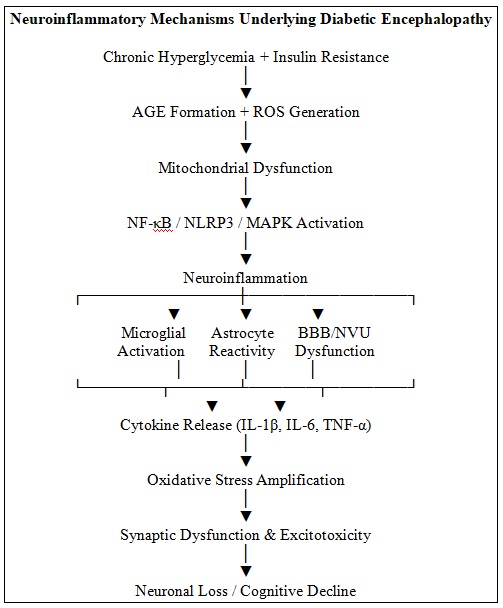
Microglial Activation: Microglial cells are the main immune cells that protect the central nervous system. They are also important for keeping neurons stable and balanced. In diabetes, high levels of glucose, oxidative stress, and the buildup of advanced glycation end products all keep microglial cells active for a long time. Ongoing stimulation induces complex activation states in microglia characterized by diverse transcriptional and functional profiles. Although the traditional M1/M2 framework has been widely used to describe pro-inflammatory and anti-inflammatory microglial responses, growing evidence indicates that microglial activation exists along a dynamic spectrum rather than as discrete phenotypic states. In diabetic and other neuroinflammatory conditions, activated microglia may simultaneously express both protective and detrimental molecular signatures depending on the local metabolic and inflammatory microenvironment. When this state is activated, it makes more inflammatory mediators, such as tumor necrosis factor alpha, interleukin 1, interleukin 6, and inducible nitric oxide synthase 22. These are all involved in neuroinflammatory processes. These alterations cause impairment of the synapses, neuronal damage, and neuro plasticity. There are also metabolic changes in microglia which are caused by diabetes and continue to propagate inflammatory signaling. Exposure to a high glucose level reprograms the microglial energy metabolism to elevated glycolysis, which favors long-term cytokine production and increases the production of reactive oxygen species 23. Lasting activation of microglia impairs the normal physiological synaptic remodeling and hastens the degeneration of neuronal cells, especially those involved in the hippocampal parts of the brain that are involved in learning and memory.
Astrocyte Reactivity: Astrocytes are critical to the control of the synaptic system, metabolic support, and preservation of the blood-brain barrier activity. Reactive astrogliosis occurs in the diabetic brain, involving cellular hypertrophy and altered transcriptional changes as well as functional dysregulation of astrocytes 24. Reactive astrocytes exhibit substantial heterogeneity in response to metabolic and inflammatory stimuli. While the A1/A2 classification has been useful in describing neurotoxic and neuroprotective astrocytic responses, recent studies suggest that astrocytes display multiple context-dependent activation states that cannot be fully captured by a binary classification system. The phenotype adopted by astrocytes depends on factors such as disease stage, metabolic status, inflammatory mediators, and interactions with neighboring neural and immune cell. The A1 type of phenotype is the most valid when facing diabetic conditions and is known to release pro-inflammatory cytokines, complement components, and neurotoxic molecules that increase the neuronal damage. Astrocytic pathology also increases the pathogenesis of diseases by the failure to clear glutamate and buffering potassium, making them susceptible to excitotoxic damage. Moreover, the astrocytes are actively involved in the amplification of the neuroinflammatory responses by the two-sided interactions with the activated microglia, which supports the networks of inflammatory signaling in the diabetic brain 25.
It is important to acknowledge that the classical M1/M2 microglial and A1/A2 astrocytic frameworks represent simplified conceptual models that may not fully reflect the complexity of glial biology in diabetic encephalopathy.
Recent advances in single-cell transcriptomics and spatial profiling have revealed diverse and highly plastic glial activation states that vary according to disease stage, local microenvironment, metabolic stress, and inflammatory signaling. Therefore, the terminology used in this review is intended to provide a simplified description of predominant functional responses rather than to imply the existence of fixed or mutually exclusive glial phenotypes 26, 27.
Cellular Infiltration on the Periphery: The damage of the blood-brain barrier by diabetes supports the infiltration of peripheral immune cells into the central nervous system. Monocytes, macrophage and T lymphocytes move across the injured barrier in response to chemokines and adhesion molecules synthesized by activated endothelial and glial cells 28. These infiltrating immune cells once in the brain parenchyma then enhance inflammatory reactions by the release of cytokines, reactive oxygen species and proteolytic enzymes. Recent articles point to an increased contribution of adaptive immune cells in diabetes neuroinflammation. CD4+ and CD8+ T lymphocytes have been observed to be present in diabetic brain tissue, and they promote long term inflammatory signaling and neuronal damage. These leads dedicate that inflammation in diabetic encephalopathy is systemic and has a strong interaction between peripheral immune activation and central nervous system pathology 29.
Inflammatory Pathways: There are a range of intracellular signaling pathways that mediate neuroinflammatory responses in diabetic encephalopathy. Experimental evidence from STZ-induced and high-fat diet diabetic rodent models has consistently demonstrated increased NF-κB activation in the hippocampus and cerebral cortex, accompanied by elevated expression of TNF-α, IL-1β, and IL-6. These findings suggest that NF-κB signaling contributes directly to diabetes-associated neuroinflammation and cognitive impairment. The sustained activation of NF-kB leads to sustained production of pro-inflammatory mediators, which participates in the chronic dysfunction of neurons 30. However, evidence from human diabetic encephalopathy studies remains limited, and the majority of mechanistic data currently originate from preclinical animal investigations.
The other critical process is the activation of NLRP3inflammasome. Activation of NLRP3 induces maturation of IL-1b and IL-18 via the action of caspase-1 and the maintenance of prolonged inflammation and neuronal damage. Several animal studies and high-glucose cellular models have demonstrated increased NLRP3 inflammasome activation, caspase-1 cleavage, and IL-1β maturation in diabetic conditions. Pharmacological inhibition of NLRP3 has been reported to improve cognitive performance and reduce neuroinflammation in diabetic rodents 31, supporting a causal role for this pathway. Nevertheless, direct clinical evidence in patients with diabetic encephalopathy remains scarce.
Activation of MAPK and JNK signaling pathways has been observed primarily in experimental cellular and animal models exposed to hyperglycemic stress. These pathways are believed to mediate inflammatory gene transcription, oxidative injury, and neuronal apoptosis. However, their specific contribution to diabetic encephalopathy in humans remains largely inferential and requires further validation 32.
Oxidative Stress-Inflammation Crosstalk: Crosstalk occurs when oxidative stress and inflammation increase protein synthesis, which is promoted by activating latent T cells of the innate immune system. Neuroinflammation and oxidative stress are highly interrelated events that support each other in diabetic encephalopathy. Due to the overproduction of reactive oxygen and nitrogen species, antioxidant defenses within the body become overwhelmed and result in the redox imbalance and the activation of inflammatory signal transduction. NF-kB and NLRP3 inflammasome activation are promoted by oxidative stress, whereas inflammatory mediators worsen mitochondrial functions and stimulate the production of reactive oxygen species 33.
The down-regulation of the NRF2 which is the major regulator of cell antioxidant response also enhances oxidative damage by down-regulating cytoprotective and detoxifying enzymes. Mitochondrial dysfunction is an essential amplifier in the process since impaired mitochondria are the cause and consequences of oxidative stress and release vicious cycle leads to a faster neuronal dysfunction and is one of the causes of gradual cognitive impairments in diabetic encephalopathy. Reduced NRF2 activity has been reported in diabetic animal models and in vitro hyperglycemic systems, where impaired antioxidant defense contributes to oxidative damage and inflammatory signaling. Although NRF2 activation improves cognitive outcomes in several preclinical studies, clinical evidence supporting NRF2 dysregulation as a therapeutic target in diabetic encephalopathy is currently limited.
It should be noted that the majority of mechanistic evidence regarding NF-κB, NLRP3 inflammasome, MAPK/JNK, and NRF2 signaling in diabetic encephalopathy is derived from experimental cellular and animal models. While these pathways are strongly implicated in diabetes-associated neuroinflammation, direct validation in human diabetic encephalopathy remains limited.
TABLE 2: NEUROINFLAMMATORY MECHANISMS IN DIABETIC ENCEPHALOPATHY
| S. no. | Mechanism | Molecular Mediators | Principal Consequences |
| 1 | Microglial Activation | IL-1β, IL-6, TNF-α, iNOS, ROS, AGEs, NF-κB | Neuroinflammation, synaptic dysfunction, neuronal injury |
| 2 | Astrocyte Reactivity | GFAP, IL-6, TNF-α, Complement Proteins, GLT-1↓ | Excitotoxicity, impaired glutamate homeostasis, neuronal damage |
| 3 | Peripheral Immune Cell Infiltration | Monocytes, Macrophages, CD4+ T cells, CD8+ T cells, ICAM-1, VCAM-1 | Sustained neuroinflammation and BBB dysfunction |
| 4 | Inflammatory Signaling Pathways | NF-κB, NLRP3 Inflammasome, Caspase-1, IL-1β, IL-18, MAPK, JNK | Cytokine production, inflammasome activation, neuronal injury |
| 5 | Oxidative Stress | ROS, RNS, NRF2, NF-κB, NLRP3, Mitochondrial Dysfunction | Redox imbalance, inflammation, neurodegeneration |
Cellular and Molecular Repercussions: Chronic neuroinflammatory stress and metabolic stress in diabetes initiates a broad spectrum of cellular and molecular abnormalities that all lead to cognitive dysfunction in diabetic encephalopathy. Such pathological alterations influence synaptic communication, neuronal viability, adult neurogenesis and white matter organization, which eventually disrupt neural circuitry and reduce brain plasticity 34.
Synaptic Dysfunction and Dendritic Spine Loss: Learning and memory rely on efficient transmission of synaptic impulses, and growing evidence indicates that synaptic damage is a pathological early sign of diabetic encephalopathy. Long-term exposure of brain neurons to the effects of inflammatory cytokines, such as tumor necrosis factor-a (TNF-a) and interleukin-1b (IL-1b), disrupts synaptic signaling by modifying the composition of neurotransmitter receptors and impairs long-term potentiation in hippocampal neurons 35. The mediators of the process also lead to the impairment of synaptic vesicle trafficking and intracellular calcium homeostasis due to inflammatory mediators and subsequently, the reduced synaptic strength. There are also noticeable structural synaptic defects in diabetes that include decreases in the dendritic spine densities and changes in the morphology of the spine. These alterations are more pronounced in hippocampal and cortical areas that are critical in cognitive processing 36. The persistent microglial activation in diabetes state conditions leads to uncontrolled and over stimulated synaptic pruning, which further enhances the rate of synaptic loss and functional deterioration.
Impaired Neurogenesis: Adult neurogenesis, which takes place most actively in dentate gyrus of the hippocampal document, is critical in memory encoding and cognitive plasticity. This process is substantially reduced in diabetic patients by its oxidative stress, chronic inflammatory, and insulin signaling 37. The pro-inflammatory cytokines repress the growth and transformation of the neural precursors, whereas the oxidative harm caused by hyperglycemia stimulates the kill of progenitor cells. Reduced neurogenesis is strongly linked with the impairment of spatial learning and memory in diabetic animal models. Notably, interventions that aid metabolic regulation or a decrease in neuroinflammation was also demonstrated to be partially reversible in recovering neurogenic activity, which suggests that a compromise of neurogenesis can be reversed in specific conditions of a therapeutic intervention 38.
Phosphorylation of Tau and Amyloid-Like Alterations: Diabetic encephalopathy has a number of molecular aspects in common with classical neurodegenerative diseases, such as aberrant tau phosphorylation and amyloid-associated changes. The resistance to insulin in the brain impairs the following critical brain signaling pathways: insulin-induced tau regulation via the glycogen synthase kinase-3b signaling pathway, which causes tau hyperphosphorylation and microtubules destabilization39. These alterations affect axonal transportation and damage synapses. Simultaneously, the chronic inflammation and oxidative stress contribute to the amyloid precursor protein processing and the deposition of the amyloid-like peptides in the diabetic brains. Even though the extent of amyloid deposition is usually not as extensive as in the case of the Alzheimer disease, the pathological alterations are believed to be involved in the dysfunction of neurons and cognitive impairment in diabetic encephalopathy 40.
Apoptosis and Pyroptosis: The programmed apoptotic paths and inflammatory types of cell death mediate neuronal loss in diabetic encephalopathy. Intrinsic apoptotic processes are started by mitochondrial dysfunction, oxidative stress, and enhanced pro-inflammatory cytokine that leads to cytochrome c release and caspase activation 41. It is also through apoptosis that neurons in susceptible parts of the brain are gradually depleted especially the hippocampus. New data points to pyroptosis, another cause of neurodegeneration of diabetes. The NOD-like receptor pyrin domain-containing 3 (NLRP3) inflammasome activation and caspase-1 leads to happen a gasderminmediated membrane pore creation, causing an inflammatory cell death, and discharge of cytokines like IL-1b42.Pyroptosis increases neuroinflammation and creates a vicious cycle of inflammation and neuronal death.
White Matter Injury: White matter integrity is what is needed to facilitate concerted neural communication and diabetes is increasingly linked with notable pathology of the white matter. Chronic hyperglycemia and microvascular impairment hinder the viability of oligodendrocytes, and myelin maintenance, which is followed by demyelination and axonal degeneration 43. Inflammatory mediators also interfere with oligodendrocyte development and survival further enhancing the destruction of white matter. The neuroimaging techniques used in patients with diabetes reveal that the white matter integrity and structural dysconnectivity is low and this is associated with cognitive impairment. There is experimental evidence that white matter damage does not necessarily result in information processing slowing but also in executive functions impairments in diabetic encephalopathy 44.
Experimental Evidence: Preclinical and in-vitro experimental studies have provided valuable evidence that elucidates the pathophysiological processes of diabetic encephalopathy, especially the role of neuroinflammation in the cognitive impairment of diabetes. Experimental models allow systematic study of metabolic dysfunctions, inflammatory responses and neurodegradation that are hard to isolate in clinical studies of human beings.
Rodent Models: The most common experimental tools used to study diabetic encephalopathy is the rodent models. One of them, streptozotocin (STZ)-induced diabetes is a well-established mouse model of type 1 diabetes and has been widely used to investigate the implications of chronic hyperglycemia on the brain. STZ leads to long-lasting hyperglycemia, insulin resistance, and cognitive impairment, and severe impairments in hippocampus-mediated learning and memory processes. The recurring pattern in these models is the improvement of microglial activation, growth of the pro-inflammatory cytokine levels, augmented oxidative stress, and progressive neuronal destruction. Rodent models of type 2 diabetes mellitus (T2DM) using high-fat diet (HFD) models and combined HFD-STZ models better represent the metabolic derangements observed in human T2DM. These are characterized by insulin resistance, disrupted lipid metabolism and sustained low-grade systemic inflammation, as well as by substantial neuroinflammatory activity and cognitive dysfunction 45. The experimental paradigms such as Morris water maze, Y-maze and novel object recognition tests continue to show a deficit in spatial learning, memory consolidation and executive functioning. The support of the connection between brain pathology and metabolic dysfunction has been further supported by genetic and transgenic models of insulin resistance such as db/db and ob/ob mice. These creatures exhibit premature disorientation of the mind, changes in the synaptic function of the hippocampus and a rise in inflammatory messaging, which frequently precedes the appearance of overt hyperglycemia 46. In general, rodent study evidence suggests that insulin deficiency and insulin resistance can act on their own and synergistically to facilitate neuroinflammation and cognitive dysfunction.
In-vitro Studies: Experimental systems in vitro offer useful complementary information as they can be thoroughly analyzed on the mechanistic level both on a cellular and molecular level. High-glucose conditions of primary neuronal cultures and neuronal cell lines cause oxidative stress, mitochondrial dysfunction, and apoptosis, which are highly reminiscent of the major characteristics of diabetes-associated neuronal damage47. Such pathological modifications are often related to a decrease in the extension of neurites and the expression of synaptic proteins. Equally, microglial and astrocyte cultures in hyperglycemic conditions also show a high inflammatory response that is reflected in a high production of TNF-a, IL-1b, IL-6, and inducible nitric oxide synthase. Microglial polarization towards pro-inflammatory phenotype also occurs under high-glucose conditions and facilitates inflammasome pathway activation 48. Co-culture of neurons and glial cells has also shown that glial cell-initiated inflammatory mediators are very critical in mediating the dysfunction in neurons under diabetic conditions. Also, in-vitro models of the blood-brain barrier (BBB), based on endothelial cell mono-culture, have demonstrated that hyperglycemia and the effect of inflammatory cytokines destroy the tight junction integrity and increase the barrier permeability. These data points are consistent with in-vivo data of BBB perturbation and mechanistic evidence of the enhanced immune cell infiltration and amplification of neuroinflammatory responses in diabetic conditions 49.
Regardless of the different methodological variations across the experimental models, there are some findings that always come out in the studies of diabetic encephalopathy. The hallmark feature is cognitive impairment (especially learning and memory), which is strongly coupled with the dysfunction of the hippocampal. Neuroinflammatory markers such as activated microglia, reactive astrocytes and high concentration of pro-inflammatory cytokines are substantially elevated in diabetic brains. Recurrent sterilization of inflammatory signaling tissue like the NF-kB and the NLRP3 inflammasome is commonly viewed and is closely pertinent to oxidative stress and mitochondrial damage. The brain structures such as the loss of synapses, neuronal degeneration, and atrophy of the hippocampal have been reported in various experimental models 47. In general, these results support the primary importance of neuroinflammation in the occurrence of diabetic encephalopathy and the usefulness of experimental models as the tools of assessing possible treatment options.
Biomarkers, Diagnostic Methodology: Although several inflammatory, oxidative stress, and neurodegeneration-related biomarkers have been associated with cognitive impairment in diabetes, none currently demonstrate sufficient specificity to serve as standalone diagnostic biomarkers for diabetic encephalopathy. Many candidate biomarkers, including IL-6, TNF-α, C-reactive protein (CRP), advanced glycation end products (AGEs), soluble receptor for advanced glycation end products (sRAGE), and neurofilament light chain (NfL), are also altered in other metabolic, inflammatory, vascular, and neurodegenerative disorders 50-52. Consequently, their clinical utility is presently limited to providing supportive information regarding disease activity, systemic inflammation, oxidative stress, or neuronal injury rather than establishing a definitive diagnosis of diabetic encephalopathy. Future studies should focus on multimodal biomarker panels integrating biochemical, neuroimaging, and cognitive measures to improve diagnostic accuracy and disease specificity.
TABLE 3: BIOMARKERS IN DIABETIC ENCEPHALOPATHY: EVIDENCE AND LIMITATIONS
| Biomarker | Biological Source | Study Population | Assay Method | Association with Cognitive Outcomes | Diagnostic Performance | Major Limitations |
| IL-6 | Serum, plasma, CSF | T1DM, T2DM, animal models | ELISA, multiplex immunoassays | Higher levels associated with cognitive decline and neuroinflammation | Not established | Elevated in numerous inflammatory disorders; poor specificity |
| TNF-α | Serum, plasma, CSF | T1DM, T2DM, experimental models | ELISA, multiplex assays | Correlates with neuroinflammatory activity and cognitive dysfunction | Not established | Non-specific marker of systemic inflammation |
| CRP | Serum | Mainly T2DM cohorts | Immunoturbidimetry, ELISA | Associated with vascular risk and cognitive decline | Low specificity | Influenced by obesity, infection, cardiovascular disease |
| Oxidative Stress Markers (MDA, ROS, 8-OHdG) | Blood, urine, tissues | Experimental and clinical studies | Spectrophotometry, HPLC, ELISA | Reflect oxidative injury linked to cognitive impairment | Not established | Not specific to CNS pathology |
| AGEs | Serum, plasma | Diabetic patients and animal models | ELISA, fluorescence assays | Associated with cognitive decline and vascular dysfunction | Moderate association only | Elevated in many diabetic complications |
| Soluble RAGE (sRAGE) | Serum, plasma | Clinical diabetes studies | ELISA | Proposed marker of AGE-RAGE pathway activation | Inconsistent findings | Limited validation and poor disease specificity |
| Neurofilament Light Chain (NfL) | Serum, plasma, CSF | Clinical cohorts | SIMOA, ELISA | Reflects neuroaxonal injury and cognitive decline | Promising but exploratory | Elevated in many neurological disorders |
| GFAP (optional addition) | Serum, CSF | Emerging studies | SIMOA | May indicate astroglial activation | Emerging evidence | Limited diabetic encephalopathy data |
| Neuroimaging Biomarkers (MRI, DTI, fMRI, PET) 53-55 | Brain imaging | Human studies | Imaging-based | Correlate with structural and functional cognitive changes | More informative than single biochemical markers | Cost, accessibility, lack of standardization |
Current evidence suggests that no single biomarker possesses adequate sensitivity and specificity for the diagnosis of diabetic encephalopathy. Most available biomarkers reflect broader pathological processes such as systemic inflammation, oxidative stress, endothelial dysfunction, or neurodegeneration and therefore lack disease specificity.
Therapeutic Strategies for Diabetic Encephalopathy: Currently, no pharmacological or biological therapy has been specifically approved for the prevention or treatment of diabetic encephalopathy. Most therapeutic strategies discussed in this review are based on evidence derived from experimental cellular studies, animal models of diabetes, or clinical investigations conducted primarily in patients with diabetes, neurodegenerative disorders, or related metabolic conditions. Consequently, the reported neuroprotective effects should be interpreted cautiously, as many findings remain preliminary and require confirmation in well-designed clinical studies specifically evaluating cognitive outcomes in diabetic populations.
TABLE 4: THERAPEUTIC STRATEGIES FOR DIABETIC ENCEPHALOPATHY: EVIDENCE, OUTCOMES, AND LIMITATIONS
| Therapeutic Strategy | Intervention | Evidence Type | Study Design / Model | Reported Outcomes | Major Limitations |
| Anti-inflammatory Therapy | NLRP3 Inflammasome Inhibitors56 | Preclinical | Diabetic rodent models; pharmacological and genetic inhibition | Reduced microglial activation, decreased IL-1β and TNF-α release, improved cognitive performance | No clinical trials specific to diabetic encephalopathy; long-term safety unknown |
| TNF-α and IL-1β Targeted Therapies57 | Preclinical | Experimental animal studies | Reduced neuroinflammation, improved synaptic integrity, decreased neuronal apoptosis | Evidence limited to preclinical studies; lack of diabetes-specific clinical data | |
| Antioxidant Therapy | NRF2 Activators58 | Preclinical | Cellular and diabetic animal models | Enhanced antioxidant defense, reduced oxidative stress, improved mitochondrial function and cognition | Human evidence lacking; optimal therapeutic agents remain uncertain |
| Mitochondria-Targeted Antioxidants (e.g., CoQ10)59 | Preclinical | Experimental diabetic models | Reduced ROS production, preserved mitochondrial function, decreased neuronal injury | Limited clinical validation and uncertain brain bioavailability | |
| Glucose-Lowering and Metabolic Therapies | Metformin60 | Preclinical and Limited Clinical Evidence | Experimental diabetic models; observational clinical studies | Improved insulin sensitivity, reduced inflammation, potential cognitive benefits | Mixed clinical findings; no approval for diabetic encephalopathy |
| GLP-1 Receptor Agonists61 | Strong Preclinical; Emerging Clinical Evidence | Animal models and limited human studies | Improved synaptic plasticity, reduced neuroinflammation, enhanced cognition | Human cognitive outcome data remain limited | |
| SGLT2 Inhibitors62 | Preclinical; Limited Clinical Evidence | Diabetic animal studies and clinical diabetes populations | Improved metabolic control, reduced systemic inflammation, potential neurovascular protection | Few studies directly evaluating cognitive outcomes | |
| Lifestyle Interventions | Physical Exercise63 | Clinical and Preclinical Evidence | Human intervention studies and animal models | Increased neurogenesis, improved insulin sensitivity, reduced inflammation, enhanced cognition | Variable adherence; heterogeneous study protocols |
| Anti-inflammatory/Antioxidant Diets64 | Clinical Observational and Interventional Studies | Human diabetes populations | Improved glycemic control and reduced cognitive decline risk | Difficult dietary standardization and long-term compliance | |
| Emerging Therapies | Stem Cell Therapy65 | Preclinical | Diabetic animal models | Reduced neuroinflammation, enhanced neurogenesis, restoration of neural networks | Limited human data; safety and regulatory concerns |
| Nanoparticle-Based Drug Delivery66 | Experimental | Cellular and animal studies | Improved brain-targeted drug delivery and reduced systemic toxicity | Early developmental stage; limited translational evidence | |
| Gene-Modulation Approaches | Experimental | Preclinical studies | Regulation of inflammatory and oxidative stress pathways | Safety, delivery efficiency, and long-term effects remain unresolved |
Future Perspectives: In spite of the fact that diabetic encephalopathy is becoming recognized as a topical neurological complication of diabetes, there are still several gaps in the existing knowledge base. One of the major problems is that there have not been universally recommended diagnostic criteria and well-verified biomarkers that can detect early cognitive impairments in diabetics. Most of the available information relies on experimental animal models, but clinical studies in humans are quite limited in number and are largely cross-sectional, which does not allow causal interpretation or prolonged disease course assessment 67.
The other substantial weakness is the limited access to human brain tissue, which limits the validation of the mechanistic understanding of preclinical experiments. The problem of heterogeneity in patient populations in terms of disease duration and glycemic control levels, coexistence and interventions complicate further the process of translational interpretation. In addition, the impact of biological sex and aging on neuroinflammation attributed to diabetes and cognitive impairments are under-investigated whereas some early findings show that they differ by sex and age 68.
Therapeutically, most intervention studies focus on short term efficacy, and aim at single isolated molecular targets. Given the multifactorial and intricate nature of diabetic encephalopathy, future studies need to be focused on long-term, integrative treatment methods, which involve a combination of optimized metabolic care and anti-inflammatory therapy and neuroprotective therapy. In this regard, extensive multi-omics technologies such as transcriptomic, proteomic and metabolomic analyses can provide promising options that can reveal new molecular patterns, and novel therapeutic targets 69.
Also, the combination of the latest neuroimaging methods and online cognitive assessment systems can facilitate its initial diagnosis and allow tracking the disease evolution with more accuracy. Future studies should also further investigate the differential effects of T1DM and T2DM on neuroinflammation and cognitive decline. Factors such as age at disease onset, duration of diabetes, glycemic variability, recurrent hypoglycemia, insulin exposure, and vascular comorbidities may influence disease progression and biomarker profiles but remain inadequately characterized.
CONCLUSION: Diabetic encephalopathy remains a frequently overlooked but progressively common neurological consequence of diabetes, exerting a substantial negative impact on cognitive performance and overall quality of life. Growing experimental and clinical evidence suggests that neuroinflammation plays a pivotal role in disease development by linking sustained hyperglycemia, insulin resistance, oxidative imbalance, and neurovascular impairment to neuronal damage and cognitive deterioration.
Ongoing activation of glial cells, compromise of blood-brain barrier integrity, stimulation of pro-inflammatory signaling cascades, and the interplay between oxidative stress and mitochondrial dysfunction act in concert to accelerate disease progression. Both preclinical and clinical investigations underscore the diagnostic and therapeutic potential of inflammatory and oxidative stress-related biomarkers, advanced neuroimaging modalities, and targeted treatment strategies. Nevertheless, substantial challenges remain in effectively translating these advances into routine clinical practice. Progress in this field will depend on a more comprehensive understanding of underlying pathogenic mechanisms, the development of sensitive early diagnostic tools, and the implementation of integrated therapeutic strategies. Targeting neuroinflammatory processes may therefore represent a crucial approach for reducing cognitive decline and preserving neurological function in individuals living with diabetes.
ACKNOWLEDGEMENT: I would like to thank the management and faculty of Karnataka College of Pharmacy, Bengaluru for providing the academic environment and facilities, that enabled the completion of this review paper.
Funding Statement: Self-funded
Ethical Clearance: Not applicable
CONFLICT OF INTEREST: No conflicts of interests in terms of the publication of this review article.
REFERENCES:
- International Diabetes Federation. IDF Diabetes Atlas. 10th ed. Brussels: IDF 2021.
- Biessels GJ, Deary IJ and Ryan CM: Cognition and diabetes: a lifespan perspective. Lancet Neurol 2008; 7(2): 184-190.
- Moheet A, Mangia S and Seaquist ER: Impact of diabetes on cognitive function and brain structure. Ann N Y Acad Sci 2015; 1353: 60-71.
- Kodl CT and Seaquist ER: Cognitive dysfunction and diabetes mellitus. Endocr Rev 2008; 29(4): 494-511.
- Brownlee M: The pathobiology of diabetic complications: a unifying mechanism. Diabetes 2005; 54(6):1615-1625.
- Kleinridders A, Ferris HA, Cai W and Kahn CR: Insulin action in brain regulates systemic metabolism and brain function. Diabetes 2014; 63(7): 2232-2243.
- Vincent AM, Russell JW, Low P and Feldman EL: Oxidative stress in the pathogenesis of diabetic neuropathy. Endocr Rev 2004; 25(4): 612-628.
- Salim S. Oxidative stress and neuroinflammation in neurodegenerative disorders. Neurochem Int 2017; 104: 20-27.
- Donath MY and Shoelson SE: Type 2 diabetes as an inflammatory disease. Nat Rev Immunol 2011; 11(2): 98-107.
- Biessels GJ and Reagan LP: Hippocampal insulin resistance and cognitive dysfunction. Nat Rev Neurosci 2015; 16(11): 660-671.
- Giacco F and Brownlee M: Oxidative stress and diabetic complications. Circ Res 2010; 107(9): 1058-1070.
- Arnold SE, Arvanitakis Z and Macauley-Rambach SL: Brain insulin resistance in type 2 diabetes and Alzheimer disease. Nat Rev Neurol 2018; 14(3): 168-181.
- Ramasamy R, Yan SF and Schmidt AM: Advanced glycation endproducts: from precursors to RAGE-mediated pathology. Ann N Y Acad Sci 2011; 1126: 1-12.
- Kothari V, Luo Y and Tornabene T: High fat diet induces brain insulin resistance and cognitive impairment. Biochim Biophys Acta Mol Basis Dis 2017; 1863(2): 499-508.
- Hawkins BT and Davis TP: The blood–brain barrier/neurovascular unit in health and disease. Pharmacol Rev 2005; 57(2): 173-185.
- Prasad S, Sajja RK, Naik P and Cucullo L: Diabetes mellitus and blood–brain barrier dysfunction. J Pharmacovigil 2014; 2(2): 125.
- Banks WA: Blood–brain barrier transport of cytokines: a mechanism for neuropathology. Curr Pharm Des 2005; 11(8): 973-984.
- Iadecola C: The neurovascular unit coming of age: a journey through neurovascular coupling in health and disease. Neuron 2017; 96(1): 17-42.
- Verkhratsky A, Parpura V, Pekna M, Pekny M and Sofroniew M: Glial asthenia and functional paralysis of astrocytes in neurodegeneration. Neurochem Res 2014; 39(1): 1-16.
- Novak V and Hajjar I: The relationship between blood pressure and cognitive function. Nat Rev Cardiol 2010; 7(12): 686-698.
- Heneka MT, Carson MJ and El Khoury J: Neuroinflammation in Alzheimer’s disease. Lancet Neurol 2015; 14(4): 388–405.
- Tang Y and Le W: Differential roles of M1 and M2 microglia in neurodegenerative diseases. Mol Neurobiol 2016; 53(2): 1181–1194.
- Orihuela R, McPherson CA and Harry GJ: Microglial M1/M2 polarization and metabolic states. Br J Pharmacol 2016; 173(4): 649–665.
- Sofroniew MV: Astrocyte reactivity: subtypes, states, and functions. Nat Rev Neurosci 2020; 21(2): 101–114.
- Liddelow SA, Barres BA. Reactive astrocytes: production, function, and therapeutic potential. Immunity 2017; 46(6): 957–967.
- Paolicelli RC, Sierra A and Stevens B: Microglia states and nomenclature: a field at its crossroads. Neuron 2022; 110(21): 3458–3483. https://doi.org/10.1016/j.neuron.2022.10.020
- Escartin C, Galea E and Lakatos A: astrocyte nomenclature, definitions, and future directions. Nat Neurosci 2021; 24: 312–325. https://doi.org/10.1038/s41593-020-00783-4
- Varatharaj A and Galea I: The blood–brain barrier in systemic inflammation. Brain Behav Immun 2017; 60: 1–12.
- Gate D, Saligrama N and Leventhal O: Clonally expanded CD8 T cells in neurodegeneration. Nature 2020; 577(7790): 399–404.
- Lawrence T: The nuclear factor NF-κB pathway in inflammation. Cold Spring HarbPerspect Biol 2009; 1(6): 001651.
- Ising C, Venegas C and Zhang S: NLRP3 inflammasome activation drives neurodegeneration. Nature 2019; 575(7784): 669–673.
- Kyriakis JM and Avruch J: Mammalian MAPK signal transduction pathways. Physiol Rev 2012; 92(2): 689–737.
- Reuter S, Gupta SC, Chaturvedi MM and Aggarwal BB: Oxidative stress and inflammation. Free Radic Biol Med 2010; 49(11): 1603–1616.
- Ma Q: Role of NRF2 in oxidative stress and toxicity. Annu Rev Pharmacol Toxicol 2013; 53: 401–426
- Viviani B, Boraso M, Valero M and Gardoni F: Cytokines and neuronal ion channels in health and disease. Int Rev Neurobiol 2014; 118: 109–127.
- Li ZG, Zhang W, Grunberger G and Sima AA: Hippocampal neuronal apoptosis in diabetes. Brain Res 2002; 946(2): 221–231.
- Stranahan AM, Arumugam TV, Cutler RG, Lee K, Egan JM and Mattson MP: Diabetes impairs hippocampal function through glucocorticoid-mediated effects. J Neurosci 2008; 28(15): 3816–3825.
- van der Borght K, Kóbor-Nyakas DE and Klauke K: Physical exercise leads to rapid adaptations in hippocampal neurogenesis. Hippocampus 2009; 19(10): 928–936.
- Talbot K, Wang HY and Kazi H: Demonstrated brain insulin resistance in Alzheimer’s disease patients. J Clin Invest 2012; 122(4): 1316–1338.
- Sims-Robinson C, Kim B, Rosko A and Feldman EL: How does diabetes accelerate Alzheimer disease pathology? Nat Rev Neurol 2010; 6(10): 551–559.
- Green DR and Reed JC: Mitochondria and apoptosis. Science 1998; 281(5381): 1309–1312.
- Shi J, Zhao Y and Wang K: Cleavage of GSDMD by inflammatory caspases determines pyroptotic cell death. Nature 2015; 526(7575): 660–665.
- Allen NJ and Lyons DA: Glia as architects of central nervous system formation. Science 2018; 362(6411): 181–185.
- Reijmer YD, van den Berg E and Ruis C: Cognitive dysfunction in patients with type 2 diabetes. Diabetes Care 2010; 33(9): 1933–1938.
- Sima AA and Kamiya H: Diabetic neuropathy differs in type 1 and type 2 diabetes. Ann N Y Acad Sci 2006; 1084: 235–249.
- Russell JW, Golovoy D and Vincent AM: High glucose-induced oxidative stress and mitochondrial dysfunction in neurons. FASEB J 2002; 16(13): 1738–1748.
- Wang S, Yuan YH, Chen NH and Wang HB: The mechanisms of NLRP3 inflammasome activation in diabetic neuroinflammation. Neuroscience 2019; 409: 42–53.
- Prasad S, Sajja RK, Naik P and Cucullo L: Diabetes mellitus and blood–brain barrier dysfunction. J Pharmacovigil 2014; 2(2): 125.
- Biessels GJ, Strachan MW, Visseren FL, Kappelle LJ and Whitmer RA: Dementia and cognitive decline in type 2 diabetes. Lancet Neurol 2014; 13(10): 1018–1035.
- Schmidt AM, Hori O and Chen JX: Advanced glycation end products interacting with their receptor induce stress responses. J Clin Invest 1995; 96(3): 1395–1403.
- Srikanth V, Westcott B and Forbes J: Advanced glycation endproducts and cognitive impairment in diabetes. Neurology 2011; 77(13): 1276–1282.
- Gaetani L, Blennow K, Calabresi P, Di Filippo M, Parnetti L and Zetterberg H: Neurofilament light chain as a biomarker in neurological disorders. J Neurol Neurosurg Psychiatry 2019; 90(8): 870–881.
- Moran C, Phan TG and Chen J: Brain atrophy in type 2 diabetes. Diabetes Care 2013; 36(12): 4036–4042.
- Musen G, Jacobson AM and Ryan CM: Resting-state brain connectivity in type 2 diabetes. Diabetes 2012; 61(9): 2249–2257.
- Calsolaro V and Edison P: Neuroinflammation in Alzheimer’s disease. Alzheimers Dement 2016; 12(6): 719–732.
- Swanson KV, Deng M and Ting JP: The NLRP3 inflammasome: molecular activation and regulation. Nat Rev Immunol 2019; 19(8): 477–489.
- Dinarello CA: Anti-inflammatory agents: present and future. Cell 2010; 140(6): 935–950.
- Cuadrado A, Rojo AI and Wells G: Therapeutic targeting of NRF2. Nat Rev Drug Discov 2019; 18(4): 295–317.
- Murphy MP and Hartley RC: Mitochondria as a therapeutic target. Annu Rev Pharmacol Toxicol 2018; 58: 247–267.
- Koenig AM, Mechanic-Hamilton D and Xie SX: Effects of metformin on cognition. J Alzheimers Dis 2017; 59(2): 633–642.
- Hölscher C: GLP-1 analogues in neurodegeneration. Neuropharmacology 2018; 136: 169–177.
- Heerspink HJL, Perkins BA, Fitchett DH, Husain M and Cherney DZ: SGLT2 inhibitors in diabetes. Lancet Diabetes Endocrinol 2016; 4(5): 389–398.
- Erickson KI, Hillman C and Stillman CM: Physical activity and brain health. Nat Rev Neurosci 2019; 20(7): 425–437.
- Morris MC, Tangney CC and Wang Y: Diet and cognitive decline. Alzheimers Dement 2015; 11(9): 1015–1022.
- Li J, Tang Y and Cai D: Stem cell therapy for neurodegenerative diseases. Stem Cell Res Ther 2018; 9(1): 1–12.
- Glass CK and Saijo K: Nuclear receptor transrepression pathways. Trends Endocrinol Metab 2010; 21(1): 13–22.
- Strachan MWJ, Reynolds RM, Marioni RE and Price JF: Cognitive function, dementia and type 2 diabetes. Lancet Diabetes Endocrinol 2011; 378(9786): 219–231.
- Kautzky-Willer A, Harreiter J and Pacini G: Sex and gender differences in risk, pathophysiology and complications of type 2 diabetes. Endocr Rev 2016; 37(3): 278–316.
- Hasin Y, Seldin M and Lusis A: Multi-omics approaches to disease. Genome Biol. 2017; 18(1): 83.
How to cite this article:
Tamang S and Jha DK: Neuroinflammation in diabetic encephalopathy: mechanistic insights, biomarkers, and emerging therapeutic targets. Int J Pharm Sci & Res 2026; 17(10): 2798-12. doi: 10.13040/IJPSR.0975-8232.17(10).2798-12.
All © 2026 are reserved by International Journal of Pharmaceutical Sciences and Research. This Journal licensed under a Creative Commons Attribution-NonCommercial-ShareAlike 3.0 Unported License.
Article Information
2
2798-2812
598 KB
9
English
IJPSR
Siddharth Tamang * and Deepak Kumar Jha
Department of Pharmacology, Karnataka College of Pharmacy, Bengaluru, Karnataka, India.
siddharthtmg7@gmail.com
12 May 2026
14 June 2026
20 June 2026
10.13040/IJPSR.0975-8232.17(10).2798-12
01 October 2026
 Download