




PHARMACOGNOSTIC STANDARDIZATION AND PHYTOCHEMICAL PROFILING OF ROOT BARK OF PAJANELIA LONGIFOLIA AND RADERMACHERA XYLOCARPA, TWO LESS-KNOWN ETHNOMEDICINAL TREES OF BIGNONIACEAE
HTML Full TextPHARMACOGNOSTIC STANDARDIZATION AND PHYTOCHEMICAL PROFILING OF ROOT BARK OF PAJANELIA LONGIFOLIA AND RADERMACHERA XYLOCARPA, TWO LESS-KNOWN ETHNOMEDICINAL TREES OF BIGNONIACEAE
C. K. Smitha *, C. H. Kavitha and S. S. Anupama
Department of Botany, Government College for Women, Thiruvananthapuram, Kerala, India.
ABSTRACT: Pharmacognostic evaluation becomes a simple and reliable method for the authentication of crude herbal drugs, once the morphological characters are lost during processing. This study presents a comprehensive pharmacognostic, physicochemical, and elemental characterization of the root bark of two ethnomedicinally important but underexplored trees- Pajanelia longifolia and Radermachera xylocarpa of Bignoniaceae family. The roots of both species share some common histological characters like stone cells and calcium oxalate crystals, but differ in odour, cork thickness, and crystal size. Minor differences in ash values indicate variation in inorganic content. For the first time, this study reports the mineral and heavy metal elemental profiles of the root bark of P. longifolia and R. xylocarpa. Calcium was identified as the predominant mineral in P. longifolia (268.54 mg Kg⁻¹) and R. xylocarpa (328.43 mg Kg⁻¹), followed by magnesium, highlighting their potential nutritional and therapeutic significance. Major toxic heavy metals such as Al, Ni, Pb, Hg and Co were found to be below detectable levels in both species, while copper was detected only in R. xylocarpa. HPTLC analysis of root bark extracts showed that P. longifolia possessed 9 resolved peaks distributed mainly in the lower and mid Rf regions (0.02–0.65), on the other hand, R. xylocarpa displayed a broader phytochemical profile extending from Rf 0.02 to 0.80 with 12 resolved peaks. The dominant HPTLC peak at Rf 0.03 in both the root barks indicates the occurrence of a major common phytoconstituent. GC–MS analysis of P. longifolia root bark extracts revealed coumaran as the major phytoconstituent, represented by the highest peak at a retention time of 11.33 min, whereas the predominance of compounds such as quinic acid, guaiacol (2-methoxy phenol), isovanillic acid, β-sitosterol, and coumaran reflects the phytochemical complexity of R. xylocarpa. The generated diagnostic standards provide a scientific basis for the authentication, quality assurance, and safe medicinal use of these underexplored ethnomedicinal roots.
Keywords: Ethnomedicinal, Pajanelia longifolia, Radermachera xylocarpa, Powder microscopy, ICP-AES, GC-MS analysis
INTRODUCTION: Medicinal plants have been an integral component of traditional healthcare systems since ancient times, serving as valuable sources of therapeutic agents.
Their medicinal applications evolved through empirical observations and the accumulated knowledge of traditional healers.
Through centuries of practice and observation, ethnomedicinal knowledge has guided the identification of medicinal plants with therapeutic potential, many of which have subsequently evolved into evidence-based medicines through modern scientific validation. Pajanelia longifolia (Willd.) K. Schum. and Radermachera xylocarpa (Roxb.) K. Schum. are two ethnomedicinally important yet scientifically underexplored trees of the family Bignoniaceae. P. longifolia, commonly known as ‘Tender wild jack’, is an inhabitant of deciduous and semi-evergreen forests of India, Myanmar, Peninsular Malaysia and Sri Lanka 1. It holds significance in Indian folklore medicine, with reported antioxidant, antimicrobial, and anthelmintic activities 2, 3. Tribal populations inhabiting various parts of India use this plant to treat arthritis, jaundice, and skin diseases 4, 5. HRLC–MS analysis revealed the presence of a diverse spectrum of bioactive phytoconstituents, several of which have been documented in the literature to possess a broad range of pharmacological activities, including anticancer, anti-arthritic, anti-inflammatory, and antiviral properties 6. Computational analysis of bioactive phytocompounds from the methanolic extract of P. longifolia revealed promising interactions with the cancer-associated targets epidermal growth factor receptor and Transforming growth factor beta, suggesting their potential anticancer activity 7. R. xylocarpa is a tree species endemic to India, confined to the dry deciduous landscapes of Central India and the Western Ghats, where it thrives at elevations of approximately 1,200 feet 8. The resinous stem exudate of this tree is traditionally used to treat cutaneous disorders 9, 10. The bark paste is traditionally used to treat rheumatoid arthritis and joint pain 11, 12. At the same time, its fruit paste is applied to snakebite wounds or administered orally for the expulsion of snake venom from the body 13-15. The classical Ayurvedic formulation Dasamoola, comprising ten medicinal roots, includes ‘Patala’ and ‘Syonaka’as key ingredients. According to Ayurvedic literature, the root of R. xylocarpa is recognised as an alternative source for Patala, which is authentically obtained from Stereospermum suaveolens 16. Conversely, the root of P. longifolia is considered as an unauthorised substitute for Syonaka, which is traditionally and authentically sourced from Oroxylum indicum.
Despite the ethnomedicinal relevance of P. longifolia and R. xylocarpa, detailed pharmacognostic characterization, mineral analysis and heavy metal profiling of these species has not been reported. Pharmacognostic evaluation remains one of the most dependable and economical approaches for the authentication and quality control of herbal drugs, even when the raw materials are reduced to powder form and lose their morphological identity 17. The present study addresses the lack of scientific documentation on these species by establishing diagnostic parameters for root identification in both raw and powdered forms, while also providing detailed phytochemical characterization to elucidate the scientific basis underlying the therapeutic potential of their root bark.
MATERIALS AND METHODS:
Collection of Plant Samples: Plant materials were collected from the Chittur region along the banks of the Bhavani River in Attappadi, Mannarkkad Taluk, Palakkad District, Kerala, during April 2023, after obtaining prior permission from the Divisional Forest Officer, Mannarkkad Forest Division. Collected materials were identified and authenticated by Dr. P.S. Udayan, Senior Consultant, National Medicinal Plants Board, Regional Cum-Facilitation Centre (Southern Region), Kerala Forest Research Institute, Thrissur District. The authenticated voucher specimens were deposited at the Calicut University Herbarium (CALI), with voucher numbers CALI359–366 for P. longifolia and CALI367–375 for R. xylocarpa. The pharmacognostic standardization studies were conducted in the laboratories of Government College for Women, Thiruvananthapuram. Inductively Coupled Plasma- Atomic Emission Spectroscopy (ICP-AES) and Gas Chromatography–Mass Spectrometry (GC–MS) profiling were carried out at the Centre for Analytical Instrumentation, Kerala Forest Research Institute (KFRI), Peechi, Thrissur, Kerala.
Preparation of Samples for Pharmacognostic Studies: The separated root barks were cut into small pieces, shade-dried and sieved through a 60 mesh size sieve to obtain a uniform powder, as per the methodology described in the Ayurvedic Pharmacopoeia of India 18. The powdered material was then preserved in airtight containers for further examination. Before drying, the organoleptic properties of both the fresh root bark and the powdered samples were noted.
Moisture Content of the Root Bark and Loss on Drying: The percentage of water content in the fresh root bark was calculated as [(Initial weight of fresh bark – final weight of fresh bark) / Initial weight] x 100, according to the procedure mentioned in Indian Standards 19. To determine the percentage loss on drying, which indicates the volatile matter in the powdered crude drug, 5 grams of crude drug powder was dried at 120 °C for 2 hours in a preheated oven and reweighed. The percentage of loss on drying is [(Initial weight of powder – Final weight of powder) / Initial weight] x 100.
Determination of Ash Value: Three grams of crude drug were incinerated in a tarred silica crucible at < 450 °C for 4 h to obtain total ash. The total ash in percentage was calculated as (Weight of total ash / Weight of crude drug) × 100. For finding water-soluble ash, the total ash was boiled in 25 mL of distilled water for 2 min, filtered, and the insoluble residue was oven-dried at 120 °C for 2 hours 20. Percentage of water-soluble ash = [(Total ash – Water-insoluble ash) / Weight of crude drug] × 100. For acid-soluble ash, the total ash was boiled with 25 mL of 2 M HCl for 5 min, filtered, washed, oven-dried at 120 °C, and weighed. The percentage of acid-insoluble ash = (Weight of acid-insoluble ash / Weight of crude drug) × 100.
Analysis of Mineral Content and Heavy Metals: One gram of powdered drug sample was boiled with 1% HNO₃, followed by the addition of perchloric acid and heating until fumes ceased. Aqua regia (HNO₃: HCl in 3:1 ratio) was then added and heated, ensuring complete decomposition of organic matter and release of elements. The solution was diluted to 100 mL with deionized water. Mineral and heavy metal content in the crude drug powder was determined using Inductively Coupled Plasma- Atomic Emission Spectrometry (ICP-AES) on a PerkinElmer Avio 200 instrument. Characteristic emission spectra of each element were subsequently recorded by the spectrophotometer.
Anatomical and Histochemical Studies: Anatomical observations were carried out using three to five root bark samples collected from authenticated plant materials, following the guidelines of WHO 21. Fresh root barks were fixed in FAA (10% Formaldehyde, 50% Absolute alcohol, 5% acetic acid and 35% distilled water). Thin free-hand transverse sections were prepared using a sharp stainless-steel razor blade. Representative sections were selected, stained with safranin, temporarily mounted in glycerine, and observed under a Leica DM1000 compound microscope at 100 x and 400 x magnification. For histochemical studies, sections were treated with respective reagents- phloroglucinol, iodine solution and ferric chloride and were observed under the microscope to detect the presence of lignin, starch and tannin, respectively.
Powder Microscopic Studies: A small amount of fine powder sample was soaked in distilled water for 1 hour, treated with chloral hydrate, and stained with phloroglucinol-HCl, safranine, and toluidine blue 22. The material was mounted on grease-free slides and examined under a Leica DM 1000 microscope, and photomicrographs were captured using a DFC 295 camera.
HPTLC Analysis: HPTLC was performed on aluminium plates precoated with silica gel 60 F254 (0.25 mm thickness). The plates were prewashed with methanol and activated at 60 °C for 5 min before sample application. The plant extract was dissolved in 1 mL of chromatographic grade methanol. 5 µL of each sample was applied as bands using a 25 µL Camag automatic TLC sampler. The chromatographic development was carried out in a Camag automatic developing chamber (ADC, 20 × 10 cm, with two tracks maintained at a start position of 15 mm from the bottom edge and 15 mm inter-track distance. After optimization, the chromatographic chamber was saturated for 20 min with 100 mL of the mobile phase consisting of toluene:ethyl acetate:methanol (7:3:1) prior to development. Development was performed using 12 mL of mobile phase until appropriate separation was achieved. Developed plates were air-dried for 4 min. followed by heating for 1 min and densitometric scanning was performed using a Camag TLC scanner at 295 nm with a slit dimension of 6.00 × 0.30 mm and a scanning speed of 20 mm/s. The developed chromatograms were visualized under white light, 254 nm, and 360 nm using a Camag visualizer, and images were documented using a digital imaging system.
GC-MS Analysis: GC–MS analysis of the methanolic root bark extracts was performed using a Shimadzu QP 2010 S instrument equipped with a 30 m × 0.25 mm × 0.25 µm column under electron impact mode (70 eV) with helium as carrier gas. The oven temperature was programmed from 80 ºC to 260 ºC at 10 ºC min⁻¹ and held for 8 min. A 1 µl sample was injected in split mode and analyzed for 50 min. Compounds were identified by comparing retention times and mass spectra with NIST 05 and Wiley 8 libraries.
Statistical Analysis: All physicochemical analyses were performed in triplicate, and the results are expressed as mean ± standard deviation (SD). Statistical analysis was carried out using Microsoft Excel
RESULTS: The physicochemical parameters, elemental analysis and histochemical characteristics of the samples are presented in Table 1, 2 and 3 respectively. Fig. 1 illustrates the plants along with their medicinally important parts. The powder microscopic characteristics of the root barks of both species are depicted in Fig. 2 & 3 and the results are explained in Table 4. Histochemical features of the root barks are presented in Fig. 4 to 7.
Fig. 8 and 9 represent the preliminary HPTLC plate of the methanolic root extracts of the two species under different wavelengths of light and their corresponding chromatograms, respectively. Root bark of P. longifolia revealed 10 resolved peaks with Rf values ranging from 0.03 to 0.65, indicating the presence of multiple phytoconstituents mainly distributed in the lower and middle Rf regions.
The major peaks were observed at Rf values of 0.03 (19.86% area), 0.07 (17.17%), and 0.09 (15.39%), suggesting the predominance of relatively polar compounds Table 5. In comparison, the root bark extract of R. xylocarpa exhibited a relatively broader phytochemical profile with 12 resolved peaks spanning an Rf range of 0.03 to 0.79, indicating greater chemical diversity. The highest peak area was recorded at Rf 0.03 (29.67%), followed by notable peaks at Rf 0.22 (11.60%) and 0.79 (10.07%), reflecting the presence of both polar and less polar constituents Table 6.
In the absence of previously reported marker compounds for these species, the present preliminary HPTLC profiles may serve as baseline chromatographic fingerprints for their identification and future phytochemical investigations.
GC-MS chromatograms of the methanolic root bark extracts of two species are presented in Fig. 10. P. longifolia revealed 9 tentative phytocompounds with coumaran as the predominant phytoconstituent, accounting for 61.07% of the total peak area, followed by guanosine and 5-hydroxymethylfurfural.
On the other hand, R. xylocarpa revealed 12 tentatively identified compounds with retention times ranging from 7.616 to 42.270 min, indicating a chemically diverse phytoconstituent profile.
Among them, guanosine (24.79% area) and quinic acid (22.02% area) were the major constituents, followed by isovanillic acid (10.26%) and β-sitosterol (9.12%), suggesting their potential contribution to the medicinal properties of the plant. The results indicate that P. longifolia is dominated mainly by heterocyclic aromatic compounds, whereas R. xylocarpa exhibited comparatively greater phytochemical diversity with phenolics, nucleosides, and sterol derivatives.
TABLE 1: ORGANOLEPTIC AND PHYSICO-CHEMICAL CHARACTERISTICS OF THE ROOT BARKS OF P. LONGIFOLIA AND R. XYLOCARPA
| Character | P. longifolia | R. xylocarpa |
| Colour of fresh root bark | Reddish-brown | Reddish brown |
| Odour of fresh root bark | Pungent | Aromatic |
| Taste of fresh root bark | Bitter | Bitter |
| Moisture content of bark (%) | 69.6±1.83 | 63.8±0.83 |
| Moisture content of crude powder (%) | 8.20±0.21 | 8.60±0.32 |
| Total ash (%) | 13.6±0.24 | 12.8±0.31 |
| Water soluble ash (%) | 3.20±0.23 | 3.10±0.52 |
| Acid insoluble ash (%) | 2.20±0.42 | 1.80±0.33 |
*values represent mean ±SD
TABLE 2: MINERAL AND HEAVY METAL COMPOSITION OF ROOT BARK POWDER OF P. LONGIFOLIA & R. XYLOCARPA
| Mineral (mg Kg-1) | |||||||
| Ca | Mg | Mn | Fe | Zn | Na | Cr | |
| P. longifolia | 268.54 | 120.68 | 0.950 | 65.399 | BDL | 0.421 | 3.428 |
| R.xylocarpa | 328.431 | 149.60 | 1.033 | 70.177 | 5.031 | 2.357 | 3.877 |
| Heavy metals (mg Kg-1) | |||||||
| Al | Cd | Cu | Ni | Pb | Hg | Co | |
| P. longifolia | BDL | 0.902 | BDL | BDL | BDL | BDL | BDL |
| R. xylocarpa | BDL | 0.583 | 21.09 | BDL | BDL | BDL | BDL |
*BDL- below detectable live
TABLE 3: HISTOLOGICAL CHARACTERS OF ROOT BARK OF P. LONGIFOLIA & R. XYLOCARPA
| Characters | P. longifolia | R. xylocarpa |
| Thickness of cork layer | 500-800 µm | 180-220 µm |
| No. of layers in the cork | 20 to 40 | 15-25 |
| No of layers of cork cambium | 3-5 layers | 7-9 layers |
| Nature of parenchyma cells in primary cortex | Round crushed | Round intact |
| Measurement of primary cortical cells | 40-70 µm | 30-50 µm |
| Medullary rays | Multiseriate | Triseriate |
| Cork cells | 40-70 µm | 30-40 µm) |
TABLE 4: POWDER MICROSCOPIC ANALYSIS OF ROOT BARK POWDERS OF R. XYLOCARPA & P. LONGIFOLIA
| Microscopic characters | P. longifolia | R. xylocarpa |
| Sclerified stone cells in secondary cortex | Present (70-80 µm) | Present (60-70µm) |
| Measurement of the stone cell group | 180 -220 µm | 100 -180 µm |
| Acicular crystals of calcium oxalate | Present (15-25 µm) | Present (25-35µm) |
| Phloem fibres with tapering end & wide lumen | Present | Present |
| Type of thickening in xylem vessels | Reticulate | Reticulate |
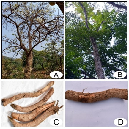
FIG. 1: THE TREES IN THEIR NATURAL HABITAT IN ATTAPPADI FOREST A. R. XYLOCARPA B. P. LONGIFOLIA, C. ROOT OF R. XYLOCARPA, D. ROOTS OF P. LONGIFOLIA
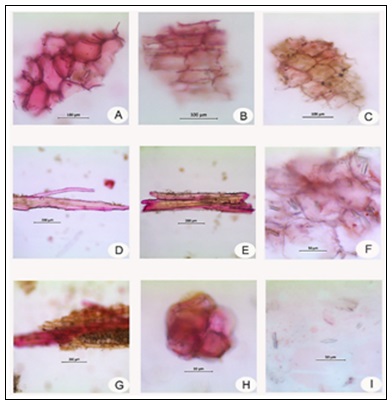
FIG. 2: POWDER MICROSCOPIC CHARACTERS OF ROOT BARK OF P. LONGIFOLIA. A. Surface view of suberized cork cells, B. Sectional view of cork cells, C. Cortical cells, D. Fragments of fibres, E. Xylem vessels, F. Cortical parenchyma embedded with acicular crystals of calcium oxalate, G. Phloem fibres with parenchyma cells. H. Group of stone cells. I. Acicular crystals of calcium oxalate.
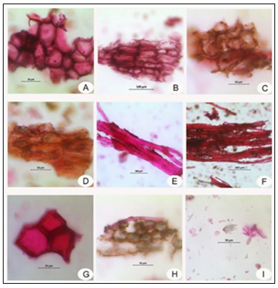
FIG. 3: POWDER MICROSCOPIC CHARACTERS OF ROOT BARK OF R. XYLOCARPA: A. Cork cells surface view, B. Cork cell sectional view, C. Cortical cells with calcium oxalate crystals, D. Sectional view of cortical cells, E. Fragments of fibres, F. Group of fibres associated with parenchyma cells. G. Hexagonal stone cells, H. Parenchyma cells embedded with calcium oxalate crystals, I. Acicular crystals of calcium oxalate.
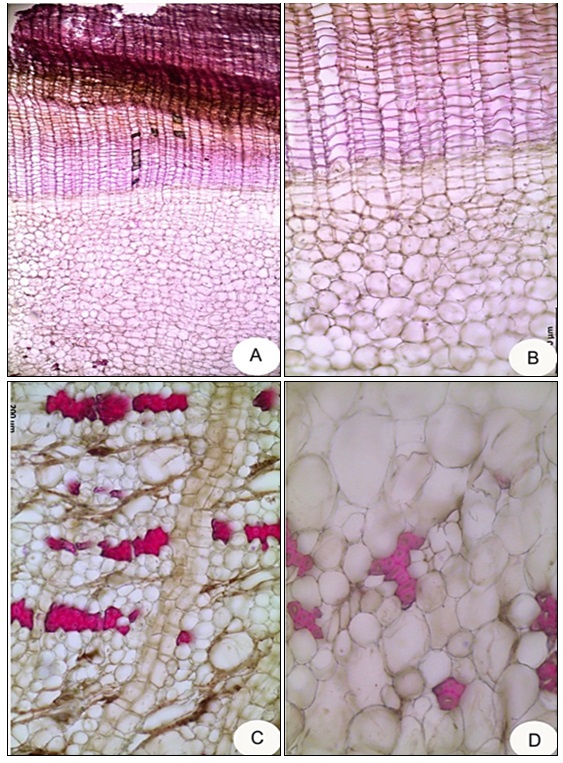
FIG. 4: ANATOMICAL FEATURES OF ROOT BARK OF P. LONGIFOLIA. A: T.S. of root bark showing cork, cork cambium and secondary cortex, B: Barrel-shaped suberized cork cells and round thin-walled parenchymatous secondary cortex, with thin-walled cambium in-between, C: Primary cortex interrupted with radial rows of medullary rays, and traversed by groups of lignified stone cells. D: Unequally sized parenchymatous cells in the primary cortex and lignified stone cells.
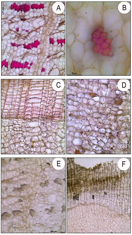
FIG. 5: HISTOCHEMICAL PECULIARITIES OF ROOT BARK OF P. LONGIFOLIA. A: Lignified stone cells in the cortical region, B: Group of stone cells, C & D: Oil droplets in the cambium and secondary cortex, E & F: Tannin deposition in the cortical cells and medullary rays.
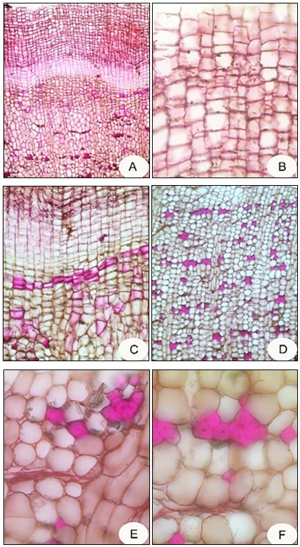
FIG. 6: ANATOMICAL FEATURES OF ROOT BARK OF R. XYLOCARPA. A. T.S of root cork, B. Suberised cork cells, C. Multi-layered cambium, D. Sclerenchymatous stone cells embedded in the isodiametric parenchyma cells in the cortex, E&F. Group of stone cells.
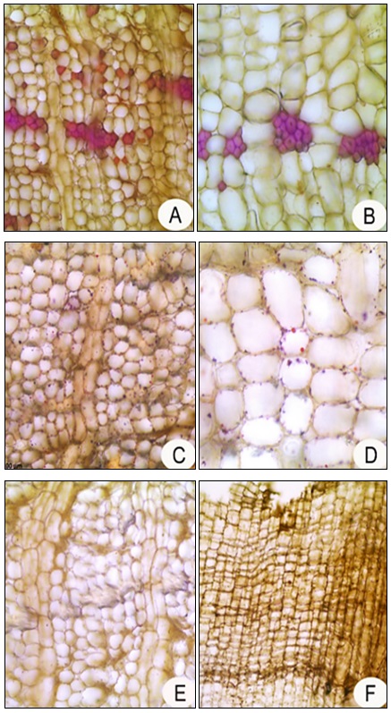
FIG. 7: HISTOCHEMICAL FEATURES OF ROOT BARK OF R. XYLOCARPA. A. Lignin detected in stone cells, B. Stone cells enlarged, C&D. Oil globules in cortical cells, E. Tannin in medullary rays, F. Tannin in cork cell.
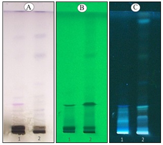
FIG. 8: HPTLC PLATE: TRACK 1: P. LONGIFOLIA, TRACK 2: R. XYLOCARPA. Viewed under A) visible light, B)254 nm, C) 366 respectively
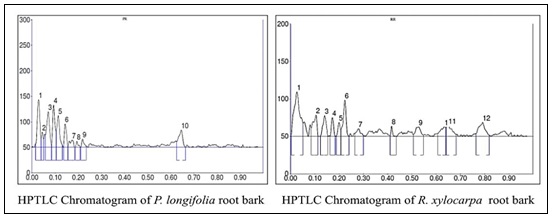
FIG. 9: HPTLC DENSITOMETRIC CHROMATOGRAM OF THE METHANOLIC ROOT BARK EXTRACTS OF P. LONGIFOLIA AND R. XYLOCARPA SHOWING DISTRIBUTION OF PHYTOCOMPOUNDS
TABLE 5: HPTLC FINGERPRINT PROFILE OF METHANOLIC EXTRACT OF P. LONGIFOLIA ROOT BARK
| Peak | Start Rf | End Rf | Max Rf | Start height | End height | Max height | Percentage | Area | Area % |
| 1 | 0.01 | 0.04 | 0.03 | 7.2 | 17.3 | 93.7 | 20.20 | 992.4 | 19.86 |
| 2 | 0.04 | 0.05 | 0.04 | 20.0 | 16.4 | 30.3 | 6.52 | 214.0 | 4.28 |
| 3 | 0.06 | 0.08 | 0.07 | 24.9 | 3.2 | 70.5 | 15.20 | 857.9 | 17.17 |
| 4 | 0.09 | 0.10 | 0.09 | 4.1 | 32.7 | 83.5 | 18.00 | 769.2 | 15.39 |
| 5 | 0.11 | 0.12 | 0.11 | 34.2 | 5.9 | 62.5 | 13.47 | 714.0 | 14.29 |
| 6 | 0.14 | 0.16 | 0.14 | 3.3 | 2.9 | 45.9 | 9.89 | 406.4 | 8.13 |
| 7 | 0.16 | 0.18 | 0.17 | 3.4 | 0.5 | 14.2 | 3.07 | 227.9 | 4.56 |
| 8 | 0.19 | 0.21 | 0.19 | 0.2 | 0.0 | 12.2 | 2.62 | 90.8 | 1.82 |
| 9 | 0.21 | 0.23 | 0.22 | 0.6 | 2.4 | 16.5 | 3.59 | 152.1 | 3.04 |
| 10 | 0.63 | 0.67 | 0.65 | 11.9 | 0.3 | 34.4 | 7.43 | 572.2 | 11.45 |
TABLE 6: HPTLC FINGERPRINT PROFILE OF METHANOLIC EXTRACT OF R. XYLOCARPA ROOT BARK
| Peak | Start Rf | End Rf | Max Rf | Start height | End height | Max height | Percentage | Area | Area % |
| 1 | 0.00 | 0.05 | 0.03 | 0.5 | 17.5 | 59.3 | 20.44 | 1340.7 | 29.67 |
| 2 | 0.09 | 0.11 | 0.11 | 11.1 | 2.0 | 28.5 | 9.81 | 425.6 | 9.42 |
| 3 | 0.12 | 0.15 | 0.14 | 0.8 | 1.4 | 27.7 | 9.54 | 338.8 | 7.50 |
| 4 | 0.16 | 0.18 | 0.17 | 0.4 | 0.2 | 25.1 | 8.64 | 211.0 | 4.67 |
| 5 | 0.19 | 0.21 | 0.20 | 1.0 | 8.3 | 18.6 | 6.40 | 164.8 | 3.65 |
| 6 | 0.21 | 0.24 | 0.22 | 9.2 | 0.2 | 48.5 | 16.73 | 524.2 | 11.60 |
| 7 | 0.26 | 0.30 | 0.28 | 3.8 | 1.1 | 10.4 | 3.59 | 170.6 | 3.77 |
| 8 | 0.42 | 0.44 | 0.42 | 0.8 | 0.2 | 13.5 | 4.65 | 76.9 | 1.70 |
| 9 | 0.51 | 0.55 | 0.53 | 2.0 | 2.0 | 12.7 | 4.39 | 249.3 | 5.52 |
| 10 | 0.61 | 0.64 | 0.64 | 3.6 | 11.6 | 12.7 | 4.36 | 245.9 | 5.44 |
| 11 | 0.64 | 0.68 | 0.65 | 11.6 | 2.1 | 14.6 | 5.03 | 315.5 | 6.96 |
| 12 | 0.77 | 0.82 | 0.79 | 4.0 | 1.9 | 18.6 | 6.42 | 455.2 | 10.07 |
TABLE 7: GC-MS COMPOUNDS TENTATIVELY IDENTIFIED FROM THE METHANOLIC ROOT BARK EXTRACT OF P. LONGIFOLIA
| Peak no. | RT (min) | Area % | Height % | Compound Tentatively Identified | Base m/z |
| 1 | 11.733 | 61.07 | 57.61 | Coumaran | 120.05 |
| 2 | 12.053 | 6.50 | 6.26 | 5-Hydroxymethylfurfural | 97.05 |
| 3 | 12.173 | 7.04 | 5.77 | Coumaran | 120.05 |
| 4 | 17.716 | 4.68 | 7.68 | Benzofuran-2-carboxaldehyde | 145.00 |
| 5 | 19.858 | 4.41 | 0.77 | 3-Propionyloxytetradecane | 57.00 |
| 6 | 20.122 | 6.51 | 5.58 | Guanosine | 57.00 |
| 7 | 31.006 | 5.15 | 7.15 | Nonylphenol (mix of isomers) | 107.05 |
| 8 | 39.340 | 2.48 | 5.27 | 1,2-Benzenedicarboxylic acid | 149.00 |
| 9 | 39.674 | 2.14 | 3.91 | Equilenin | 223.00 |
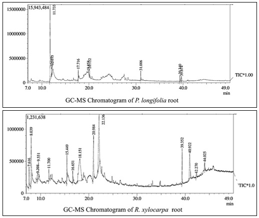
FIG. 10: GC-MS CHROMATOGRAM OF THE METHANOLIC ROOT BARK EXTRACT OF P. LONGIFOLIAAND R. XYLOCARPA
TABLE 8: GC-MS COMPOUNDS TENTATIVELY IDENTIFIED FROM THE METHANOLIC ROOT BARK EXTRACT OF R. XYLOCARPA
| Peak no. | RT (min) | Area % | Height % | Compound tentatively identified | Base m/z |
| 1 | 7.616 | 3.78 | 6.02 | Thymine | 126.00 |
| 2 | 8.039 | 10.33 | 14.35 | Guaiacol | 109.00 |
| 3 | 9.398 | 2.30 | 1.92 | Erythritol (D) | 61.00 |
| 4 | 9.551 | 3.66 | 5.18 | Pyranone | 144.00 |
| 5 | 11.700 | 2.11 | 3.62 | Coumaran | 120.05 |
| 6 | 15.449 | 3.43 | 7.49 | Phenol, 2,6-dimethoxy- | 154.00 |
| 7 | 16.651 | 2.07 | 3.57 | 3-[N'-(3H-Indol-3-ylmethylene)-hydrazino]-5-methyl-[1,2,4]triazol-4-ylamine | 142.00 |
| 8 | 18.151 | 24.79 | 6.84 | Guanosine | 57.00 |
| 9 | 20.984 | 10.26 | 13.34 | Isovanillic acid | 168.00 |
| 10 | 22.136 | 22.02 | 15.59 | Quinic acid | 60.00 |
| 11 | 40.922 | 9.12 | 7.72 | β-Sitosterol | 57.05 |
| 12 | 42.270 | 1.12 | 1.14 | Methyl commate C | 218.10 |
DISCUSSION: Accurate identification of crude herbal drugs becomes increasingly challenging once diagnostic macromorphological characters are lost during processing and powdering. In the two species studied here, organoleptic differences, particularly the pungent odour of P. longifolia and the aromatic odour of R. xylocarpa, provide useful preliminary cues for crude drug identification. Although fresh bark showed high moisture content, the powdered samples (~8%) were within acceptable limits as mentioned in the Ayurvedic pharmacopoeia, indicating suitability for storage after proper drying. It was mentioned that a moisture content below 14% is good for herbal drug powder 23, 24. Slightly higher total, water-soluble, and acid-insoluble ash values in P. longifolia indicate marginally greater inorganic content, which correlates with the abundant calcium oxalate crystals observed microscopically in both species. The therapeutic efficacy of many Ayurvedic drugs is often attributed to their mineral constituents 25.
However, adequate screening of herbal medicines is essential to prevent heavy metal contamination and ensure their safety and quality 26. Elemental profiling carried out in P. longifolia and R. xylocarpa revealed appreciable levels of calcium, magnesium, and iron, supporting their traditional therapeutic relevance. Most heavy metals analyzed, including aluminium, nickel, lead, mercury, and cobalt, were below detectable limits; however, cadmium levels in both species slightly exceeded the WHO permissible limit of 0.33 mg/kg.
Anatomical and histochemical studies revealed several shared features like barrel-shaped cork cells, lignified stone cells, tannin deposits, and acicular calcium oxalate crystals, reflecting their taxonomic affinity. Distinguishing characters were also evident: P. longifolia possessed a thicker, easily detachable cork, while R. xylocarpa exhibited abundant pigmented cortical oil globules and longer calcium oxalate crystals. Powder microscopy further confirmed consistent markers like cork fragments, stone cell groups, phloem fibres, reticulate xylem vessels, and crystal-bearing parenchyma, which are critical for identifying the drug in powdered form.
Since the phytochemistry of both species is poorly documented and no specific marker compounds have been established, the present HPTLC analysis was undertaken as a preliminary fingerprinting study to provide baseline chromatographic profiles for future investigations. Such species-specific chromatographic fingerprints are valuable for the authentication, differentiation, and quality control of medicinal plant materials and have been widely recognized as reliable tools for herbal drug standardization 27.
Among the GC-MS compounds tentatively identified from the root bark of P. longifolia, coumarans, 5-hydroxymethyl furfural, and 1,2-benzenedicarboxylic acid were reported to possess notable biological activities 28-30. Guaiacol, a prominent phenolic constituent identified from the root bark of R. xylocarpa, has been widely reported to exhibit significant antioxidant and antimicrobial activities 31. Likewise, β-sitosterol is well known for its apoptotic, antidiabetic, and anticancer properties 32-34, while isovanillic acid, a phenolic acid derivative, has been associated with gastrointestinal stimulatory effects 35.
CONCLUSION: P. longifolia and R. xylocarpa exhibited distinct pharmacognostic and powder microscopic characteristics. GC–MS profile of both root bark extracts demonstrated the presence of several pharmacologically important bioactive compounds, highlighting their potential therapeutic significance and supporting their traditional medicinal applications. Their mineral composition further supports their significance in ethnomedicinal practices. The diagnostic standards developed in this study, together with the elemental parameters, provide a practical framework for the authentication, quality control, and safe medicinal use of these underexplored ethnomedicinal roots.
ACKNOWLEDGEMENTS: The author is thankful to the Centre for Analytical Instrumentation, Kerala Forest Research Institute, Peechi, Thrissur, Kerala, for providing instrument facilities and to Dr. Udayan P.S., Senior Consultant, National Medicinal Plants Board, Regional Cum-Facilitation Centre, Kerala Forest Research Institute, Peechi, for giving the necessary information regarding the plants.
CONFLICT OF INTEREST: The authors declare that, there are no conflicts of interest.
REFERENCES:
- Royal Botanic Gardens Kew: Pajanelia longifolia (Willd.) K. Schum. Plants of the World Online 2025. https://powo.science.kew.org/taxon/110192-1
- Zainab A, Bhat R, Acharya S, Yende A and Padyana S: Studies on antioxidant and antimicrobial activities of Pajanelia longifolia (Willd.) Schumann, Obesity Research Journal 2013; 2: 1-13.
- Gini EJ, Sivakkumar T and Kuppuswami S: Determination of antioxidant activity of various extracts of Pajanelia longifolia (Willd.) K. Schum, isolation and characterization of flavonoid from ethanol extract by column chromatography, Research Journal of Pharmacy and Technology 2017; 10(10): 3391-3397.
- Choudhury, Prakash Roy, Manabendra Dutta Choudhury, Sanjoy Singh Ningthoujam, Dipan Das, Deepa Nath and Anupam Das Talukdar: Ethnomedicinal plants used by traditional healers of North Tripura district, Tripura, North East India. Journal of Ethnopharmacology 2015; 166: 135-148.
- Zainab A, Bhat R, Acharya S, Yende A and Padyana S: Studies on antioxidant and antimicrobial activities of Pajanelia longifolia (Willd.) Schumann. Obesity Research Journal 2013; 2: 1-13.
- Smitha CK, Udayan PS and Vaishnavi M: Unravelling the phytochemistry of ethnomedicinal tree Pajanelia longifolia (Willd.) K. Schum. Natural Product Experiments in Drug Discovery. Springer Publishers, Humana, New York 2023; 121-130.
- Nath P, Das BN, Ahmed T, Das M, Nath R, Talukdar AD, Nath D, Nahar L and Kityania S: Computational Analysis of Bioactive Phytocompounds from Methanolic Extract of Pajanelia longifolia (Willd.) K. Schum against EGFR and TGF-β Cancer Targets. Natural Products Analysis 2025; 1(1): 100007.
- Gamble JS: A Manual of Indian Timbers.BS Mahendra Pal Singh Publishers, Dehra Dun 1972; 313.
- Kshirsagar RD and Singh NP: Some less known ethnomedicinal uses from Mysore and Coorg districts, Karnataka state, India. Journal of Ethnopharmacology 2001; 75(2-3): 231-238.
- Duggal RR, Masih SK and Mishra RP: Medicinally valuable plants form sacred groves of Jabalpur Forest Division (M.P). Asian Journal of Plant Science and Research 2017; 7(2): 37-44.
- Aniel Kumar O, Mallikarjuna K and Mutyala Naidu L: Ethnomedicine for Rheumatoid arthritis by tribes of Papikondalu forest, Andhra Pradesh, India. American Journal of Ethnomedicine 2015; 2(6): 373-378.
- Manjula RR, Rao JK and Reddi TS: Ethnomedicine for rheumatism by the tribals of Khammam District, Andhra Pradesh. J of Natural Remedies 2013; 13(2): 138-141.
- Jain SP, Srivastava S, Singh J and Singh SC: Traditional phytotherapy of Balaghat district, Madhya Pradesh, India, Indian J of Traditional Knowledge 2011; 10(2): 334-338.
- Misra RC: Therapeutic uses of some seeds among the tribals of Gandhamardan hill range, Orissa. Indian Journal of Traditional Knowledge 2004; 3(1): 105-115.
- Srivastava A, Patel SP, Mishra RK, Vashistha RK, Singh A and Puskar AK: Ethnomedicinal importance of the plants of Amarkantak region, Madhya Pradesh, India. International Journal of Medicinal and Aromatic Plants 2012; 2(1): 53-59.
- Sumanth MV, Yellanthoor MB, Ravikumar K and Ravichandran P: Comparative physicochemical, phytochemical and HPTLC studies on root species used as Patala in Ayurvedic system of medicine. Journal of Pharmacy Research 2013; 7(9): 810-816.
- Evans WC: Trease and Evans’ pharmacognosy. Elsevier, 2009; 121.
- Government of India: The Ayurvedic Pharmacopoeia of India Volumes I-IV. Ministry of Health and Family Welfare, Department of AYUSH 2004.
- Indian Standard specification No.1797–1985. Moisture in Spices / Methods of Test for Spices and Condiments. Bureau of Indian Standards, 17th edition2000. 154.
- Saklani S, Mishra AP, Sati B and Sati H: Pharmacognostic, phytochemical and antimicrobial screening of Aphanamixis polystachya, an endangered medicinal tree. International Journal of Pharmacy and Pharmaceutical Sciences 2013; 4(3): 235-240.
- World Health Organization. WHO Guidelines for Assessing Quality of Herbal Medicines with Reference to Contaminants and Residues. Geneva: WHO; 2007.
- O’Brien TP, Feder N and Mc Cull ME: Polychromatic staining of plant cell walls by Toluidine blue–O. Protoplasma 1964; 59: 364-373.
- Derbal Said: Microbial contamination of medicinal plants. Journal of Molecular and Pharmaceutical Sciences 2023; 2(1): 33-44.
- Khandelwal KR: Practical pharmacognosy-techniques and experiments. Nirali Prakashan, Edition 32nd 2010; 23-25.
- Jagtap C, Galib R, Bedarkar P, Patgiri B and Prajapati PK: Therapeutic potentials of minerals in ancient India: a review through Charaka Samhita. Journal of Research and Education in Indian Medicine 2014; 20(1): 9-20.
- Philips CA, Paramaguru R, Joy AK, Antony KL and Augustine P: Clinical outcomes, histopathological patterns, and chemical analysis of Ayurveda and herbal medicine associated with severe liver injury- A single-center experience from southern India. Indian Journal of Gastroenterology 2018; 37(1): 9-17.
- Balekundri A and Mannur V: Quality control of the traditional herbs and herbal products: a review. Future Journal of Pharmaceutical Sciences 2020; 6(1): 67.
- Ilya E, Kulikova L, Van der Eycken EV and Voskressensky L: Recent advances in phthalan and coumaran chemistry. Chem Open 2018; 7(11): 914-917.
- Krishnan K, Mani A and Jasmine S: Cytotoxic activity of bioactive compound 1, 2-benzene dicarboxylic acid, mono 2-ethylhexyl ester extracted from a marine derived Streptomyces sp. VITSJK8. International Journal of Molecular and Cellular Medicine 2014; 3(4): 246-252.
- Zhao L, Chen J, Su J, Li L, Hu S, Li B, Zhang X, Xu Z and Chen T: In-vitro antioxidant and antiproliferative activities of 5-hydroxymethylfurfural. Journal of Agriculture and Food Chemistry2013; 61(44): 10604-10611.
- Mika A and Luthje S: Properties of guaiacol peroxidase activities isolated from corn root plasma membranes. Plant Physiology 2003; 132(3): 1489-98.
- Chai JW, Kuppusamy UR and Kanthimathi MS: Beta-sitosterol induces apoptosis in MCF-7 cells. Malaysian J of Biochemistry and Molecular Biology 2008; 16(2): 28-30.
- Ponnulakshmi R, Shyamaladevi B, Vijayalakshmi P and Selvaraj J: In-silico and in-vivo analysis to identify the antidiabetic activity of beta-sitosterol in adipose tissue of high-fat diet and sucrose-induced type-2 diabetic experimental rats. Toxicology Mechanisms and Methods 2019; 29(4): 276-90.
- Cabeza M, Bratoef E, Heuze I, Ramírez E, Sanchez M and Flores E: Effect of beta-sitosterol as inhibitor of 5alpha-reductase in Hamster prostate. Proceedings-Western Pharmacology Society 2003; 46: 153-155.
- Sadraei H, Ghanadian M, Asghari G and Azali N: Antidiarrheal activities of isovanillin, iso-acetovanillin and Pycnocycla spinosa Decneex. Boiss extract in mice. Research in Pharmaceutical Sciences 2014; 9(2): 83-85.
How to cite this article:
Smitha CK, Kavitha CH and Anupama SS: Pharmacognostic standardization and phytochemical profiling of root bark of Pajanelia longifolia and Radermachera xylocarpa, two less-known ethnomedicinal trees of bignoniaceae. Int J Pharm Sci & Res 2026; 17(10): 3021-35. doi: 10.13040/IJPSR.0975-8232.17(10).3021-35.
All © 2026 are reserved by International Journal of Pharmaceutical Sciences and Research. This Journal licensed under a Creative Commons Attribution-NonCommercial-ShareAlike 3.0 Unported License.
Article Information
16
3021-3035
4179 KB
6
English
IJPSR
C. K. Smitha *, C. H. Kavitha and S. S. Anupama
Department of Botany, Government College for Women, Thiruvananthapuram, Kerala, India.
smithack.ck@gmail.com
02 June 2026
18 June 2026
19 June 2026
10.13040/IJPSR.0975-8232.17(10).3021-35
01 October 2026
 Download