



PHARMACOGNOSTICAL STANDARDIZATION OF TERMINALIA CHEBULARETZ. AND WRIGHTIA TINCTORIA (ROXB.) R. BR.
HTML Full TextPHARMACOGNOSTICAL STANDARDIZATION OF TERMINALIA CHEBULARETZ. AND WRIGHTIA TINCTORIA (ROXB.) R. BR.
J. Jayapriya, K. Keerthana and G. Prakash Yoganandam*
Department of Pharmacognosy, College of Pharmacy, Mother Theresa Post Graduate and Research Institute of Health Sciences, Gorimedu, Puducherry, India.
ABSTRACT: The present study was undertaken to establish pharmacognostical standards for the fruit of Terminalia chebula and the leaf of Wrightia tinctoria, the major medicinal plant used in many Ayurvedic preparation, in order to ensure their proper identification, authentication, and quality assessment. There is a lacuna in the comprehensive standardization profile for the selected drug. The research gap was addressed through this current research. Authentic plant materials were subjected to macroscopic and microscopic evaluation, including transverse-section and powder microscopy, revealing diagnostic characters such as longitudinal ridges, stone cells and tannin cells in the fruit of T. chebula, and paracytic stomata, calcium oxalate crystals and latex cells in the leaf of W. tinctoria. The total ash values, water extractive value, alcohol soluble extractive value and loss on drying of the T. chebula and Wrightia tinctoria found to be 5.2% and 6.2%, 18% and 15%, 12% and 11% and 6.5% and 5.8% respectively. The preliminary phytochemical screening indicated the presence of alkaloids, flavonoids, tannins, glycosides and saponins, consistent with the traditional therapeutic use of both plants. The findings of the present investigation provide scientifically validated pharmacognostical standards for Terminalia chebula and Wrightia tinctoria which can serve as reliable reference parameters for quality control, standardization and future research involving these medicinal plants.
Keywords: Pharmacognostical standardization, Terminalia chebula, Wrightia tinctoria, Microscopy, Physicochemical evaluation, Quality control
INTRODUCTION: Medicinal plants continue to play a significant role in both traditional and modern systems of medicine, serving as valuable sources of bioactive compounds with diverse therapeutic applications. Among the medicinally important plants, Terminalia chebula and Wrightia tinctoria have gained considerable attention due to their wide range of pharmacological activities and extensive use in traditional medicine 1-6. Terminalia chebula commonly known as the “King of Medicines,” is widely utilized in Ayurvedic medicine for its antimicrobial, antioxidant, anti-inflammatory, wound healing, and rejuvenating properties.
The fruits of the plant are rich in tannins, phenolic compounds, and flavonoids, which contribute to its therapeutic potential 7-12. Similarly, Wrightia tinctoria belonging to the family Apocynaceae and commonly known as “Sweet Indrajao,” has been traditionally used in the treatment of skin diseases, inflammation, microbial infections, wounds, and bone-related disorders. The leaves of the plant contain various phytoconstituents such as flavonoids, alkaloids, phenolic compounds, steroids, and triterpenoids, which are responsible for its antimicrobial, antioxidant, anti-inflammatory, and antibiofilm activities 13, 14.
Despite their extensive medicinal importance, the quality, purity, and efficacy of crude herbal drugs may vary significantly due to differences in geographical source, environmental conditions, collection period, harvesting practices, and processing methods. In addition, adulteration and substitution of plant materials remain major concerns affecting the safety and therapeutic effectiveness of herbal formulations. Therefore, pharmacognostical standardization is essential to establish reliable identification parameters and ensure the authenticity and quality control of medicinal plants 1, 4-5.
Pharmacognostical evaluation involves macroscopic, microscopic, powder microscopic, and physicochemical studies, which together provide comprehensive diagnostic characteristics of crude plant materials. These parameters serve as important tools for the proper identification, authentication, and standardization of herbal drugs. Hence, the present study was undertaken to establish detailed pharmacognostical standards for Terminalia chebula and Wrightia tinctoria, which may serve as reference data for future research, quality assessmentand formulation development involving these medicinal plants. Although both species are individually well documented, standardized pharmacognostical data generated on the same fruit and leaf samples under a single, directly comparable protocol are limited; this study was therefore designed to generate such directly comparable diagnostic and physicochemical data for T. chebula fruit and W. tinctoria leaf, addressing this gap 1-4.
MATERIALS AND METHODS:
Collection and Authentication: The fruit of Terminalia chebula was collected from Poorveegam Herbology Research and Training Centre, Poothurai village Tamil Nadu and Fresh leaves of Wrightia tinctoria were collected from healthy plants growing in suitable natural habitat during the flowering season, in 13.04.2026 from Mailam temple road. The collected samples were thoroughly cleaned to remove adhering soil, dust, and other extraneous matter. The plant material was authenticated based on its macroscopic and pharmacognostical characters with the help of standard literature and expert consultation. The identification was confirmed by Dr. Kumaresan V, Assistant Professor of Botany, Kanchi Mamunivar Govt. college for Arts and Sciences and Research, Puducherry. After authentication, the plant material was shade dried at room temperature to prevent degradation of active constituents and then coarsely powdered using a mechanical grinder.
The powdered sample was stored in an airtight container for further analysis.
Macroscopic Evaluation: The macroscopic evaluation of Terminalia chebula and Wrightia tinctoria was carried out to determine their morphological characteristics, which are essential for preliminary identification and authentication of the crude drug. The freshly collected fruit of T. chebula and the fresh leaf of W. tinctoria were each thoroughly cleaned to remove any adhering impurities and observed separately under normal daylight conditions 1, 3.
The evaluation was performed by examining the external features of the sample, including size, shape, color, surface characteristics, texture, odor, and taste. The size of the fruit was measured using a standard measuring scale, while the shape and surface features were assessed visually. The color was noted under natural light to avoid variation due to artificial illumination. The texture was determined by tactile examination, and the odor was evaluated by gently smelling the sample. The taste was assessed carefully by placing a small portion of the sample on the tongue to identify its characteristic astringent nature 1, 2.
All observations were recorded systematically and compared with standard pharmacognostical descriptions available in the literature to confirm the identity of the plant material. The macroscopic characteristics thus obtained serve as important diagnostic features for distinguishing Terminalia chebula and Wrightia tinctoria from related species and possible adulterants 1, 3.
Microscopic Evaluation: The microscopic evaluation of Terminalia chebula and Wrightia tinctoria was carried out to study the internal anatomical characteristics, which serve as reliable diagnostic features for authentication of the crude drug. For T. chebula, fresh as well as dried fruit pericarp was used for the preparation of transverse sections (T.S.); for W. tinctoria, fresh mature leaves (mid-lamina region, including the midrib) were used for the preparation of transverse sections 1, 3. Thin sections were obtained manually using a sharp blade and transferred into a watch glass containing water. The sections were then cleared with chloral hydrate solution to remove colouring matter and enhance visibility of cellular structures. The cleared sections were stained using suitable staining agents such as phloroglucinol and hydrochloric acid to identify lignified tissues. After staining, the sections were mounted on a glass slide using glycerine and covered with a cover slip 2.
The prepared slides were observed under a compound microscope at different magnifications. Various anatomical features such as epidermis, parenchymatous cells, vascular bundles, sclerenchymatousfibers, and tannin-containing cells were carefully examined and recorded. Photomicrographs were taken, wherever possible, to support the observations 1, 2-3. The observed microscopic characters were compared with standard descriptions available in pharmacognostical literature to confirm the identity and purity of Terminalia chebula and Wrightia tinctoria. These features provide definitive evidence for distinguishing the plant material from adulterants and ensure quality control 1, 3.
Physicochemical Parameters: Physicochemical evaluation of Terminalia chebula and Wrightia tinctoria was carried out to determine the quality, purity, and suitability of the crude drug for further formulation. Standard procedures were followed to estimate parameters such as loss on drying, ash values, and extractive values; all determinations were carried out in triplicate (n = 3) and results are expressed as mean ± SD 4, 5.
Determination of Loss on Drying (LOD): A known quantity of the powdered plant material was accurately weighed and placed in a previously dried and weighed dish. The sample was dried in a hot air oven at 105°C until a constant weight was obtained. The loss in weight was calculated as a percentage of the initial weight to determine the moisture content present in the sample 4, 5.
Loss on drying (%) = Initial weight – Final weight / Initial weight × 100
Determination of Ash Values:
Total Ash: A known weight of the powdered drug was incinerated in a silica crucible at a temperature not exceeding 600°C until the sample became white, indicating the absence of carbon. The residue obtained was weighed and calculated as total ash percentage 4, 5.
Total ash (%) = Weight of ash / Weight of sample × 100
Acid Insoluble Ash: The total ash obtained was boiled with dilute hydrochloric acid in an ashless filter paper and the insoluble matter was collected in an ashless filter paper and incinerated again. The residue was weighed to determine the acid insoluble ash,which indicates the presence of siliceous matter.
Acid insoluble ash (%) = Weight of insoluble ash / Weight of sample × 100
Water Soluble Ash: The total ash obtained was boiled with distilled water for about 5 minutes. The insoluble matter was collected on an ashless filter paper and washed with hot water. The insoluble residue along with filter paper was incinerated again in a silica crucible at about 450°C until constant weight was obtained. The weight of total ash to obtain the water soluble ash, which indicates the presence of water soluble inorganic salts.
% Water soluble ash = Weight of water soluble ash / Weight of air-dried drug × 100
Determination of Extractive Values:
Water Soluble Extractive Value: A known quantity of powdered drug was macerated with distilled water for 24 hours with intermittent shaking. The solution was filtered, and a measured volume of filtrate was evaporated to dryness. The residue obtained was weighed to calculate the water soluble extractive value 4, 5.
Water soluble extractive (%) = Weight of extract / Weight of sample × 100
Alcohol Soluble Extractive Value: Similarly, the powdered drug was macerated with alcohol (99%v/v) for 24 hours, filtered, and evaporated to dryness. The residue was weighed to determine the alcohol soluble extractive value.
Alcohol soluble extractive (%) = Weight of extract / Weight of sample × 100
Preliminary Phytochemical Screening: Powdered T. chebula fruit and W. tinctoria leafwere separately extracted with methanol maceration, to obtain extracts at a working concentration of 1% w/v. Each extract was subjected to qualitative phytochemical screening, performed separately for the two plants, using standard test procedures to identify the presence of various bioactive constituent classes.
Test for Alkaloids: A small quantity of the extract was treated with Dragendorff’s reagent. The formation of an orange precipitate indicated the presence of alkaloids.
Test for Flavonoids: The extract was treated with magnesium turnings followed by concentrated hydrochloric acid (Shinoda test).
The appearance of a pink or red color confirmed the presence of flavonoids.
Test for Tannins and Phenols: The extract was treated with a few drops of ferric chloride solution. The formation of a blue-black or green coloration indicated the presence of tannins and phenolic compounds.
Test for Saponins: The extract was shaken vigorously with distilled water. The formation of stable and persistent foam indicated the presence of saponins.
Test for Terpenoids: The extract was mixed with chloroform and concentrated sulfuric acid (Salkowski test). A reddish-brown coloration at the interface confirmed the presence of terpenoids.
Test for Steroids: The extract was treated with acetic anhydride followed by concentrated sulfuric acid (Liebermann–Burchard test). The development of a green color indicated the presence of steroids.
Test for Glycosides: The extract was treated with glacial acetic acid, ferric chloride, and concentrated sulfuric acid (Keller–Killiani test). The formation of a brown ring indicated the presence of glycosides.
Test for Carbohydrates: The extract was treated with Molisch’s reagent followed by concentrated sulfuric acid. The formation of a violet ring at the junction confirmed the presence of carbohydrates.
Test for Proteins: The extract was treated with sodium hydroxide and copper sulfate solution (Biuret test). The appearance of a violet color indicated the presence of proteins.
RESULTS:
Macroscopic Evaluation:
Macroscopic Evaluation of Terminalia chebula:
TABLE 1: MACROSCOPIC EVALUATION OF TERMINALIA CHEBULA
| S. no. | Parameter | Observation |
| 1 | Size | 2–4 cm length |
| 2 | Shape | Ovoid / oblong |
| 3 | Color | Yellowish-brown to dark brown |
| 4 | Surface | Rough, wrinkled with longitudinal ridges |
| 5 | Texture | Hard when dry |
| 6 | Odor | Characteristic |
| 7 | Taste | Astringent |
| 8 | External appearance | Five distinct longitudinal ridges present |
| 9 | Pericarp | Thick, hard and wrinkled; yellowish brown |
| 10 | Fracture | Short, hard and granular |
| 11 | Seed | Single, hard, ovoid seed; pale yellow to light brown in colour |
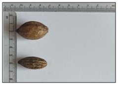
FIG. 1: MACROSCOPIC CHARACTERISTICS OF TERMINALIA CHEBULA FRUITS SHOWING OVOID SHAPE, WRINKLED SURFACE, AND SIZE MEASUREMENT USING A SCALE
Macroscopic Evaluation of Wrightia tinctoria:
TABLE 2: MACROSCOPIC EVALUATION OF WRIGHTIA TINCTORIA
| S. no. | Parameter | Observation |
| 1 | Size | 6–12 cm length |
| 2 | Shape | Elliptic to lanceolate |
| 3 | Color | Green |
| 4 | Surface | Smooth |
| 5 | Texture | Soft and leathery |
| 6 | Odor | Characteristic |
| 7 | Taste | Slightly bitter |
| 8 | Apex | Acuminate |
| 9 | Margin | Entire |
| 10 | Venation | Reticulate |
| 11 | Leaf base | Acute (Tapering) |
| 12 | Petiole | Short (0.5–1.0 cm), slender, green and smooth |
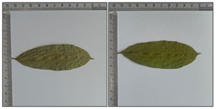
FIG. 2: DORSAL VIEW OF FIG. 3: VENTRAL VIEW OF WRIGHTIA TINCTORIA LEAF WRIGHTIA TINCTORIA LEAF
Microscopic Evaluation:
Microscopic Evaluation of Terminalia chebula:
Transverse Section of Fruit of Terminalia chebula:
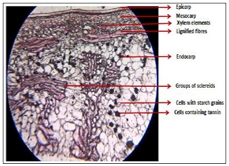
FIG. 3: TRANSVERSE SECTION OF TERMINALIA CHEBULA FRUIT PERICARP (MAGNIFICATION: 10X)
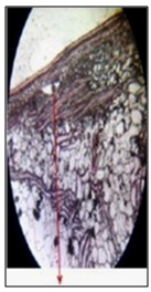
FIG 4: ROSETTE CRYSTALS OF CALCIUM OXALATE (MAGNIFICATION: 40X)
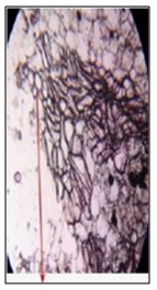
FIG. 5: XYLEM ELEMENTS (MAGNIFICATION: 40X)
Powder Microscopic Analysis of Terminalia chebula Fruit Powder:
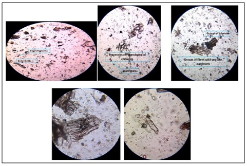
FIG. 6: FRAGMENTS OF XYLEM VESSELS WITH SIMPLE PITTED THICKENINGS (MAGNIFICATION: 40X)
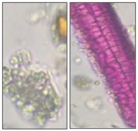
FIG. 7: TANNIN CONTENTS OF HARITAKI (MAGNIFICATION: 40X)
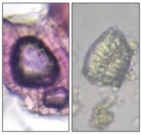
FIG. 8: STONE CELLS OF HARITAKI (MAGNIFICATION: 40X)
The structure consists of a single-layered epidermis made up of compactly arranged cells, covered by a thick and well-developed cuticle. Beneath this, the parenchyma is composed of thin-walled, loosely arranged cells. Sclerenchyma tissue is also present, characterized by thick-walled, lignified cells. The vascular bundles are well-developed and scattered within the tissue. Elongated, thick-walled fibers are observed, along with abundant dark-stained tannin cells. Stone cells (sclereids) are present and appear irregular in shape. The medullary region consists of parenchymatous cells.
TABLE 3: QUANTITATIVE MICROSCOPY OF T. CHEBULA LEAF
| S. no. | Parameter | Observed value (Mean ± SD) |
| 1 | Stone cells | 38 ± 3 /mm² |
| 2 | Calcium oxalate crystals | 22 ± 2 /mm² |
| 3 | Tannin cells | 31 ± 2 /mm² |
| 4 | Xylem vessels | 18 ± 1 /mm² |
| 5 | Fibre length | 486 ± 21 µm |
Microscopic Evaluation of Wrightia tinctoria:
Transverse Section of Wrightia Tinctoria Leaf: The transverse section (T.S.) of the leaf shows distinct anatomical features. It includes the upper and lower epidermis along with well-developed vascular bundles and collenchyma tissue. Starch grains and simple trichomes are also observed. In addition, latex droplets and calcium oxalate crystals are present within the tissue. The leaf further exhibits paracytic type stomata.
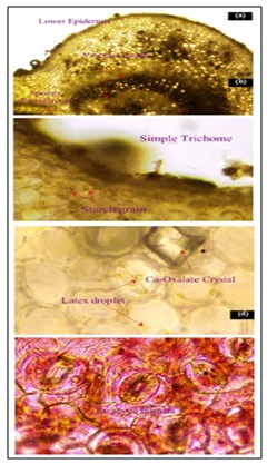
FIG. 9: T.S. OF LEAF SHOWING (A) LOWER EPIDERMIS, UPPER EPIDERMIS, VASCULAR BUNDLE & COLLENCHYMA; (B) STARCH GRAIN & SIMPLE TRICHOME; (C) LATEX DROPLET & CALCIUM OXALATE CRYSTAL; (D) PARACYTIC STOMATA. (MAGNIFICATION: 40X)
Powder Microscopy:
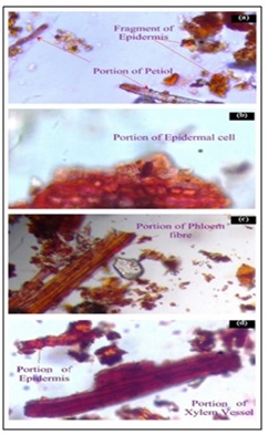
FIG. 10: LEAF POWDER MICROSCOPY SHOWING (A) FRAGMENT OF EPIDERMIS & PORTION OF PETIOLE; (B) PORTION EPIDERMAL CELL; (C) PORTION OF PHLOEM FIBRE; (D) PORTION OF EPIDERMIS & PORTION OF XYLEM VESSEL. (MAGNIFICATION: 40X)
The powder characteristics reveal the presence of lignified fibers. Xylem vessels with spiral and pitted thickening are observed. Paracytic type stomata are present. Unicellular trichomes are also observed. Calcium oxalate crystals are present within the powder. In addition, thin-walled parenchyma cells are observed.
TABLE 4: QUANTITATIVE MICROSCOPY OF WRIGHTIA TINCTORIA LEAF
| S. no. | Parameter | Mean ± SD |
| 1 | Stomatal number | 182 ± 6 /mm² |
| 2 | Stomatal index | 21.4 ± 0.8 % |
| 3 | Vein islet number | 18 ± 1 /mm² |
| 4 | Vein termination | 13 ± 1 /mm² |
| 5 | Palisade ratio | 6.8 ± 0.4 |
Physicochemical Evaluation:
TABLE 5: PHYSICOCHEMICAL EVALUATION OF TERMINALIA CHEBULA AND WRIGHTIA TINCTORI
| S. no. | Parameter | Terminaliachebula | Wrightia tinctoria |
| 1 | Loss on drying | 6.5±0.08% | 5.8±0.12% |
| 2 | Total ash | 5.2±0.06% | 6.2±0.08% |
| 3 | Acid insoluble ash | 1.1±0.02% | 1.3±0.04% |
| 3 | Water soluble ash | 3.5±0.05% | 2.6±0.06% |
| 4 | Water soluble extractive | 18±0.12% | 15±0.15% |
Preliminary Phytochemical Screening:
Preliminary Phytochemical Screening of Terminalia Chebula:
TABLE 6: PRELIMINARY PHYTOCHEMICAL SCREENING OF TERMINALIA CHEBULA
| S. no. | Phytochemical | Test | Observation | Terminaliachebula | Wrightia tinctoria |
| 1 | Alkaloids | Dragendorff’s test | Orange precipitate | Present | Present |
| 2 | Glycosides | Keller–Killiani test | Reddish brown ring | Present | Present |
| 3 | Flavonoids | Shinoda test | Pink/red color | Present | Present |
| 4 | Tannins | Ferric chloride test | Blue-black/green color | Strongly Present | Present |
| 5 | Saponins | Foam test | Persistent foam | Present | Present |
| 6 | Phenols | Ferric chloride | Deep blue/green | Present | Present |
| 7 | Terpenoids | Salkowski test | Reddish brown layer | Present | Present |
| 8 | Steroids | Liebermann–Burchard | Green color | Present | Present |
| 9 | Carbohydrates | Molisch’s test | Violet ring | Present | Present |
| 10 | Proteins | Biuret test | Violet color | Present (mild) | Present |
DISCUSSION: The present pharmacognostical evaluation of Terminalia chebula and Wrightia tinctoria established important diagnostic parameters useful for the identification, authentication, and quality assessment of the crude drugs. The macroscopic characteristics observed in both plants were found to be consistent with standard pharmacognostical descriptions reported in the literature. Terminalia chebula exhibited characteristic features such as ovoid shape, wrinkled surface, yellowish-brown to dark coloration, and an astringent taste, whereas Wrightia tinctoria showed distinct leaf morphology with entire margin, reticulate venation, and characteristic bitter taste. These morphological features serve as important preliminary indicators for distinguishing the genuine plant materials from adulterants or substitutes 1, 2. Microscopic studies further confirmed the authenticity of both plants by revealing characteristic anatomical structures. In Terminalia chebula, the presence of sclerenchymatous cells, parenchymatous tissues, vascular bundles, and tannin-containing cells were observed, while Wrightia tinctoria showed well-defined epidermal layers, palisade cells, vascular bundles, trichomes, and calcium oxalate crystals. These anatomical markers are considered highly significant for pharmacognostical authentication and quality control of crude herbal drugs 1, 3, 13, 14.
Powder microscopic analysis also provided valuable diagnostic characters for both plant materials. Terminalia chebula powder exhibited fibers, starch grains, and brown-colored tannin masses, whereas Wrightia tinctoria powder showed characteristic fibers, vessels, crystals, and stomata. Such powder characteristics are particularly useful for the identification of powdered crude drugs and for detecting adulteration when the plant material is not available in intact form 2, 4, 13. The physicochemical parameters evaluated, including loss on drying, ash values, and extractive values, were compared with the corresponding limits prescribed in the Indian Pharmacopoeia and WHO quality-control guidelines [specific limit values to be cited] and were broadly consistent with these references for both plants, indicating no evidence of significant adulteration in the samples analysed. The observed moisture content is comparable to values reported for T. chebula fruit and W. tinctoria leaf in earlier pharmacognostical studies 13, 14, supporting good storage stability. Ash values fell within the range expected for genuine plant material, while extractive values reflected the presence of appreciable amounts of water- and alcohol-soluble phytoconstituents; taken together with the diagnostic macroscopic and microscopic characters described above, these parameters can serve as a practical basis for distinguishing authentic T. chebula and W. tinctoria material from adulterants or substitutes 4, 5. Overall, the combined pharmacognostical and physicochemical evaluations support the authenticity and quality of the analysed samples of Terminalia chebula and Wrightia tinctoria. The findings of the present study provide standard reference data that can be effectively utilized for the identification, quality control, and future phytopharmaceutical research involving these medicinal plants 1, 2, 4, 5, 13, 14.
CONCLUSION: The present study established detailed pharmacognostical standards for Terminalia chebula and Wrightia tinctoria through comprehensive macroscopic, microscopic, powder microscopic, and physicochemical evaluations. The diagnostic characteristics obtained from the study can be effectively utilized for the identification, authentication, and quality control of these crude drugs in future research and herbal formulations. The established standard parameters may also serve as valuable reference data for ensuring the purity, consistency, and therapeutic efficacy of plant-based medicinal preparations.
The established standards may also support future herbal monograph development and industrial quality assurance of medicinal plant materials.
ACKNOWLEDGEMENTS: Nil
CONFLICTS OF INTEREST: Nil
REFERENCES:
- Kokate CK, Purohit AP and Gokhale SB: Pharmacognosy. 49th Ed. Pune: Nirali Prakashan 2014.
- Khandelwal KR: Practical Pharmacognosy: Techniques and Experiments. 23rd ed. Pune: Nirali Prakashan 2008.
- Evans WC: Trease and Evans Pharmacognosy. 16th Ed London: Elsevier 2009.
- World Health Organization. Quality Control Methods for Herbal Materials. Geneva: WHO 2011.
- Indian Pharmacopoeia Commission. Indian Pharmacopoeia. Ghaziabad: IPC 2022.
- Harborne JB: Phytochemical Methods: A Guide to Modern Techniques of Plant Analysis. 3rd ed. London: Springer 1998.
- Nadkarni KM: Indian Materia Medica. Mumbai: Popular Prakashan 2007; 1.
- Kirtikar KR and Basu BD: Indian Medicinal Plants. 2nd ed. Dehradun: International Book Distributors 2005.
- Chang CLand Lin CS: Phytochemical composition and antimicrobial activity of Terminalia chebula. Evid Based Complement Alternat Med 2012; 2012: 125247.
- Singh R, Singh SK and Arora S: Evaluation of antibacterial activity of Terminalia chebula fruit extracts. Int J Pharm Sci Res 2011; 2(8): 1984–1988.
- Bellamkonda P, Kothari RK and Bee A: Antimicrobial activity of Terminalia chebula extract against Streptococcus mutans. J Pharm Res Int 2021; 33(36A): 218–224.
- Sarabhai S, Sharma P and Capalash N: Ellagic acid derivatives from Terminalia chebula inhibit quorum sensing genes. PLoS One 2013; 8(1): 53441.
- Reddy YSR, Venkatesh S and Ravichandran V: Pharmacognostical studies on Wrightia tinctoria Anc Sci Life 2008; 27(4): 18–22.
- Rajalakshmi Rand Mohan VR: Preliminary phytochemical and pharmacognostical studies on Wrightia tinctoria. J Chem Pharm Res 2011; 3(3): 798–803.
- Yoganandam GP, Ilango K and Biswas D: Pharmacognostical and preliminary phytochemical studies on the leaves of Wedelia biflora (Linn) D.C. Journal of Pharmacy Research 2009; 2(6): 1113–1115.
How to cite this article:
Jayapriya J, Keerthana K and Yoganandam GP: Pharmacognostical standardization of Terminalia chebula Retz. and Wrightia tinctoria (Roxb.) R. BR. Int J Pharm Sci & Res 2026; 17(11): 3533-42. doi: 10.13040/IJPSR.0975-8232.17(11).3533-42.
All © 2026 are reserved by International Journal of Pharmaceutical Sciences and Research. This Journal licensed under a Creative Commons Attribution-NonCommercial-ShareAlike 3.0 Unported License.
Article Information
20
3533-3542
1194 KB
8
English
IJPSR
J. Jayapriya, K. Keerthana and G. Prakash Yoganandam*
Department of Pharmacognosy, College of Pharmacy, Mother Theresa Post Graduate and Research Institute of Health Sciences, Gorimedu, Puducherry, India.
prakashyoganandam@mtpgrihs.ac.in
22 June 2026
10 September 2026
23 October 2026
10.13040/IJPSR.0975-8232.17(11).3533-42
01 November 2026
 Download