



PHYTO SCREENING, ANTIBACTERIAL AND ANTI-INFLAMMATORY ACTIVITIES, AND ACUTE ORAL TOXICITY OF ERYTHRINA ABYSSINICA LAM. EX DC. (FABACEAE) HYDROETHANOLIC LEAF AND BARK EXTRACTS
HTML Full TextPHYTO SCREENING, ANTIBACTERIAL AND ANTI-INFLAMMATORY ACTIVITIES, AND ACUTE ORAL TOXICITY OF ERYTHRINA ABYSSINICA LAM. EX DC. (FABACEAE) HYDROETHANOLIC LEAF AND BARK EXTRACTS
P. Fireyi, J. Chifamba, S. Zengeni and H. Michael
Department of Chemistry and Biochemistry, University of California, Los Angeles, USA.
ABSTRACT: Objective: To evaluate the phytochemical profile, antibacterial and anti-inflammatory activities, and acute oral toxicity of Erythrina abyssinica hydroethanolic leaf and bark extracts. Methods: Leaves (177 g) and bark (216 g) were collected from Lake Chivero, Zimbabwe, air-dried, powdered, combined (1:1 w/w), and extracted with 70% ethanol by maceration (72 h). The extract was concentrated under vacuum, lyophilized (yield 9.8% w/w), and subjected to qualitative phytochemical screening. Antibacterial activity against Staphylococcus aureus (ATCC 25923) and Escherichia coli (ATCC 25922) was assessed by agar well diffusion (60–180 mg/mL) and macro broth dilution for minimum inhibitory concentration (MIC). Anti-inflammatory activity was evaluated by egg albumin denaturation inhibition (100–500 µg/mL). Acute oral toxicity was tested in Sprague-Dawley rats following OECD Guideline 425 (up-and-down procedure, 250, 2000 mg/kg). Results: Phytochemical screening revealed strong presence (+++) of alkaloids, flavonoids, phenolics, tannins, saponins, steroids, coumarins, glycosides, and terpenoids. The extract inhibited protein denaturation dose-dependently, reaching 77.82 ± 0.54% at 500 µg/mL (IC₅₀ = 147.6 µg/mL), comparable to diclofenac (81.13 ± 0.37%). Against S. aureus, the corrected zone of inhibition at 180 mg/mL was 16 mm (MIC = 15.6 µg/mL); against E. coli, MIC was 7.8 µg/mL. No mortality or clinical signs occurred up to 2000 mg/kg; the estimated oral LD₅₀ exceeded 2000 mg/kg. Conclusion: E. abyssinica hydroethanolic leaf and bark extracts are rich in bioactive phytochemicals, exhibit significant antibacterial and anti-inflammatory activity, and show low acute oral toxicity in rats, supporting their potential as a safe, plant-based therapeutic for inflammatory and infectious conditions.
Keywords: Erythrina abyssinica, Phytochemical screening, Anti-inflammatory activity, Antibacterial efficacy, Acute oral toxicity
INTRODUCTION:
Erythrina abyssinica: Erythrina abyssinica (E. abyssinica) is a deciduous tree.
Its common names include red-hot-poker tree, lucky bean tree, Abyssinian coral tree or flame tree (Eng.), it’s also known in different dialects as umkhamba, umsinsi (isiZulu), umgqogqogqo (isiNdebele), umtholo, umgqwashu (isiXhosa), mudzwiri, Mutiti (Shona) mophethe (Sesotho).
It can be a medium‑sized tree or large shrub and it belongs to the Fabaceae family 1. It is widely distributed across East, Central, and Southern Africa 2.
It grows up to 15–20 m tall. It has a stout, fluted trunk and stout branches with thorns. It has bright green leaves and they are made up of three leaflets. The leaves are broad and slightly rounded. The leaves become smooth and glossy as they mature, and they appear soon after the flowers 3. The tree produces scarlet–orange flowers just before the commencement of the rainy season. These flowers are rich in nectar and as a result they attract sunbirds and bees for the commencement of the pollination process 4. The velvety black seed pods contain seeds that can be mildly toxic 5. This toxicity is due to erythrina‑type alkaloids found in them, but regardless the plant is deeply used especially in Africa for both medicinal and non‑medicinal purposes 6. Different parts of E. abyssinica are shown in Fig. 1 below:
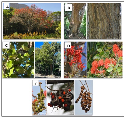
FIG. 1: (A) ERYTHRINA ABYSSINICA TREE, (B) ERYTHRINA ABYSSINICA TRUNK, (C) ERYTHRINA ABYSSINICA TREE AND LEAVES, (D) ERYTHRINA ABYSSINICA FLOWERS (E) ERYTHRINA ABYSSINICA PODS WITH FRESH MATURE SEEDS. SOURCE: HTTPS://PZA.SANBI.ORG/ERYTHRINA-ABYSSINICA
In African traditional systems, E. abyssinica is used to prepare remedies or concotions for a wide range of human and livestock diseases, including wounds, malaria, bacterial and fungal infections, tuberculosis, HIV‑related opportunistic infections, diarrhoea, inflammatory diseases, urinary tract infections, diabetes mellitus, and skin and soft‑tissue injuries. Bark decoctions and root infusions, are commonly used to treat pain, fever, respiratory conditions, gastrointestinal complaints, and wound infections 7. The leaves can also be used to make tea 8 E. abyssinica is also valued for its light and soft timber which is used in making stools and small tools. Its spiny branches and dense canopy provide shelter and nesting sites for birds and small mammals. In agro‑ecological landscapes it can act as a living barrier combining ecological, cultural, and pharmacological significance across the continent 3. Despite the widespread ethnomedicinal use of E. abyssinica for inflammation and infections 9, limited scientific validation exists for its leaf and bark hydroethanolic extracts. While root preparations are common in traditional practice 3, leaves and bark were selected for this study to enable sustainable harvesting without tree falling. Plant secondary metabolites, including flavonoids, alkaloids, tannins, and terpenoids, are known to possess antibacterial and anti-inflammatory properties 10. This study aimed to: (1) perform phytochemical screening of hydroethanolic leaf and bark extracts of E. abyssinica; (2) evaluate in-vitro antibacterial activity against wound pathogens S. aureus and E. coli; (3) assess in-vitro anti inflammatory activity via protein denaturation inhibition; and (4) determine acute oral toxicity in a rodent model following OECD guidelines.
MATERIALS AND METHODS:
Chemicals, Reagents, and Equipment: All chemicals and reagents used in this study were of analytical grade or higher. Ethanol (70% v/v, AnalaR NORMAPUR grade, 99.8% purity) was obtained from VWR International (USA), while dimethyl sulfoxide (HPLC grade, ≥99.9%), ferric chloride hexahydrate (ACS reagent, ≥99%), acetic anhydride (ACS reagent), concentrated sulfuric acid (98%), sodium hydroxide pellets (ACS reagent), phosphate-buffered saline tablets (pH 7.4), diclofenac sodium (Sigma-Aldrich D6899, ≥98%), and doxycycline hyclate (Sigma-Aldrich D9891, ≥98%) were purchased from Sigma Aldrich (USA). Iodine solution (0.1 N) was obtained from Himedia (India), chloroform (HPLC grade) from Fisher Scientific (UK), ammonia solution (25%) from BDH (UK), and sulfuric acid (98%) from Merck (Germany). Microbiological media including nutrient agar (Himedia M001), Mueller-Hinton broth (Himedia M391), and Mueller-Hinton agar (Himedia M108) were procured from HiMedia Laboratories (India). Bacterial reference strains Staphylococcus aureus (ATCC 25923) and Escherichia coli (ATCC 25922) were obtained from the American Type Culture Collection (USA). Fresh hen eggs (Gallus gallus domesticus, White Leghorn breed) used for egg albumin were sourced from the Department of Animal Science, University of Zimbabwe, and were verified for albumin integrity within 24 hours of laying.
Key instruments included a Rotavapor R-300 rotary evaporator (Büchi, Switzerland), a FreeZone 2.5L lyophilizer (Labconco, USA) operating at −50°C and 140 Pa, a UV-1800 UV/Vis spectrophotometer with quartz cuvettes (Shimadzu, Japan), a Hirayama HVE-50 autoclave (Hirayama, Japan), a Memmert INE 400 incubator (Memmert, Germany), a Stuart SL162 magnetic stirrer (Cole-Parmer, UK), a Mettler Toledo ME204 analytical balance (Mettler Toledo, Switzerland), a Mettler Toledo FE20 pH meter (Mettler Toledo, Switzerland), an ESCO Airstream laminar airflow cabinet (ESCO, Singapore), and a Grant JB1 water bath (Grant Instruments, UK). All assays were conducted in the Pharmacology and Microbiology Laboratories, Department of Pharmacy and Pharmaceutical Sciences, Faculty of Medicine and Health Sciences, University of Zimbabwe, Harare.
Plant Material Collection and Authentication: The barks and leaves of Erythrina abyssinica (locally known as Mutiti) were collected during the end of the rainy season from March 15–20, 2025, from the shore area adjacent to Lake Chivero, approximately 35 km southwest of Harare, Zimbabwe. The collection site is located within the Lake Chivero Recreational Park at GPS coordinates 17°53'42"S, 30°49'18"E (elevation 1,380 m). The area is classified as highveld woodland, dominated by Brachystegia and miombo tree species. The plant material was officially authenticated by Ms. Mutasa K.B., research officer at the National Herbarium and Botanical Garden of Zimbabwe, Harare. No endangered or protected species were involved.
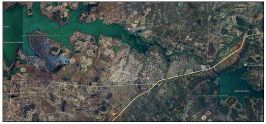
FIG. 2: MAP SHOWING THE COLLECTION SITE OF ERYTHRINA ABYSSINICA LEAVES AND BARK AT LAKE CHIVERO RECREATIONAL PARK, NEAR NORTON, ZIMBABWE (GPS COORDINATES 17°53'42"S, 30°49'18"E).
Leaves and bark were collected separately, processed in parallel, and combined in equal proportions (1:1 w/w) for all extractions and assays described herein. While traditional uses often cite roots (Obakiro et al., 2021), leaves and bark were prioritized to enable sustainable harvesting without plant destruction and to reflect topical wound applications where aerial parts are commonly used 12.
To remove surface contaminants and debris, the collected leaves (177g) and bark (216g) were thoroughly washed using clean water. After washing, they were spread out and shade dried at room temperature until they reached a constant, stable weight and a low moisture content of about 4%. This drying period took approximately 2.5 weeks. The dried material was then crash-grounded to a fine powder using a mortar and pestle.
The leaf powder (177 g) and bark powder (216 g) were combined (total 393 g) before extraction. The combined powder was added to 1000 mL of 70% (v/v) hydroethanolic mixture in a 2-litre sterile amber bottle. The mixture was macerated for 72 hours at room temperature (25 ± 2°C) with manual shaking for 3 minutes twice daily to ensure thorough mixing.
The liquid extract was separated from solids by straining through a muslin cloth, followed by fine filtration using Whatman No. 1 filter paper. The resulting filtrate was concentrated under reduced pressure at 40°C using a Rotavapor R-300 (Büchi, Switzerland) to approximately one-third of its original volume. The concentrated liquid was then subjected to freeze-drying (lyophilization) at −50°C under 140 Pa pressure using a Labconco FreeZone 2.5L lyophilizer. The final lyophilized extract yielded 38.5 g of a dark brown, amorphous, hygroscopic powder with a characteristic aromatic odour.
The percentage yield was calculated as:
Yield (%) = (38.5 g / 393 g) × 100 = 9.8% w/w
The dried extract was stored in an airtight amber glass bottle with desiccant at 4°C ± 2°C until further use. For all subsequent assays, the extract was reconstituted freshly in sterile distilled water or phosphate-buffered saline as appropriate.
Phytochemical Screening of Erythrina abyssinica: In a 200ml flask, 5g of the freeze-dried hydro-ethanolic extracts from Erthyrina abyssinica were mixed with 50ml of distilled water. This resulting solution was then tested using various chemical methods to check for the presence or absence of specific plant compounds that might have medicinal benefits. Several specific tests were performed on the liquid extract to identify these compounds.
All procedures were carried out as per guidelines by Chifamba et al. 13 with slight modifications. For comparative purposes, a separate distilled-water extract was prepared by macerating 350 g of the combined leaf-bark powder in 1000 mL of distilled water for 72 hours at room temperature, followed by filtration, evaporation, and lyophilization using identical parameters as the hydroethanolic extract.
Detection for Alkaloids by the Iodine Test: The Iodine test was used to determine the presence of alkaloids. In this test, a few drops of iodine solution were slowly added along the sides of the test tube to 3ml of the lyophilized extract solution. The presence of alkaloids was identified by the appearance of a blue colour, which characteristically disappears on boiling and reappears on cooling 14.
Detection of Tannins: The Braymer’s test was used to detect the presence of tannins. To 1ml lyophilized extract solution, 2mls of distilled water was added followed by 4 drops of a 10% Ferric chloride solution. The presence of tannins is confirmed by the formation of a blue-green colour 15.
Detection of Flavonoids by the Ammonia Test: Flavonoids were detected by means of the Ammonia test where 5 ml dilute ammonia solution was added to 5ml of the lyophilized extract solution, followed by a few drops of concentrated sulfuric acid. The emergence of a yellow colour indicates the presence of flavonoids 16.
Detection of Glycosides: To 2mls of the extract, 3ml of chloroform was added followed by 3 drops of ammonia in a test-tube. The formation of a pink color indicates the presence of the glycosides 17.
Detection of Phenolic Compounds: To 1 ml of the extract, 2 m of distilled water was added followed by a few drops of ferric chloride. The formation of a blue-green color indicates the presence of phenols 18.
Detection of Saponins by the Simplified Foam Test: The simplified foam test was used to determine the presence of saponins. In this assay, 2ml of the extract was added to 20ml distilled water. The mixture was shaken in a graduated cylinder for 15 minutes. The presence of saponins was confirmed by the formation of foam with a head height of at least 1cm 19.
Detection of Phytosterols (Salkowski Test): The Salkowski test was used to detect the presence of phytosterols. To 2 mL of extract, 2 mL of chloroform and 2 mL of concentrated sulfuric acid were added along the test tube wall. A red coloration in the chloroform layer indicated phytosterols 20.
Detection of Steroids: The Liebermann-Burchard test was used to detect the presence of steroids. To 2 mL of extract, 2 mL of acetic anhydride followed by 2 drops of concentrated sulfuric acid were added. A blue-green color indicated steroids 21.
Detection of Coumarins: To 2 mL of extract, 3 mL of 10% sodium hydroxide was added. Formation of a yellow color indicated coumarins 21.
Detection of Terpenoids: The Salkowski test was used to detect the presence of tepernoids. To 2 mL of extract, 2 mL of chloroform and 2 mL of concentrated sulfuric acid were carefully added. A reddish-brown interface indicated terpenoids 21. The intensity of colour or precipitate formation was scored semi-quantitatively as: (+++) strong presence, (++) moderate presence, (+) detected, (−) absent, based on comparison with positive controls and literature descriptions.
Anti-Inflammatory Activity of Erythrina abyssinica using the Egg Albumin Denaturation Test: The anti-inflammatory potential of Erythrina abyssinica crude extracts was evaluated using an in-vitro assay focused on the inhibition of egg albumin denaturation. This method was employed to determine the ability of the plant’s secondary metabolites to stabilize proteins, a biological process that mirrors the stabilization of lysosomal membranes during an inflammatory response. The experimental procedure began with the preparation of a series of reaction mixtures, each reaching a total volume of 5 mL. For each test, 0.2 mL of a 1–2% egg albumin solution, sourced from fresh hen’s eggs, was combined with 2.8 mL of phosphate-buffered saline (pH 7.4). To this mixture, 2 mL of the E. abyssinica sample extract was added. To assess the dose-dependent response of the plant, the extracts were prepared and tested at a concentration gradient of 100, 200, 300, 400, and 500µg/mL. To provide a comparative baseline for therapeutic efficacy, a standard reference was prepared using Diclofenac sodium across the same concentration range. A control solution was also established by mixing 0.2 mL of the egg albumin solution and 2.8 mL of phosphate-buffered saline with 2 mL of triple-distilled water in place of the plant extract.
Once the mixtures were prepared, they were subjected to a controlled thermal environment to induce protein denaturation. The reaction tubes were first incubated at 37±2°C for 30 minutes. Following this initial incubation, the tubes were transferred to a water bath and heated at 70±2°C for 15 minutes. This heating phase provided the thermal stress necessary to challenge the stability of the albumin protein, with the presence of E. abyssinica metabolites intended to prevent the unfolding of the protein chains. After the heating process was complete, the mixtures were allowed to cool to room temperature. The degree of protein stabilization was quantified by measuring the absorbance of each sample at 280 nm using a UV/Vis spectrophotometer, with triple-distilled water utilized as the blank. By comparing the absorbance of the test samples to the control, the percentage of inhibition was calculated for each concentration through the use of the following equation:
Equation 1: the percentage inhibition formula.
In𝑓𝑙𝑎𝑚𝑎𝑡𝑖𝑜𝑛 𝑖𝑛ℎ𝑖𝑏𝑖𝑡𝑖𝑜𝑛 𝑝𝑒𝑟𝑐𝑒𝑛𝑡𝑎𝑔𝑒 𝑒𝑓𝑓𝑒𝑐𝑡 = 𝐴𝐵𝑆_𝑐𝑜𝑛𝑡𝑟𝑜𝑙−𝐴𝐵𝑆_𝑠𝑎𝑚𝑝𝑙 / 𝐴𝐵𝑆_𝑐𝑜𝑛𝑡𝑟𝑜𝑙 × 100
Where, ABS-control = absorbance of the control reaction (egg albumin + PBS + distilled water instead of extract) at 280 nm, after subtracting the blank absorbance ABS-sample = absorbance of the test reaction (egg albumin + PBS + extract) at 280 nm, after subtracting the blank absorbance Blank = triple-distilled water (used to zero the spectrophotometer before each measurement).
All reactions were prepared in triplicate (n = 3) for each concentration of both the extract and the standard drug (diclofenac sodium). Concentrations tested were 100, 200, 300, 400, and 500 µg/mL for both the extract and diclofenac. Diclofenac sodium was used as the positive control at the same concentration range.
Absorbance Measurement: After cooling to room temperature, the absorbance of each sample was measured at 280 nm using a Shimadzu UV-1800 UV/Vis spectrophotometer with quartz cuvettes. Triple-distilled water was used as the blank reference and was measured between each sample to ensure baseline stability.
IC₅₀ Determination: The concentration required to inhibit 50% of protein denaturation (IC₅₀) was calculated by non-linear regression analysis using GraphPad Prism version 9.0 (GraphPad Software, San Diego, CA, USA). A four-parameter logistic (4PL) curve was fitted to the log-transformed concentration versus percentage inhibition data. The IC₅₀ values are reported in the results section.
Antibacterial Activities of Erythrina abyssinica: Evaluation of the antibacterial potential of Erythrina abyssinica extracts involved measuring zones of inhibition against Escherichia coli and Staphylococcus aureus using the agar well diffusion method. This qualitative screening facilitated the direct observation of the inhibitory effects exerted by the extracts, which provided clear evidence regarding their antimicrobial properties.
Preparation of Nutrient Agar Culture Media: Preparation of the nutrient agar involved weighing 6.24g of the medium and dissolving it into 450ml of distilled water within a borosilicate glass beaker. A magnetic stirrer and a Bunsen burner facilitated the mixing and heating process until the powder reached full dissolution. This solution was then transferred to a glass container for sterilization in an autoclave at 121°C for a duration of 15 minutes. Following the sterilization cycle, the medium cooled within a workspace previously disinfected with 70% ethanol. Once the temperature dropped sufficiently, 20ml portions of the sterile agar were dispensed into 12 sterile Petri dishes and left to solidify.
Bacterial Strains and Authentication: The bacterial strains used were Staphylococcus aureus (ATCC 25923) and Escherichia coli (ATCC 25922), obtained from the American Type Culture Collection (Manassas, VA, USA). Stock cultures were maintained on nutrient agar slants at 4°C and subcultured monthly. Working cultures were prepared by transferring a loopful of stock culture into 5 mL of nutrient broth and incubating at 37°C for 18–24 hours.
Inoculum Standardization: Overnight bacterial cultures were adjusted to a 0.5 McFarland standard (equivalent to approximately 1.5×10⁸ CFU/mL) using sterile saline. The adjusted inoculum was further diluted 1:10 in sterile saline to achieve a final concentration of approximately 1.5×10⁷ CFU/mL for lawn plating. This standardization was verified by spectrophotometry at 625 nm (optical density of 0.08–0.13).
Preparation of Nutrient Agar Plates: Nutrient agar (6.24 g in 450 mL distilled water) was dissolved with heating and stirring, then sterilized by autoclaving at 121°C for 15 minutes. After cooling to approximately 45°C, 20 mL of sterile agar was poured into each sterile Petri dish (90 mm diameter) and allowed to solidify on a level surface. Plates were dried in a laminar flow cabinet for 30 minutes before use.
Inoculation of Plates: Using a sterile cotton swab, the standardized bacterial suspension was evenly spread across the entire surface of each nutrient agar plate in three directions (60° rotation between streaks) to ensure a confluent lawn. Plates were allowed to dry for 10 minutes at room temperature before well boring.
Preparation of Test Extract And Controls: The lyophilized E. abyssinica extract was dissolved in sterile distilled water containing 5% DMSO (to enhance solubility of hydrophobic compounds) to achieve final concentrations of 180 mg/mL, 130 mg/mL, and 60 mg/mL. The positive control, doxycycline hyclate (Sigma-Aldrich D9891, ≥98%), was dissolved in sterile distilled water to the same concentrations (180, 130, and 60 mg/mL). The negative control was sterile distilled water containing 5% DMSO (solvent control). A separate negative control of distilled water without DMSO was also included to assess any DMSO-related inhibition.
Well Diffusion Assay: Wells of 4 mm diameter were bored into the inoculated agar plates using a sterile stainless steel cork borer. Each plate contained four wells: three for the extract concentrations and one for the positive control. A separate set of plates was used for negative controls. Using a micropipette, 50 µL of each test solution was dispensed into the respective wells. The plates were left at room temperature for 30 minutes to allow pre-diffusion of the extracts, then incubated at 37°C for 24 hours. The measured zone of inhibition (ZOI) included the well diameter. The corrected ZOI was calculated by subtracting the well diameter (4 mm) from the measured diameter: Corrected ZOI = Measured ZOI − Well diameter. This correction accounts for the physical space of the well and allows direct comparison with other studies.
Measurement and Statistical Analysis: After incubation, zones of inhibition (ZOI) were measured to the nearest millimeter using a digital Vernier caliper. The corrected ZOI was calculated by subtracting the well diameter (4 mm) from the measured diameter. All assays were performed in triplicate (three independent experiments), and for each experiment, each concentration was tested in duplicate plates. Results are expressed as mean ± standard deviation (SD). Percentage inhibition relative to doxycycline was calculated using:
Equation 2:
% inhibition= 𝐶𝑜𝑟𝑟𝑒𝑐𝑡𝑒𝑑 𝑍𝑂𝐼 𝑜𝑓 𝑒𝑥𝑡𝑟𝑎𝑐𝑡 𝑎𝑡 𝑐𝑜𝑛𝑐𝑒𝑛𝑡𝑟𝑎𝑡𝑖𝑜𝑛 𝑋 / C𝑜𝑟𝑟𝑒𝑐𝑡𝑒𝑑 𝑍𝑂𝐼 𝑜𝑓 𝑑𝑜𝑥𝑦𝑐𝑦𝑐𝑙𝑖𝑛𝑒 𝑎𝑡 𝑐𝑜𝑛𝑐𝑒𝑛𝑡𝑟𝑎𝑡𝑖𝑜𝑛 × 100
Statistical comparisons between extract and doxycycline at each concentration were performed using an independent t-test (GraphPad Prism 9.0). A p-value < 0.05 was considered statistically significant.
Determination of Minimum Inhibitory Concentration (MIC): The Minimum Inhibitory Concentration (MIC) of lyophilized hydroethanolic extract of Erythrina abyssinica against Staphylococcus aureus and Escherichia coli was determined using the macro broth dilution method according to CLSI guidelines, with slight modifications.
Preparation of Extract Stock Solution: The lyophilized extract was dissolved in sterile distilled water containing 5% DMSO (to enhance solubility) to obtain a stock concentration of 2000 µg/mL. This stock was then serially diluted two‑fold in sterile Mueller‑Hinton broth (MHB) to achieve final test concentrations of 1000, 500, 250, 125, 62.5, 31.25, 15.6, 7.8, 3.9, and 1.95 µg/mL.
Bacterial Inoculum Preparation: Fresh overnight cultures of S. aureus and E. coli were adjusted to 0.5 McFarland standard (~1.5 × 10⁸ CFU/mL) and then diluted in MHB to obtain a final inoculum of approximately 5 × 10⁵ CFU/mL in each tube.
Macro Dilution Procedure: Sterile glass test tubes (13 × 100 mm) were arranged in racks. To each tube, 1.0 mL of the respective extract dilution was added, followed by 1.0 mL of the standardized bacterial suspension. The final volume in each tube was 2.0 mL, with extract concentrations as listed above. Control tubes included:
- Positive control: Doxycycline (final concentration 160 µg/mL) + bacterial suspension.
- Negative control (growth control): MHB + bacterial suspension (no extract).
- Sterility control: MHB + extract (highest concentration) without bacteria.
All tubes were loosely capped and incubated aerobically at 37°C for 24 hours.
MIC Determination: After incubation, each tube was examined for visible turbidity. The MIC was defined as the lowest concentration of extract that completely inhibited visible bacterial growth (no turbidity). Tubes showing no growth were then subcultured onto Mueller‑Hinton agar plates and incubated for another 24 hours to determine the Minimum Bactericidal Concentration (MBC) if desired. All tests were performed in triplicate.
Acute Oral Toxicity Evaluation of E. abyssinica: The acute oral toxicity of lyophilized Erythrina abyssinica hydroethanolic leaf and bark extract was evaluated following the OECD Guideline 425 (Up-and-Down Procedure, UDP). The study protocol was approved by the Joint Parirenyatwa Research Ethics Committee (JREC) under approval number JREC/582/2025 dated December 18, 2025.
Animals and Housing: Eight adult Sprague-Dawley rats (4 males, 4 nulliparous non-pregnant females), aged 8–12 weeks and weighing 180–220 g, were obtained from the University of Zimbabwe Animal House. Animals were acclimatized for 7 days under standard laboratory conditions: temperature 22 ± 3°C, relative humidity 50 ± 10%, 12:12 h light/dark cycle, with ad libitum access to standard pellet diet and reverse osmosis water. All animals were confirmed healthy by a veterinary specialist prior to dosing.
Dose Selection and Administration: The starting dose of 250 mg/kg was selected based on a limit test approach, as no toxicity was expected from traditional use reports. Animals were fasted for 18 hours (water ad libitum) before oral gavage. Each animal received a single dose, with 48-hour intervals between successive doses. The dose progression was: 250 mg/kg → 500 mg/kg → 1000 mg/kg → 2000 mg/kg (one animal per dose level, if the previous animal survived). A control group (n=3, 1 male + 2 females) received distilled water (10 mL/kg) only.
Clinical Observations: Animals were observed individually for the first 30 minutes, then periodically during the first 24 hours (at 1, 2, 4, 6, and 12 hours), and then once daily for 14 days post-dosing. Observations included: general appearance, skin and fur, eyes and mucous membranes, respiratory pattern, motor activity, posture, tremors, salivation, diarrhoea, urination, and feeding behavior. Body weight was recorded on days 0, 7, and 14.
Humane Endpoints: Animals meeting any of the following criteria were immediately euthanized via intraperitoneal pentobarbital (150 mg/kg): ≥20% weight loss in 24 hours, hunched posture with piloerection, inability to access food/water, moribund status, or severe respiratory distress. No animals met these endpoints during the study.
Determination of LD₅₀ and NOAEL: The LD₅₀ was estimated based on the up-and-down procedure using the OECD 425 computational approach (AOT425StatPgm). The No-Observed-Adverse-Effect Level (NOAEL) was defined as the highest dose at which no treatment-related clinical signs or mortality were observed. Statistical analysis of body weight changes was performed using two-way ANOVA with repeated measures (GraphPad Prism 9.0).
RESULTS AND DISCUSSIONS:
Phytochemical Analysis: Through the phytochemical screening assay, it was successfully validated that the plant contains a significant number of secondary metabolites, which are highly relevant for biomedical and pharmaceutical applications, thus validating its use in traditional medicine. Among the detected compounds, phenolics were the most dominant group, with a notably high prevalence of flavonoids. These findings align with previous research by Obakiro S and colleagues 3, which also identified a range of bioactive compounds in Erythrina abyssinica, including alkaloids, terpenes, tannins, flavonoids, and coumarins. The abundance of these medically significant phytochemicals provides a strong scientific basis for the plant's established use in traditional medicine, particularly for treating infections and inflammatory conditions 22.
The table below details the specific phytoconstituents identified in Erythrina abyssinica.
TABLE 1: BIOACTIVE COMPOUNDS IN E. ABYSSINICA
| Test | Presence in hydro-ethanolic extract | Presence in distilled water extract | Observed color change (hydro‑ethanolic) |
| Alkaloids | +++ | + | Blue color |
| Phytosterols | +++ | ++ | Red coloration in chloroform layer |
| Flavonoids | +++ | + | Yellow color |
| Saponins | +++ | + | Foam head >1 cm |
| Steroids | +++ | + | Blue‑green color |
| Coumarins | +++ | + | Yellow color |
| Phenolic compounds | +++ | + | Blue‑green color |
| Tannins | +++ | + | Blue‑green color |
| Glycosides | +++ | + | Pink color |
| Terpenoids | +++ | + | Reddish‑brown interface |
Key: (-): Indicates the absence of the phytochemical, (+): Indicates the presence of the phytochemical, (++): Indicates moderate presence of the phytochemical, (+++): Indicates strong presence of the phytochemical.
Phenolics are a major group of these metabolites, primarily produced by the plant as a defensive response to adverse environmental conditions 23, such as microbial invasion or stress. Chemically, these plant phenolics are predominantly acid derivatives of hydroxybenzoic and hydroxycinnamic acids 24. There has been growing interest in their potential medicinal applications, particularly as antioxidants, anti-inflammatory agents and anti-bacterial agents 25. Flavonoids are a key category within the phenolics group. They are a class of polyphenolic compounds with subgroups like 26, 27 flavones, flavanols, and flavanones. Notably, these flavonoids are recognized for exhibiting potent antioxidant activities. By neutralizing the excessive Reactive Oxygen Species (ROS) prevalent in chronic wound beds, flavonoids help reduce the overall oxidative stress burden, reducing oxidative stress and inflammation 28. It is important to note that the phytochemical screening employed semi-quantitative colorimetric tests that indicate the presence of compound classes but do not identify specific active constituents, quantify absolute concentrations, or establish structure-activity relationships. While the detection of alkaloids, flavonoids, and tannins is consistent with known antibacterial and anti-inflammatory mechanisms reported in the literature 26, 27, these preliminary findings require confirmation through chromatographic isolation (e.g., HPLC, GC-MS), quantitative analysis, and bioactivity-guided fractionation.
Acute Oral Toxicity Assay of E. abyssinica:
TABLE 2: SHOWING THE BEHAVIOURAL PATTERNS OF THE RATS IN RESPONSE TO DIFFERENT DOSAGES
| Observed parameter | Dose of E. abyssinica in mg/kg body weight | ||||
| 250 | 500 | 1000 | 2000 | Control | |
| Food intake | Normal | Normal | Normal | Normal | Normal |
| Water intake | Normal | Normal | Normal | Normal | Normal |
| Death | No deaths | No deaths | No deaths | No deaths | No deaths |
| Breathing | Normal | Normal | Normal | Normal | Normal |
| Diarrhea | None | None | None | None | None |
| Urination | Normal | Normal | Normal | Normal | Normal |
| skin color | Normal | Normal | Normal | Normal | Normal |
| Drowsiness | None | None | None | None | None |
The toxicological evaluation of Erythrina abyssinica provides a necessary bridge between ancient traditional wisdom and the rigorous requirements of modern clinical science. Under the conditions of this acute oral toxicity study in rats, the E. abyssinica extract showed no mortality or clinical signs up to 2000 mg/kg, with an estimated LD₅₀ > 2000 mg/kg. This suggests a low acute toxicity profile in this rodent model. However, acute toxicity data alone are insufficient to predict human safety. Chronic toxicity studies (e.g., 28‑day or 90‑day repeated dose), genotoxicity assessment, and pharmacokinetic studies are necessary before any human use recommendation can be made. The NOAEL observed here (2000 mg/kg) serves as a reference for future subchronic studies, not as a direct safety threshold for humans. The physiological health of the subjects remained stable throughout the observation window, suggesting that the plant metabolites do not interfere with the natural homeostatic balance of the body. Rats in the experimental cohorts, maintained growth trajectories and behavioral patterns consistent with the untreated control group. Detailed monitoring of metabolic indicators by a veterinary specialist confirmed that food intake, water consumption, and urinary characteristics remained within normal ranges across all dosage levels. These results are consistent with broader literature on the Erythrina genus, such as studies on Erythrina senegalensis, which also reported high safety margins in rodent models 29.
Comparing these findings to recent literature reveals that discrepancies in plant toxicity are often linked to the specific methods used for extraction and the environmental conditions of the habitat 30. This study utilized lyophilized extracts, a process that preserves the delicate integrity of secondary metabolites better than crude heat-based extractions. Literature suggests that variations in the phytochemical constituents, often caused by genetic traits or environmental stressors, can lead to aberrations in bioactivity and safety 31. For example, plants harvested in different geographical locations may produce varying concentrations of alkaloids as a response to existential threats in their habitat, a phenomenon also observed in Terminalia sericea. Despite these natural variations, the high safety ceiling of 2000 mg/kg observed here provides a robust foundation for pharmaceutical applications. By confirming that the phytochemical symphony of E. abyssinica protects rather than harms the biological system, this research justifies the use of these metabolites in mediating the biosynthesis of silver and gold nanoparticles for the treatment of recalcitrant chronic wounds
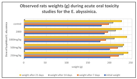
FIG. 3: SHOWING OBSERVED RAT WEIGHTS DURING THE PROCEDURE
In alignment with established protocols for toxicological investigations, the body mass of the Sprague Dawley rats was systematically monitored and documented throughout the duration of the study. Weight measurements for all experimental cohorts, including the control group, were captured at weekly intervals, with data points established on day 0, 7, 14, and 21. Comparative analysis of these metrics revealed that the four groups treated with the Erythrina abyssinica extracts-maintained growth trajectories consistent with those of the untreated controls. No statistically significant aberrations or irregular fluctuations in body weight were detected across any of the dosage levels, further suggesting that the extracts did not interfere with the metabolic health or developmental progress of the subjects during the observation window.
Anti-Inflammatory Activity of Erythrina abyssinica using the Egg Albumin Denaturation Test Results:
TABLE 3: ANTI-INFLAMMATORY ACTIVITY OF E. ABYSSINICA HYDROETHANOLIC LEAF AND BARK EXTRACT COMPARED TO DICLOFENAC SODIUM USING THE EGG ALBUMIN DENATURATION TEST. VALUES ARE MEAN ± STANDARD DEVIATION OF THREE INDEPENDENT REPLICATES
| Concentration (µg/mL) | Diclofenac (% inhibition, mean ± SD) | E. abyssinica extract (% inhibition, mean ± SD) | p-value* |
| 100 | 39.70 ± 0.29 | 38.04 ± 0.35 | 0.065 |
| 200 | 49.96 ± 0.43 | 43.99 ± 0.35 | 0.012 |
| 300 | 57.78 ± 0.47 | 54.06 ± 0.27 | 0.018 |
| 400 | 64.07 ± 0.62 | 62.01 ± 0.43 | 0.081 |
| 500 | 81.13 ± 0.37 | 77.82 ± 0.54 | 0.042 |
*p-value from independent two-tailed t-test comparing diclofenac vs. extract at each concentration. Significance set at p < 0.05. (*Average value of 3 replicates).
Shown below is a graphical presentation showing the anti-inflammatory activity of E. abyssinica.
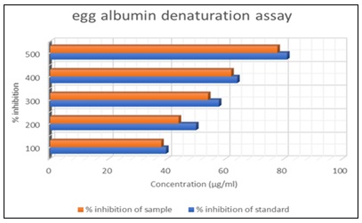
FIG. 4: PROTEIN DENATURATION ASSAY
The anti-inflammatory potential of Erythrina abyssinica is clearly evidenced by the laboratory data, as shown in Table 3 and the corresponding trends visualized in Fig. 4. At an initial concentration of 100µ/mL, the extract achieved a 38.04 ± 0.35% inhibition of protein denaturation. This protective effect climbed significantly as the dosage increased, eventually reaching 77.82 ± 0.54% at 500 µg/mL. While there is a minor difference between these values and the standard reference drug, which reached 81.13 ± 0.37%, the narrowness of this gap suggests that the plant extract is a remarkably potent biological agent in its own right. These findings essentially mean that the secondary metabolites in the plant are capable of preventing the structural breakdown of proteins, which is a fundamental requirement for controlling the inflammatory response. Persistent inflammation underlies many chronic disease states, making the anti-inflammatory properties of plant extracts clinically relevant. As shown in Fig. 5, the dose dependent upward trend indicates that E. abyssinica could act as a biochemical shield within a topical cream. By preventing protein denaturation, the extract stabilizes protein structures, which is mechanistically relevant to controlling inflammatory processes. The effectiveness of E. abyssinica in this study is highly competitive when compared with numerical results from similar recent investigations. For instance, studies on related Erythrina species have reported slightly lower inhibition values, such as 64.2% at 500µg/mL for Erythrina variegata 32. Conversely, other high potency extracts in literature have shown figures as high as 82.5% at similar concentrations 33. The minor deviations from these benchmarks, specifically the 77.82% achieved here, are likely rooted in the life history of the plant material. Factors such as altitude, soil quality, and rainfall create a unique chemical profile for each sample, while the choice of extraction solvent determines the final concentration of active metabolites 34. Additionally, the biological sensitivity of using egg albumin as a model for inflammation can lead to subtle shifts in data across different laboratory environments. Research has shown that the protein is sensitive to local pH levels and freshness, with some studies reporting baseline denaturation variances of 5 to 8% 35. Nevertheless, the consistent performance as shown in both the table and the graph confirms that E. abyssinica provides a reliable and robust defense against the protein degradation that fuels chronic inflammation. The ability of the extract to reach nearly 78% inhibition demonstrates its potential as an anti-inflammatory agent worthy of further investigation. While the egg albumin denaturation assay provides preliminary evidence of anti-inflammatory potential, it does not directly measure wound healing. Confirmatory studies using in-vivo models (e.g., carrageenan-induced paw edema for inflammation, excision wound models for healing) are required.
Antibacterial assay of E. abyssinica: The following table shows the zone of inhibition of Erythrina abyssinica in relation to its different concentrations.
For the agar well diffusion assay, concentrations of 180, 130, and 60 mg/mL were used to achieve detectable zones of inhibition. The minimum inhibitory concentration (MIC) was determined separately using macro broth dilution at concentrations ranging from 1.95 to 1000 µg/mL.
TABLE 4: ANTIBACTERIAL ACTIVITY OF ERYTHRINA ABYSSINICA EXTRACT AGAINST STAPHYLOCOCCUS AUREUS (ATCC 25923) USING THE AGAR WELL DIFFUSION METHOD
| Sample | Concentration (mg/mL) | Measured ZOI (mm, mean ± SD) | Corrected ZOI (mm, mean ± SD) | % Inhibition relative to doxycycline |
| Positive control (Doxycycline) | 180 | 27.0 ± 0.6 | 23.0 ± 0.6 | – |
| 130 | 22.0 ± 0.5 | 18.0 ± 0.5 | – | |
| 60 | 14.0 ± 0.4 | 10.0 ± 0.4 | – | |
| Negative control (Distilled water) | – | 0.0 ± 0.0 | 0.0 ± 0.0 | 0.0 |
| Erythrina abyssinica | 180 | 20.0 ± 0.8 | 16.0 ± 0.8 | 69.6 |
| 130 | 14.0 ± 0.7 | 10.0 ± 0.7 | 55.6 | |
| 60 | 10.0 ± 0.5 | 6.0 ± 0.5 | 60.0 |
Well diameter = 4 mm. Corrected ZOI = Measured ZOI − 4 mm. *All values are mean of three independent experiments (n=3), each performed in duplicate.
The figure below shows the agar well diffusion assay results: corrected zones of inhibition of E. abyssinica hydroethanolic extract vs. doxycycline against S. aureus.
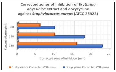
FIG. 6: CORRECTED ZONES OF INHIBITION (MM) OF ERYTHRINA ABYSSINICA LEAF AND BARK EXTRACT COMPARED TO DOXYCYCLINE AGAINST ESCHERICHIA COLI AT THREE CONCENTRATIONS (60, 130, AND 180 MG/ML)
The following table shows the zone of inhibition of Erythrina abyssinica in relation to its different concentrations.
TABLE 5: ANTIBACTERIAL ACTIVITY OF ERYTHRINA ABYSSINICA EXTRACT AGAINST E. COLI (ATCC 25922) USING AGAR WELL DIFFUSION METHOD
| Sample | Concentration (mg/mL) | Measured ZOI (mm, mean ± SD) | Corrected ZOI (mm, mean ± SD) | % Inhibition relative to doxycycline |
| Positive control (Doxycycline) | 180 | 32.0 ± 0.7 | 28.0 ± 0.7 | – |
| 130 | 23.0 ± 0.6 | 19.0 ± 0.6 | – | |
| 60 | 14.0 ± 0.4 | 10.0 ± 0.4 | – | |
| Negative control (Distilled water) | – | 0.0 ± 0.0 | 0.0 ± 0.0 | 0.0 |
| Erythrina abyssinica | 180 | 22.0 ± 0.9 | 18.0 ± 0.9 | 64.3 |
| 130 | 17.0 ± 0.6 | 13.0 ± 0.6 | 68.4 | |
| 60 | 12.0 ± 0.5 | 8.0 ± 0.5 | 80.0 |
Well diameter = 4 mm. Corrected ZOI = Measured ZOI − 4 mm. *All values are mean of three independent experiments (n=3), each performed in duplicate.
The figure below shows agar well diffusion assay results: corrected zones of inhibition of E. abyssinica hydroethanolic extract vs. doxycycline against E. coli.
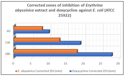
FIG. 7: CORRECTED ZONES OF INHIBITION (MM) OF ERYTHRINA ABYSSINICA LEAF AND BARK EXTRACT COMPARED TO DOXYCYCLINE AGAINST STAPHYLOCOCCUS AUREUS AT THREE CONCENTRATIONS (60, 130, AND 180 MG/ML)
Data analysis indicated that the Erythrina abyssinica extracts possessed significant antibacterial properties against both tested strains, showing a clear correlation between extract concentration and the size of the inhibition zones. The most robust activity occurred at the highest concentration of 180 mg/ml, where the inhibition levels nearly matched those of the doxycycline positive control. While both bacteria were susceptible, the inhibitory effects were notably more pronounced against S. aureus than E. coli, suggesting a higher sensitivity in the Gram-positive organism. These findings confirmed the effectiveness of the plant extracts as potential antimicrobial agents, particularly at elevated concentrations.
Minimum Inhibitory Concentration (MIC) – Macro Broth Dilution: The MIC of E. abyssinica hydroethanolic extract was determined using the macro broth dilution method (concentration range 1.95–1000 µg/mL). As shown in Table 6, the extract exhibited an MIC of 15.6 µg/mL against Staphylococcus aureus and 7.8 µg/mL against Escherichia coli. These values indicate that the extract possesses bacteriostatic activity against both Gram‑positive and Gram‑negative wound pathogens, with slightly greater potency against E. coli in this broth‑based assay. The positive control (doxycycline, 160 µg/mL) completely inhibited growth of both organisms, while negative control tubes (no extract) showed dense turbidity. Sterility controls remained clear. All tests were performed in triplicate with consistent results.
TABLE 6: MINIMUM INHIBITORY CONCENTRATION (MIC) OF ERYTHRINA ABYSSINICA HYDROETHANOLIC EXTRACT USING MACRO BROTH DILUTION METHOD
| Concentration (µg/mL) | E. coli (turbidity) | S. aureus (turbidity) |
| 1000 | – (no growth) | – (no growth) |
| 500 | – | – |
| 250 | – | – |
| 125 | – | – |
| 62.5 | – | – |
| 31.25 | – | – |
| 15.6 | - | – (MIC) |
| 7.8 | – (MIC) | +(growth) |
| 3.9 | +(growth) | + |
| 1.95 | + | + |
| Positive control (doxycycline 160 µg/mL) | – | – |
| Negative control (no extract) | + | + |
| Sterility control (extract only) | – | – |
Key: – = no visible turbidity (growth inhibited); + = visible turbidity (growth present). *All tests performed in triplicate; results were consistent across replicates.
The lower MIC against E. coli (7.8 µg/mL) compared to S. aureus (15.6 µg/mL) suggests that certain active constituents in the extract may be more effective against this Gram‑negative organism in liquid medium. This contrasts with the agar well diffusion results, where S. aureus showed larger zones of inhibition, a discrepancy that may reflect differences in compound diffusion rates, solubility, or medium composition between broth and agar.
The macro broth dilution assay revealed that E. abyssinica extract exhibited an MIC of 15.6 µg/mL against Staphylococcus aureus and 7.8 µg/mL against Escherichia coli as shown in table 6 above. The extract showed greater potency against the Gram‑negative E. coli compared to Gram‑positive S. aureus in this broth‑based assay. This is consistent with the well diffusion results where E. coli also showed sensitivity at higher concentrations. The positive control (doxycycline, 160 µg/mL) completely inhibited both organisms, while negative control tubes showed visible turbidity.
The results of this study provided evidence that Erythrina abyssinica extracts possessed substantial antibacterial potential, which directly supports its traditional application in the management of bacterial infections. Observations confirmed that the extracts inhibited the growth of both Staphylococcus aureus and Escherichia coli, which was significant because these two organisms are common pathogens in both community-acquired and nosocomial infections. Specifically, the 180 mg/ml concentration yielded a corrected zone of inhibition of 16 mm against S. aureus, representing a 69.6% efficacy relative to the standard antibiotic doxycycline. This high level of activity against Gram-positive bacteria is clinically relevant as S. aureus is a frequent cause of skin and soft tissue infections 36. The choice to utilize S. aureus and E. coli as test organisms was strategic as these represent Gram-positive and Gram-negative bacteria commonly implicated in infectious diseases. The extract achieved an 18 mm corrected zone of inhibition at 180 mg/ml against E. coli, suggesting that the plant has a compound capable of addressing the complex bacterial load found in various diseases. Literature indicates that Erythrina species contain specific flavonoids and chalcones that interfere with bacterial cell walls, which helps explain the observed inhibitory effects 37. These results implied that the plant could reduce microbial burden in infectious states.
A comparison with existing literature revealed that the zones of inhibition recorded in this study were slightly more pronounced than those found in previous evaluations of similar medicinal plants. For instance, prior research on related species often showed moderate zones ranging between 11 mm and 15 mm for methanol preparations 38. The efficacy reaching up to 18 mm in this trial likely stemmed from the lyophilization process, which preserved the bioactive compounds more effectively than air drying. This potency validates Erythrina abyssinica as a candidate for developing treatments against antibiotic-resistant bacterial strains 39. The dose dependent nature of the results, with inhibition dropping to 6 mm at 60 mg/ml for S. aureus, highlighted the necessity of using concentrated extracts for therapeutic antibacterial efficacy. The ability of Erythrina abyssinica to inhibit multiple bacterial types strengthens its profile as a broad-spectrum antibacterial agent. The data establish a clear scientific basis for the plant's antibacterial and anti-inflammatory activities.
CONCLUSION: This study validates Erythrina abyssinica as a potent and safe agent with antibacterial and anti-inflammatory properties. The experiments conducted also confirmed there are number of significant therapeutic bioactive compounds in E. abyssinica. It also showed significant anti-inflammatory and antibacterial activities, bridging the gap between traditional medicine and clinical evidence. Based on the foregoing conclusions, E. abyssinica is a promising candidate for the development of therapeutic products targeting bacterial infections and inflammatory conditions.
ACKNOWLEDGEMENTS: We wish to acknowledge the provision of laboratory facilities, equipment and study animals by the University of Zimbabwe, department of pharmacy and pharmaceutical sciences.
Disclaimer (Artificial Intelligence): The Authors hereby declare that NO generative AI technologies such as Large Language Models (ChatGPT, COPILOT, etc.) and text-to-image generators have been used during the writing or editing of this manuscript
Ethical Approval: Prior to the investigations, animal use and research ethics approvals were obtained from the Joint Parirenyatwa Research Ethics Committee (JREC) which is the local research Institutional Review board for the University of Zimbabwe.
Authors’ Contributions: This work was carried out in collaboration among all authors. Author PF led the study’s conception, coordination, and design, and took the primary role in drafting the original manuscript. Authors JC and SZ contributed to the conceptual framework and supervised the study, with Author PF and HM also performed the specific methodology of the manuscript. Data collection and draft preparation were conducted by Authors PF, JC, HM, and SZ. All authors reviewed and approved the final manuscript
CONFLICTS OF INTERESTS: Authors have declared that no competing interests exist.
- Obakiro SB, Kiprop A, Kigondu E, K’Owino I, Odero MP, Manyim S, Omara T, Namukobe J, Owor RO, Gavamukulya Y & Bunalema L: Traditional medicinal uses, phytoconstituents, bioactivities, and toxicities of Erythrina abyssinica Lam. ex DC. (Fabaceae): A Systematic Review. In Evidence-based Complementary and Alternative Medicine 2021. Hindawi Limited. https://doi.org/10.1155/2021/5513484
- Waktole G, Nemomissa S, Lemessa D, Kebede M, Jarso A & Tsegaye S: Modeling the distribution of Festuca abyssinica grass species in East African mountain ecosystems. Israel Journal of Ecology and Evolution 2026; 72: 75–90. https://doi.org/10.1163/22244662-bja10124
- Obakiro SB, Kiprop A, Kigondu E, K’Owino I, Odero MP, Manyim S, Omara T, Namukobe J, Owor RO, Gavamukulya Y & Bunalema L: Traditional Medicinal Uses, Phytoconstituents, Bioactivities, and Toxicities of Erythrina abyssinica Lam. ex DC. (Fabaceae): A Systematic Review. In Evidence-based Complementary and Alternative Medicine (Vol. 2021). Hindawi Limited. https://doi.org/10.1155/2021/5513484
- Roba Jilo K & Abeshu Erena Y: Proximate composition and mineral analysis of bee pollen from three monofloral sources: Eucalyptus camaldulensis, Guizotia abyssinica, and Acacia abyssinica. International Journal of Tropical Insect Science 2026. https://doi.org/10.1007/s42690-025-01733-y
- Hyde M: (n.d.). Erythrina abyssinica Lam. ex DC. Https://Www.Zimbabweflora.Co.Zw/Speciesdata/Species.Php?Species_id=131450.
- Abebe A: Nitrogen release dynamics of Erythrina abyssinica and Erythrina brucei litters as influenced by their biochemical composition. African Journal of Plant Science 2018; 12: 331–340. https://doi.org/10.5897/ajps2018.1717
- Mballow AE, Kahwa I, Muganga G, Ajayi CO, Sekagya Y, Sesaazi D, Fofana AK & Olet EA: Ethnobotanical and phytochemical investigation of medicinal plants used for epilepsy management by traditional healers in Mpigi District, Uganda. Next Research 2026; 4: 101260. https://doi.org/10.1016/j.nexres.2025.101260
- Kacholi DS: Ethnomedicinal Practices of the Fabaceae Family in Tanzania: A Systematic Review. In Scientific World Journal (Vol. 2026). John Wiley and Sons Ltd. https://doi.org/10.1155/tswj/6918847
- Adebayo MA, Menkir A, Blay E, Gracen V & Danquah E: Combining ability and heterosis of elite drought-tolerant maize inbred lines evaluated in diverse environments of lowland tropics. Euphytica 2017; 213. https://doi.org/10.1007/s10681-017-1840-5
- Bhatla SC & Lal MA: Secondary Metabolites. In Plant Physiology, Development and Metabolism 2023; 765–808. Springer Nature Singapore. https://doi.org/10.1007/978-981-99-5736-1_33
- Obakiro SB, Kiprop A, Kigondu E, K’Owino I, Odero MP, Manyim S, Omara T, Namukobe J, Owor RO, Gavamukulya Y & Bunalema L: Traditional Medicinal Uses, Phytoconstituents, Bioactivities, and Toxicities of Erythrina abyssinica Lam. ex DC. (Fabaceae): A Systematic Review. Evidence-Based Complementary and Alternative Medicine : ECAM 2021; 5513484. https://doi.org/10.1155/2021/5513484
- Mukaratirwa-Muchanyereyi N, Gusha C, Mujuru M, Guyo U & Nyoni S: Synthesis of silver nanoparticles using plant extracts from Erythrina abyssinica aerial parts and assessment of their anti-bacterial and anti-oxidant activities. Results in Chemistry 2022; 4. https://doi.org/10.1016/j.rechem.2022.100402
- Chifamba J, Zhou T, Michael H & Chitambira P: Phytochemical Screening and Evaluation of Pharmaceutical Activity of Nymphaea nouchali. South Asian Research Journal of Natural Products 2025.
- Balkrishna A, Sakshi, Chauhan M, Dabas A & Arya V: A comprehensive insight into the phytochemical, pharmacological potential, and traditional medicinal uses of Albizia lebbeck (L.) Benth. In Evidence-based Complementary and Alternative Medicine 2022; Hindawi Limited. https://doi.org/10.1155/2022/5359669
- Daboor SM & Haroon AM: In-vitro: Antimicrobial potential and phytochemical screening of some egyptian aquatic plants. Egyptian Journal of Aquatic Research 2012; 38: 233–239. https://doi.org/10.1016/j.ejar.2013.01.005
- Parimala M & Shoba FG: Phytochemical analysis and in-vitro antioxidant acitivity of hydroalcoholic seed extract of Nymphaea nouchali Burm. f. Asian Pacific Journal of Tropical Biomedicine 2013; 3: 887–895. https://doi.org/10.1016/S2221-1691(13)60174-4
- Daboor SM & Haroon AM: In-vitro: Antimicrobial potential and phytochemical screening of some egyptian aquatic plants. Egyptian Journal of Aquatic Research 2012; 38: 233–239. https://doi.org/10.1016/j.ejar.2013.01.005
- Sarma H, Sahariah G, Bharadwaj A, Sharma D, Porasar P, & Dutta KN: A systematic review on botanical background, phytochemical and pharmacological properties of Nymphaea nouchali. Journal of Applied Pharmaceutical Research 2024; 12: 21–36. https://doi.org/10.69857/joapr.v12i6.714
- Agidew MG: Phytochemical analysis of some selected traditional medicinal plants in Ethiopia. Bulletin of the National Research Centre 2022; 46. https://doi.org/10.1186/s42269-022-00770-8
- Addae AJ, Uyanga J, Chifamba J & Weiss PS: Advances in enhancing photoprotection of sunscreens using hydrocarbon film formers and carbon nanomaterials. In Advanced Drug Delivery Reviews 2025; 222. Elsevier B.V. https://doi.org/10.1016/j.addr.2025.115607
- Maheshwaran L, Nadarajah L, Senadeera SPNN, Ranaweera CB, Chandana AK & Pathirana RN: Phytochemical testing methodologies and principles for preliminary screening/ qualitative testing. Asian Plant Research Journal 2024; 12: 11–38. https://doi.org/10.9734/aprj/2024/v12i5267
- Obakiro SB, Kiprop A, Kigondu E, K’Owino I, Odero MP, Manyim S, Omara T, Namukobe J, Owor RO, Gavamukulya Y & Bunalema L: Traditional medicinal uses, phytoconstituents, bioactivities, and toxicities of Erythrina abyssinica Lam. ex DC. (Fabaceae): A Systematic Review. In Evidence-based Complementary and Alternative Medicine 2021; Hindawi Limited. https://doi.org/10.1155/2021/5513484
- Saini N, Anmol A, Kumar S, Wani AW, Bakshi M & Dhiman Z: Exploring phenolic compounds as natural stress alleviators in plants- a comprehensive review. In Physiological and Molecular Plant Pathology 2024; 133. Academic Press. https://doi.org/10.1016/j.pmpp.2024.102383
- Matulja D, Vranješević F, Markovic MK, Pavelić SK & Marković D: Anticancer Activities of Marine-Derived Phenolic Compounds and Their Derivatives. In Molecules 2022; 27. MDPI. https://doi.org/10.3390/molecules27041449
- Riaz M, Khalid R, Afzal M, Anjum F, Fatima H, Zia S, Rasool G, Egbuna C, Mtewa AG, Uche CZ & Aslam MA: Phytobioactive compounds as therapeutic agents for human diseases: A review. In Food Science and Nutrition 2023; 11: 2500–2529). John Wiley and Sons Inc. https://doi.org/10.1002/fsn3.3308
- Matotoka MM, Mashabela GT & Masoko P: Phytochemical content, antibacterial activity, and antioxidant, anti-inflammatory, and cytotoxic effects of traditional medicinal plants against respiratory tract bacterial pathogens. Evidence-Based Complementary and Alternative Medicine 2023. https://doi.org/10.1155/2023/1243438
- Kováč J, Slobodníková L, Trajčíková E, Rendeková K, Mučaji P, Sychrová A & Bittner Fialová S: Therapeutic Potential of Flavonoids and Tannins in Management of Oral Infectious Diseases—A Review. In Molecules 2023; 28. MDPI. https://doi.org/10.3390/molecules28010158
- Slika H, Mansour H, Wehbe N, Nasser SA, Iratni R, Nasrallah G, Shaito A, Ghaddar T, Kobeissy F & Eid AH: Therapeutic potential of flavonoids in cancer: ROS-mediated mechanisms. In Biomedicine and Pharmacotherapy 2022; 146. Elsevier Masson S.R.L. https://doi.org/10.1016/j.biopha.2021.112442
- Agyare C, Boakye YD, Bekoe EO, Hensel A, Dapaah SO & Appiah T: Review: African medicinal plants with wound healing properties. In Journal of Ethnopharmacology 2016; 177: 85–100. Elsevier Ireland Ltd. https://doi.org/10.1016/j.jep.2015.11.008
- Yenesew A, Induli M, Derese S, Midiwo JO, Heydenreich M, Peter MG, Akala H, Wangui J, Liyala P & Waters NC: Anti-plasmodial flavonoids from the stem bark of Erythrina abyssinica. Phytochemistry 2004; 65: 3029–3032. https://doi.org/10.1016/j.phytochem.2004.08.050
- Nyila MA, Leonard CM, Hussein AA & Lall N: Activity of South African medicinal plants against Listeria monocytogenes biofilms, and isolation of active compounds from Acacia karroo. South African Journal of Botany 2012; 78: 220–227. https://doi.org/10.1016/j.sajb.2011.09.001
- Carlini IC, Bertão MR & Moraes MC: Evaluation of scarification and in-vitro culture methods for callus production in the genus Erythrina. Acta Scientiarum - Biological Sciences 2025; 47. https://doi.org/10.4025/actascibiolsci.v47i1.71360
- Pandur E, Micalizzi G, Mondello L, Horváth A, Sipos K & Horváth G: Antioxidant and Anti-Inflammatory Effects of Thyme (Thymus vulgaris L.) Essential Oils Prepared at Different Plant Phenophases on Pseudomonas aeruginosa LPS-Activated THP-1 Macrophages. Antioxidants 2022; 11. https://doi.org/10.3390/antiox11071330
- Pant P, Pandey S & Dall’Acqua S: The Influence of Environmental Conditions on Secondary Metabolites in Medicinal Plants: A Literature Review. In Chemistry and Biodiversity 2021; 18. John Wiley and Sons Inc. https://doi.org/10.1002/cbdv.202100345
- Gunathilake KDPP, Ranaweera KKDS & Rupasinghe HPV: In-vitro anti-inflammatory properties of selected green leafy vegetables. Biomedicines 2018; 6. https://doi.org/10.3390/biomedicines6040107
- Bowler PG, Duerden BI & Armstrong DG: Wound microbiology and associated approaches to wound management. In Clinical Microbiology Reviews 2001; 14: 244–269. https://doi.org/10.1128/CMR.14.2.244-269.2001
- Yenesew A, Derese S, Irungu B, Midiwo JO, Waters NC, Liyala P, Akala H, Heydenreich M & Peter MG: Flavonoids and isoflavonoids with antiplasmodial activities from the root bark of Erythrina abyssinica. Planta Medica 2003; 69: 658–661. https://doi.org/10.1055/s-2003-41119
- Lagu C & Frederick KIB: (n.d.). In-vitro antimicrobial activity of crude extracts of Erythrina abyssinica and capsicum annum in poultry diseases Control in the South Western Agro-Ecological Zone of Uganda. Retrieved www.intechopen.com
- Enupe OJ, Umar CM, Philip M, Musa E, Oti VB & Khaliq A: Evaluation of the Antibacterial and Antibiofilm Activity of Erythrina senegalensis Leaf Extract Against Multidrug-Resistant Bacteria. Acta Microbiologica Hellenica (Switzerland) 2024; 69: 258–273. https://doi.org/10.3390/amh69040024
How to cite this article:
Fireyi P, Chifamba J, Zengeni S and Michael H: Phyto screening, antibacterial and anti-inflammatory activities, and acute oral toxicity of Erythrina abyssinica Lam. Ex Dc. (Fabaceae) hydroethanolic leaf and bark extracts. Int J Pharm Sci & Res 2026; 17(11): 3506-22. doi: 10.13040/IJPSR.0975-8232.17(11).3506-22.
All © 2026 are reserved by International Journal of Pharmaceutical Sciences and Research. This Journal licensed under a Creative Commons Attribution-NonCommercial-ShareAlike 3.0 Unported License.
Article Information
18
3506-3522
1370 KB
8
English
IJPSR
P. Fireyi, J. Chifamba, S. Zengeni and H. Michael
Department of Chemistry and Biochemistry, University of California, Los Angeles, USA.
pfireyi@gmail.com
15 May 2026
02 September 2026
22 October 2026
10.13040/IJPSR.0975-8232.17(11).3506-22
01 November 2026
 Download