




PHYTOCHEMICAL COMPOSITION AND ANTIOXIDANT POTENTIAL OF LANTANA CAMARA L. AND PARTHENIUM HYSTEROPHORUS L. FROM THE ARAVALLI HILLS OF RAJASTHAN
HTML Full TextPHYTOCHEMICAL COMPOSITION AND ANTIOXIDANT POTENTIAL OF LANTANA CAMARA L. AND PARTHENIUM HYSTEROPHORUS L. FROM THE ARAVALLI HILLS OF RAJASTHAN
Sanju Jatav, Vaishnavi Singh and Rekha Vijayvergia *
Department of Botany, University of Rajasthan, Jaipur, Rajasthan, India.
ABSTRACT: Lantana camara Linn. and Parthenium hysterophorus Linn. are among the invasive species of Arvalii Hills. The present study documents the comparison between the phytochemical constituents and antioxidant activity of these Invasive alien species (IAS). Leaf and flower samples of the plants were subjected to different solvents for extraction. The qualitative phytochemical creening of the plant samples indicated the presence of bioactive compounds, such as carbohydrates, proteins, flavonoids, phenols and tannins. The antioxidant activity of the crude extracts was also investigated by quantification of total phenolic content, total flavonoid content, total tannin content, ferric reducing antioxidant power (FRAP), and DPPH assay. Out of all the investigated crude extracts, L. camara leaves extract possessed the highest antioxidant activity with highest TFC, 175.62 ± 10.32 mg mg RE/g dried extract, TTC, 430.42 ± 20.70 mg GAE/g dried extract; FRAP, 2001.33 ± 41.46 mg/g dried extract Fe²⁺ eq; and the minimum IC₅₀ value of 30.3 µg ml⁻¹ in DPPH assay, while the flower extract of L. camara possessed the highest TPC of 193 ± 54.98 mg GAE/g dried extract. The crude extracts of P. hysterophorus possessed relatively low antioxidant activity for all investigated parameters. Results of this study suggested that IAS found in Aravalli region may be of considerable interest for their antioxidant activity and may be useful for future pharmacological studies.
Keywords: Phytochemical screening, Antioxidant activity, Invasive plants, DPPH assay, Aravalli Hills, Lantana camara L., Parthenium hysterophorus L
INTRODUCTION: Invasive plant species have the unique ability to accumulate high amounts of secondary metabolites that improve their ecological competitiveness and enable them to adapt to different environmental conditions. Although invasive plant species often pose ecological problems, the chemical constituents of these species can be valuable sources of bioactive compounds with pharmacological relevance 1. Two invasive plant species found in most parts of India are L. camara and P. hysterophorus.
These two plant species have been reported to contain a broad spectrum of phytochemicals that improve their biological activities 2, 3. The Aravalli hill range is known for its oldest geological formation in the Indian subcontinent and harbors a rich variety of flora with many invasive species. Studying the phytochemical content and antioxidant activities of the flora in this area would throw light on the biochemical aspects and potential use of the species.
Hence, the present study was carried out to examine the phytochemical content and antioxidant potential of L. camara and P. hysterophorus growing in the Aravalli hills in the state of Rajasthan. Invasive plant species produce a variety of metabolites that control plant physiology and ecological interactions with the environment.
Carbohydrates and proteins, which are primary metabolites, are essential in plant development and metabolic activities, whereas phenolics, flavonoids and tannins, which are secondary metabolites, have critical functions in plant defense mechanisms and adaptation to environmental stresses. These plant metabolites possess potent antioxidant activities and have received significant interest in phytochemical and pharmacological studies 4, 5.
Among these compounds, phenolic compounds are one of the most important natural antioxidants present in plants. The presence of hydroxyl groups in phenolic compounds allows them to donate a hydrogen atom or an electron, thus scavenging reactive oxygen species and stabilizing free radicals 6, 7. An excess of reactive oxygen species can cause oxidative stress, which has been linked to cell damage and the onset of different diseases. In this regard, natural antioxidants of plant origin have been of great scientific interest as they can provide protection against oxidative damage 8. The most commonly used method is the DPPH scavenging test due to its simplicity, sensitivity, and rapid response 9, 10.
This method is based on the reduction of the stable free radical 2,2-diphenyl-1-picrylhydrazyl (DPPH) into the corresponding hydrazine by hydrogen-donating antioxidant compounds. The reduction is accompanied by the change in the color of the test solution from violet to yellow, which can be quantitatively determined using spectrophotometric methods 11.
MATERIALS AND METHODS:
Collection of Plant Material and Authentification: The disease free plants of Lantana camara L. and Parthenium hysterophorus L. were collected from the Aravalli Hill region of Rajasthan, India under semi-arid climatic conditions, open scrub vegetation, high sunlight exposure, and well-drained sandy-loam soils. Herbarium sheets of the collected plant species were prepared and deposited for authentication to the department of botany, University of Rajasthan. The leaves and flowers from the collected plant of each species were separated and further left for shade drying after washing with distilled water. Thereafter, the dried parts were finely powdered using a grinder and stored in airtight containers.
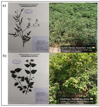
FIG. 1: PLANT COLLECTION AND AUTHENTICATION DETAILS; A) PARTHENIUM HYSTEROPHORUS WAS COLLECTED FROM RUNDH MANCH (27.599177° N, 76.576813° E), VOUCHER NO. RUBL 22051 B) LANTANA CAMARA WAS COLLECTED FROM DADHIKAR (27.598918° N, 76.576349° E), VOUCHER NO. RUBL 22050
Preparation of Plant Extracts: The powdered samples were used for further extraction. For extraction 5gm of dry powder was used on each plant part and 50ml solvents used for extraction include methanol, distilled water, benzene, hexane, and acetonitrile. The mixture was subjected to probe sonication for 1 hour and kept on an Orbital shaker for 24 hours at 31°C temperature with continued shaking. After that the extracts were filtered using Whatman filter paper 1 and the resulting filtrate was dried and all extractions were prepared 1mg/ml in triplicates.
Qualitative Phytochemical Screening: Qualitative tests of the plant extracts for presence of phytochemical compounds were done by standard procedures 12, 13, 14.
Detection of Carbohydrates:
Fehling’s Test: Fehling’s test was used for the determination of reducing sugars in plant extracts. The formation of brick red precipitates indicates the presence of reducing sugar.
Detection of Proteins:
Biuret Test: Presence of proteins in the plant extracts was detected using the Biuret test. The appearance of a violet or purple color indicated the presence of proteins.
Detection of Flavonoids:
Shinoda Test: Approximately 2 mL of the plant extract was mixed with a small amount of magnesium powder, followed by the addition of a few drops of concentrated hydrochloric acid (HCl). The appearance of pink or magenta colour indicated the presence of flavonoids.
Detection of Tannins:
The Ferric Chloride Test was carried out to detect the presence of tannins in the plant extract. A few drops of the 5% ferric chloride solution were added to the plant extract. The appearance of blue or green colors indicated the presence of tannin compounds.
Detection of Phenol:
Ferric Chloride Test: 2ml of plant extract mixed with glacial acetic acid and 5% of sodium nitrate solution to the test tube. Formation of muddy brown precipitate showcases presence of phenolic compounds in samples.
Quantitative Determination of Secondary Metabolites: The quantitative analysis of secondary metabolites including phenols, flavonoids and tannins were carried out in triplicates using following standard methods.
Determination of Total Phenolic Content (TPC): The TPC of the plant extracts was determined by the Folin-Ciocalteu colorimetric method 15, 16 Folin-Ciocalteu reagent was mixed with plant extracts and left to react for 30 minutes. Subsequently, the sodium carbonate solution was added to the mixture. The absorbance was taken at 765 nm using UV- Vis spectrophotometer. A calibration curve was prepared using gallic acid (1mg/ml) as standard and TPC was expressed as mg GAE/g dried extract of the plant extracts.
Determination of Total Flavonoid Content (TFC): Aluminum chloride colorimetric method was used for the estimation of TFC 17. 1 ml plant extract of 1mg/ml was mixed with 1ml of 10% aluminium chloride solution, 1ml of 1M sodium acetate and make up to 5ml with distilled water. The mixture was then incubated for 30 min at room temperature and the absorbance was measured at 415nm. The calibration curve was obtained using rutin (1mg/ml) in a range of concentrations (20 – 100 μg/ml) in methanol following the same protocol. The TFC was computed from this curve and represented as mg RE/g dried extract.
Determination of Total Tannin Content (TTC): The Folin–Ciocalteu reagent (FCR) method, was used to determine the TTC of the plant extracts. 5 mL of distilled water, 0.5 mL of FCR, and 0.5 mL (1mg/ml) of plant extract or standard were mixed, and left to stand for 5 minutes. After adding 1 mL of a 20% sodium carbonate solution, the mixture was allowed to develop color for 30 minutes at room temperature. A UV-Vis spectrophotometer was used to detect the absorbance at 725 nm. A calibration curve has been plotted using gallic acid as standard and TTC was expressed as mg GAE/g dried extract in the plant extracts.
Determination of Antioxidant Activity:
DPPH Assay (2,2-diphenyl-1-picrylhydrazyl): The antioxidant activity of the extracts was tested using the 2,2-diphenyl-1-picrylhydrazyl (DPPH) radical scavenging assay. This followed the method outlined by Blois et. al 18, with some minor changes. DPPH solution and plant extract solution or standard, ascorbic acid of 1mg/ml were taken in 1:1 ratio of different concentrations. Mix well and incubate for 30 min in dark after incubation the absorbance was measured using UV-Vis Spectrometer at 517 nm wavelength. Methanol was used as a blank. The IC50 value was determined from the concentration response curve.
FRAP Assay (Ferric Reducing Antioxidant Power): Antioxidant activity determination was also carried out by the FRAP assay described by Benzie & Strain 19. Briefly, the FRAP working solution was freshly prepared by mixing Acetate Buffer, TPTZ solution and FeCl3 in a ratio of 10:1:1. For the assay 2900µl of FRAP working solution was added in 100µl of the 1mg/ml standard solution or plant extract and kept for incubation for 30 min, then absorbance was measured at 593nm using UV-Vis Spectrometer. A Standard calibration curve was prepared using FeSo4 at different conc. ranging from 200-1000 µg/ml. The antioxidant potential of plant extract was expressed in mg/g Fe²⁺ equivalent.
RESULTS:
Qualitative Phytochemical Screening: Qualitative phytochemical screening of the leaf and flower of L. camara and P. hysterophorus extracts revealed the presence of bioactive compounds. The tests in both the plants confirms the presence of reducing sugars, proteins, flavonoids, phenols and tannins. Whereas, the intensity in both plants varies accordingly. Table 1. The plant extracts of L. camara contain a relatively richer and more diverse composition of bioactive compounds as compared to the P. hysterophorus extracts.
TABLE 1: RESULTS OF QUALITATIVE PHYTOCHEMICAL SCREENING TESTS (+++: STRONGLY PRESENT, ++: MODERATELY PRESENT, +: SLIGHTLY PRESENT, –: ABSENT)
| Test Name | Sample name | Extracts | |||||
| Methanolic | H2O | Benzene | Hexane | ACN | |||
| Fehling's test for (carbohydrates) | Lantana camara | Leaf | +++ | + | - | - | + |
| Flower | +++ | + | + | + | ++ | ||
| Parthenium hysterophorus | Leaf | ++ | + | - | - | + | |
| Flower | ++ | + | + | - | + | ||
| Biuret test (proteins) | Lantana camara | Leaf | +++ | + | + | ++ | + |
| Flower | ++ | + | +++ | +++ | + | ||
| Parthenium hysterophorus | Leaf | +++ | ++ | + | + | +++ | |
| Flower | +++ | ++ | + | ++ | + | ||
| Ferric Chloride Test (Phenols) | Lantana camara | Leaf | ++ | - | ++ | ++ | - |
| Flower | +++ | -- | + | + | - | ||
| Parthenium hysterophorus | Leaf | ++ | ++ | - | - | - | |
| Flower | ++ | ++ | ++ | ++ | - | ||
| Shinoda Test (Flavonoids) | Lantana camara | Leaf | +++ | - | ++ | ++ | - |
| Flower | +++ | -- | + | + | - | ||
| Parthenium hysterophorus | Leaf | ++ | ++ | - | - | - | |
| Flower | ++ | ++ | ++ | ++ | - | ||
| FeCl3 Test (Tannin) | Lantana camara
|
Leaf | +++ | - | ++ | - | ++ |
| Flower | +++ | - | - | + | ++ | ||
| Parthenium hysterophorus | Leaf | + | + | ++ | - | ++ | |
| Flower | ++ | - | - | ++ | ++ | ||
Quantitative Phytochemicals Tests:
Total Phenolic Content (TPC):
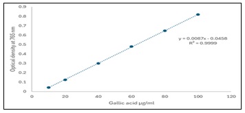
FIG. 2: CALIBRATION CURVE FOR TOTAL PHENOL CONTENT
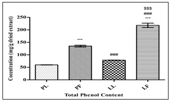
FIG. 3: QUANTIFICATION OF TOTAL PHENOLIC CONTENT IN ALL THE PLANT SAMPLES, STATISTICAL SIGNIFICANCE WAS ASSESSED BY ANOVA WITH POST HOC TUKEY’S TEST. Data were represented as mean ± SD, n = 3. ***p<0.001 vs PL, ###p<0.001 vs PF, $$$p<0.001 vs LL.
Among all the samples, LF is found to have a significantly higher TPC i.e., 193 ± 54.98 mg GAE/g dried extract in comparison with other extracts. However, a rise in the amount of TPC has been observed in PF and LF in contrast with PL. Whereas, statistically insignificant decrease has been noted in LL comparable with PF. These findings indicate the presence of phenols in all the extracts highlighting their antioxidant potential Table 2, Fig. 3.
Total Flavonoid Content (TFC): The highest amount of TFC has been marked in LL i.e, 175.62 ± 10.32 mg RE/g dried in contrast with other extracts. Whereas, in comparison with PL and PF both LL and LF significantly increased. However, TFC decreased significantly in LF when compared with LL. The presence of flavonoids in all the extracts demonstrates the potency of these plants in pharmaceuticals Table 2, Fig. 5.
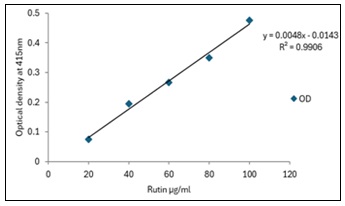
FIG. 4: CALIBRATION CURVE FOR TOTAL FLAVONOID CONTENT
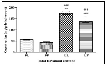
FIG. 5: QUANTIFICATION OF TOTAL FLAVONOID CONTENT IN ALL THE PLANT SAMPLES, STATISTICAL SIGNIFICANCE WAS ASSESSED BY ANOVA WITH POST HOC TUKEY’S TEST. Data were represented as mean ± SD, n = 3. ***p<0.001 vs PL, ###p<0.001 vs PF, $$$p<0.001 vs LL.
Total Tannin Content (TTC): Among the tested samples LL showcased the highest amount of tannins, with a TTC of 430.42 ± 20.70 mg GAE/g dried extract. A significant rise in TTC of LL compared to PL and PF has been noted. However, the TTC of LF has been significantly decreased in relation with LL. Whereas, PL contained lower amounts of TTC compared to other extracts. The high amount of tannins in LL suggests the presence of significant amounts of polyphenolic compounds in this plant, which could contribute significantly to its antioxidant activity.
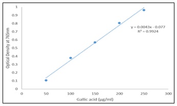
FIG. 6: CALIBRATION CURVE FOR TOTAL TANNIN CONTENT
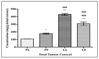
FIG. 7: QUANTIFICATION OF TOTAL TANNIN CONTENT IN ALL THE PLANT SAMPLES, STATISTICAL SIGNIFICANCE WAS ASSESSED BY ANOVA WITH POST HOC TUKEY’S TEST. Data were represented as mean ± SD, n = 3. *p<0.05 vs PL, ###p<0.001 vs PF, $$$p<0.001 vs LL.
TABLE 2: SECONDARY METABOLITES IN PLANT EXTRACTS, VALUES ARE REPRESENTED AS MEAN ± SD, INDICATING VARIATION IN SECONDARY METABOLITE CONCENTRATIONS
| Extract | TPC mg GAE/g dried extract | TFC mg RE/g dried extract | TTC mg GAE/g dried extract | |
| Parthenium hysterophorus | Leaf | 59.86 ± 1.00 | 57.22 ± 3.51 | 107.52 ± 0.94 |
| Flower | 135.03 ± 6.28 | 44.78 ± 4.40 | 177.91 ± 12.81 | |
| Lantana camara | Leaf | 78.71 ± 1.10 | 175.62 ± 10.32 | 430.42 ± 20.70 |
| Flower | 193 ± 54.98 | 137.22 ± 5.53 | 308.90 ± 36.06 | |
Antioxidant Activity:
DPPH Radical Scavenging Activity: The antioxidant potential of the plant extracts was investigated using the DPPH method of free radical scavenging activity. It was observed that the extracts showed varying degrees of radical scavenging activity Table 3. The LL showed the highest antioxidant activity with a IC₅₀ value of 30.3 µg ml⁻¹. In contrast, the PF showed moderate antioxidant activity with a IC₅₀ value of around 69.6 µg ml⁻¹. However, the PL was less inhibited at the studied concentration. The dose-response relationship in the inhibition curve also showed that the scavenging activity of the radicals increases with increasing concentrations of the plant extracts. The low IC₅₀ value of LL indicated that a smaller amount of this plant’s extract is sufficient to scavenge the free radicals, showing that this plant has stronger antioxidant potential than the plant extracts of P. hysterophorus.
TABLE 3: INHIBITION PERCENTAGE OF PLANT EXTRACTS AT DIFFERENT CONCENTRATIONS OBTAINED BY DPPH ASSAY
| S. no. | Conc. | Percentage inhibition (%) | |||
| PL | PF | LL | LF | ||
| 1 | 10 | 11.54 | 13.96 | 17.81 | 12.11 |
| 2 | 20 | 18.66 | 28.49 | 37.46 | 19.09 |
| 3 | 40 | 23.79 | 61.11 | 77.92 | 30.91 |
| 4 | 60 | 35.46 | 66.38 | 28.77 | 45.16 |
| 5 | 80 | 48.09 | 93.16 | 33.05 | 56.98 |
| 6 | 100 | 53.69 | 93.3 | 44.44 | 62.82 |
Ferric Reducing Antioxidant Power (FRAP): The ferric reducing antioxidant power assay was used to measure the reducing ability of the plant extracts. Among all the extracts, LL extract showed the highest ferric reducing antioxidant power, i.e., 2001.33 ± 41.46 mg/g dried extract Fe²⁺ eq. All the extract showcased a significant rise in comparison with PL. However, an increase in FRAP activity of LL in relation with PF and a decrease in LF has been noted in contrast with both the PF and LL. The results obtained from the FRAP activity highlights antioxidant potential among the extracts.
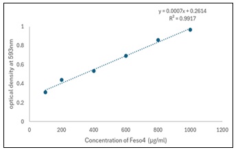
FIG. 8: CALIBRATION CURVE FOR FESO4 CONCENTRATION
TABLE 4: FERRIC REDUCING ANTIOXIDANT POWER OF PLANT EXTRACTS HERE - FE²⁺ EQ. = FERROUS EQUIVALENT, VALUES REPRESENT AS MEAN ± SD, INDICATING VARIATION IN ANTIOXIDANT POTENTIAL AMONG DIFFERENT PLANT PARTS
| Extract | FRAP (mg/g dried extract Fe²⁺ eq.) | |
| Parthenium hysterophorus | Leaf | 508 ± 18.74 |
| Flower | 1360.38 ± 7.04 | |
| Lantana camara | Leaf | 2001.33 ± 41.46 |
| Flower | 760.86 ± 40 | |
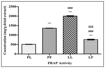
FIG. 9: COMPARISON OF FRAP ACTIVITY BETWEEN ALL PLANT SAMPLES, STATISTICAL SIGNIFICANCE WAS ANALYZED BY ANOVA WITH POST HOC TUKEY’S TEST. Data were represented as mean ± SD, n = 3. ***p<0.001 vs PL, ###p<0.001 vs PF, $$$p<0.001 vs LL
DISCUSSION: The current study explores the phytochemical composition and antioxidant potential of two invasive plant species which are L. camara and P. hysterophorus, found in most parts of India. It has been observed that both invasive plant species show occurrence of several primary and secondary plant metabolites. Presence of these metabolites shows that invasive plant species posseses phytochemical richness. Their presence reflects the physiological activity and metabolic integrity of the plant tissues. Studies on invasive plant species indicate that these plants produce a number of secondary metabolites primarily related to ecological competition 16.
The stronger qualitative reactions observed in L. camara, particularly for reducing sugars, proteins, phenol, tannins, and flavonoids, suggest that this species may possess a greater diversity of metabolically active compounds. Such phytochemical richness may be associated with its well-known invasive success, ecological competitiveness and medicinal significance.
Flavonoids are capable of directly scavenging free oxygen radicals and chelating metal ions, which are involved in redox reactions due to the presence of hydroxyl groups 6, 7. Flavonoids are also known to stabilize free radicals due to resonance structures, as well as inhibit oxidative chain reactions in biological systems 5, 25. The flavonoid content found in the leaves of L. camara, which was at a high level, might have contributed significantly to the excellent antioxidant activity of the leaves, as revealed in both the DPPH and FRAP assays. Reports revealed that plant extracts rich in flavonoids exhibit excellent radical scavenging activity as well as reduction potential in antioxidant assays 8, 21.
Interestingly, while L. camara flower extract possessed the maximum amount of phenolic content, its antioxidant activity was lower than that recorded for the leaf extract. This indicates that antioxidant activity is not only related to the total amount of phenolic content in a particular plant extract but is also related to its structural characteristics 5, 22. Several studies carried out on medicinal plants have shown the relationship between the total phenolic content and the antioxidant capacity of the extracts 8, 9, 22.
The number and positions of hydroxyl groups in phenolic molecules can greatly affect their antioxidant activity 23, 24.
The tannins have been used as an active component in medicine and beverages because of their antioxidant properties 25, 26. They function by donating hydrogen and chelating metal ions such as Fe2+, and interfering with the fenton process 27. In investigated samples of leaf extract of L. camara shows the highest amount of tannins 430.42 ± 20.70 mg GAE/g dried extract. The high amount of tannins in L. camera leaf suggests the presence of significant amounts of polyphenolic compounds in this plant, which could contribute significantly to its antioxidant activity. These findings are consistent with previous reports indicating that L. camara leaves are rich sources of phenolic compounds and possess strong antioxidant activity 28.
The antioxidant potential of the investigated plant extracts is highly correlated with the phenolic, flavonoid, and tannin contents present in the extracts. All these are highly effective in scavenging free radicals and protecting the human body from oxidative stress 4, 5, 8, 24. In several studies the high DPPH radical scavenging capacity (60.24%) was noted in aqueous methanol extract (60.24%), of L. camara flower, while the lowest in absolute methanol extract (31.32%). The variation in DPPH° scavenging ability of L. camara flower extracts in relation to different extraction solvents has been seen 29. Furthermore, the evaluation of antioxidant potential of methanolic extract of L. camara leaf reflects its good hydroxyl scavenging activity with IC50 between 0.2 mg/ml to 0.8 mg/ml 30. Whereas, the total phenolics content of leaf extract was found in considerable amounts (245.5±3.54 mg gallic acid/g) which is attributed to the antioxidant activity of L. camara’s leaf extract 31. The current analysis showcases that among all the extracts, the strongest antioxidant potential was recorded in the L. camara leaf extract, indicated by high levels of flavonoids and tannins, high FRAP value, and low IC50 value in the DPPH assay.
Thus, the high FRAP value in the L. camara leaf extract may indicate the presence of a high amount of antioxidants in this plant, which can donate an electron in a chemical reaction. Similar results have been documented in the previously tested extracts of acetone, displayed the strongest ferric reducing activity with a value of 429.55 ± 12.73 µmol trolox equivalents/g, followed by chloroform (238.61 ± 9.64 µmol TE/g) and petroleum ether (105.47 ± 5.84 µmol TE/g) 32.
In this investigation, the antioxidant activity in P. hysterophorus plant extracts was found to be lower than in L. camara leaf extract. Although this plant contains a number of bioactive secondary metabolites, these are more related to its allelopathic or antimicrobial activity 33. The results obtained in the finding revealed a positive relationship between phenolic concentration and antioxidant capacity, especially in the leaf extracts of L. camara.
CONCLUSION: The present study has shown that invasive plant species from the Aravalli region have different types of phytochemical constituents with antioxidant activity. Among the studied plant species, the methanolic extract of the leaves of L. camara showed the highest antioxidant activity with the highest content of flavonoids and tannins, as well as the highest ferric reducing power and the least IC₅₀ value. However, the methanolic extracts of the plant species P. hysterophorus showed the least antioxidant activity. This study has shown the potential importance of invasive plant species as a source of antioxidant compounds. In particular, the leaves of the plant species L. camara have the potential to be used as candidates for the study of the pharmacological importance of different types of phytoconstituents.
ACKNOWLEDGEMENT: The authors would like to thank the laboratory facilities used for the study.
Funding Source: We are thankful to the National Scheduled Castes Finance and Development Corporation (NSFDC) for providing fellowships to the authors.
CONFLICT OF INTEREST: The authors have declared no conflict of interest.
REFERENCES:
- Patel S: Harmful and beneficial aspects of Parthenium hysterophorus: An update. 3 Biotech 2011; 1(1): 1-9. doi:10.1007/s13205-011-0007-7.
- Begum S, Ayub A, Zehra S and Siddiqui BS: Chemical constituents of the aerial parts of Lantana camara. Chem Nat Compd 2013; 49(2): 313-315. doi:10.1007/s10600-013-0674-1.
- Adkins S and Shabbir A: Biology, ecology and management of the invasive Parthenium weed (Parthenium hysterophorus). Pest Manag Sci 2014; 70(7): 1023-1029. doi:10.1002/ps.3708.
- Zhang Y, Cai P, Cheng G and Zhang Y: A brief review of phenolic compounds identified from plants: Their extraction, analysis and biological activity. Nat Prod Commun 2022; 17(1): 1934578X211069721. doi:10.1177/1934578X211069721.
- Shi L, Zhao W, Yang Z, Subbiah V and Suleria HAR: Extraction and characterization of phenolic compounds and their potential antioxidant activities. Environ Sci Pollut Res 2022; 29: 81112-81129. doi:10.1007/s11356-022-23337-6.
- Twaij BM and Hasan MN: Bioactive secondary metabolites from plant sources: Types, synthesis and their therapeutic uses. Int J Plant Biol 2022; 13(1): 4-14. doi:10.3390/ijpb13010003.
- Mańkowska D and Dems-Rudnicka K: Antioxidant activity and phenolic compounds in medicinal plants: A comparison of organic and conventional Mentha piperita, Melissa officinalis, Salvia officinalis and Urtica dioica. Molecules 2025; 30(24): 4812. doi:10.3390/molecules30244812.
- Sytar O, Hemmerich I, Zivcak M, Rauh C and Brestic M: Comparative analysis of bioactive phenolic compounds composition from 26 medicinal plants. Saudi J Biol Sci 2018; 25(4): 631-641. doi:10.1016/j.sjbs.2016.01.036.
- Baliyan S, Mukherjee R, Priyadarshini A, Vibhuti A, Gupta A, Pandey RP and Chang CM: Determination of antioxidants by DPPH radical scavenging activity and quantitative phytochemical analysis of Ficus religiosa. Molecules 2022; 27(4): 1326. doi:10.3390/molecules27041326.
- Silva FV, Veiga F, Cardoso C, Dias F, Cerqueira F, Medeiros R and Paiva-Santos AC: A rapid and simplified DPPH assay for analysis of antioxidant interactions in binary combinations. Microchem J 2024; 202: 110801. doi:10.1016/j.microc.2024.110801.
- Blois MS: Antioxidant determinations by the use of a stable free radical. Nature 1958; 181(4617): 1199-1200. doi:10.1038/1811199a0.
- Harborne JB: Phytochemical Methods: A Guide to Modern Techniques of Plant Analysis. 3rd ed. Chapman & Hall, London, 1998.
- Evans WC: Trease and Evans Pharmacognosy. 16th ed. London: Elsevier 2009.
- Singleton VL and Rossi JA: Colorimetry of total phenolics with phosphomolybdic-phosphotungstic acid reagents. Am J Enol Vitic 1965; 16(3): 144-158.doi: 10.5344/ajev.1965.16.3.14
- Singleton VL, Orthofer R and Lamuela-Raventós RM: Analysis of total phenols and other oxidation substrates and antioxidants by means of Folin-Ciocalteu reagent. Methods Enzymol 1999; 299: 152-178. doi:10.1016/S0076-6879(99)99017-1.
- Woisky RG and Salatino A: Analysis of propolis: Some parameters and procedures for chemical quality control. J Apic Res 1998; 37(2): 99-105. doi:10.1080/00218839.1998.11100961
- BLOIS M: Antioxidant determinations by the use of a stable free radical. Nature 1958; 181: 1199–1200. https://doi.org/10.1038/1811199a0
- Benzie IFF and Strain JJ: The ferric reducing ability of plasma (FRAP) as a measure of antioxidant power: The FRAP assay. Anal Biochem 1996; 239(1): 70-76. doi:10.1006/abio.1996.0292.
- Ahmed ZSO, Khan E, Elias N, Elshebiny A and Dou QP: Updated review on natural polyphenols: Molecular mechanisms, biological effects, and clinical applications for cancer management. Biomolecules 2025; 15(5): 629. doi:10.3390/biom15050629.
- Susanti I, Pratiwi R, Rosandi Y and Hasanah AN: Separation methods of phenolic compounds from plant extract as antioxidant agents candidate. Plants 2024; 13(7): 965. doi:10.3390/plants13070965.
- Wilczyńska A and Żak N: Polyphenols as the main compounds influencing the antioxidant effect of honey A review. Int J Mol Sci 2024; 25(19): 10606. doi:10.3390/ijms251910606.
- Rice-Evans C, Miller N and Paganga G: Structure-antioxidant activity relationships of flavonoids and phenolic acids. Free Radic Biol Med 1996; 20(7): 933-956. doi:10.1016/0891-5849(95)02227-9.
- Sun W & Shahrajabian MH: Therapeutic potential of phenolic compounds in medicinal plants natural health products for human health. Molecules 2023; 28(4): 1845.doi.org/10.3390/molecules28041845
- Amarowicz R & Troszyńska A: Antioxidant activity of extract of pea and its fractions of low molecular phenolics and tannins. Polish Journal of Food and Nutrition Sciences 2003; 53(1): 10-15.
- Castro-Mun˜oz R, Boczkaj G and Cabezas R: A perspective on missing aspects in ongoing purification research towards Melissa officinalis. Foods 2023a; 12(9):1916. doi.org/10.3390/foods12091916
- Karamać M: Chelation of Cu (II), Zn (II), and Fe (II) by tannin constituents of selected edible nuts. International Journal of Molecular Sciences 2009; 10(12): 5485-5497. doi.org/10.3390/ijms10125485
- Sharma OP: Evaluation of antioxidant activity and total phenol in different varieties of Lantana camara BMC Research Notes 2014; 7: 560. https://doi.org/10.1186/1756-0500-7-560.
- Manzoor M, Anwar F, Sultana B & Mushtaq M: Variation in antioxidant and antimicrobial activities in Lantana camara flowers in relation to extraction methods. Acta Scientiarum Polonorum Technologia Alimentaria 2013; 12(3): 283-294.
- Naz R and Bano A: Phytochemical screening anti-oxidants and anti microbial potential of Lantana camara in different solvents. Asian Pac J Trop Dis 2013; 3: 480-6.
- Mahdi-Pour B, Jothy SL, Latha LY, Chen Y & Sasidharan S: Antioxidant activity of methanol extracts of different parts of Lantana camara. Asian Pacific Journal of Tropical Biomedicine 2012; 2(12): 960-965.doi: 10.1016/S2221-1691(13)60007-6.
- SG, AG: Lantana camara L.: a natural treasure trove of antioxidants and anti-inflammatory agents. The Trout Journal of Atatürk University 2025; 3(1): 1-9.
- Akbari B, Baghaei-Yazdi N, Bahmaie M and Mahdavi Abhari F: The role of plant-derived natural antioxidants in reduction of oxidative stress. Biofactors 2022; 48(3): 611-633. doi:10.1002/biof.183.
How to cite this article:
Jatav S, Singh V and Vijayvergia R: Phytochemical composition and antioxidant potential of Lantana camara L. and Parthenium hysterophorus L. from the Aravalli Hills of Rajasthan. Int J Pharm Sci & Res 2026; 17(10): 3002-11. doi: 10.13040/IJPSR.0975-8232.17(10).3002-11.
All © 2026 are reserved by International Journal of Pharmaceutical Sciences and Research. This Journal licensed under a Creative Commons Attribution-NonCommercial-ShareAlike 3.0 Unported License.
Article Information
14
3002-3011
1506 KB
7
English
IJPSR
Sanju Jatav, Vaishnavi Singh and Rekha Vijayvergia *
Department of Botany, University of Rajasthan, Jaipur, Rajasthan, India.
rekhavijay1367@gmail.com
25 May 2026
15 July 2026
25 July 2026
10.13040/IJPSR.0975-8232.17(10).3002-11
01 October 2026
 Download