




PHYTOCHEMICAL SCREENING, ANTIBACTERIAL AND ANTI-INFLAMMATORY ACTIVITIES, AND ACUTE ORAL TOXICITY OF ALBIZIA ANTUNESIANA HARMS: A ZIMBABWEAN MEDICINAL PLANT TRADITIONALLY USED AGAINST INFECTIONS AND INFLAMMATION
HTML Full TextPHYTOCHEMICAL SCREENING, ANTIBACTERIAL AND ANTI-INFLAMMATORY ACTIVITIES, AND ACUTE ORAL TOXICITY OF ALBIZIA ANTUNESIANA HARMS: A ZIMBABWEAN MEDICINAL PLANT TRADITIONALLY USED AGAINST INFECTIONS AND INFLAMMATION
Nicholas Muparadzi *, Joey Chifamba and Shingirayi Zengeni
Department of Pharmacy and Pharmaceutical Sciences, Faculty of Medicine and Health Sciences, University of Zimbabwe, P. O. Box MP167, Mt Pleasant, Harare, Zimbabwe.
ABSTRACT: For many generations in Zimbabwe, Albizia antunesiana tree has been used in traditional healing. It has been widely used to treat infections, inflammation and other chronic illnesses such as diabetes. This study sought to provide scientific evidence to this ancient wisdom by testing the bark and leaves against modern biological markers. Phytochemical analysis revealed the presence of bioactive compounds like alkaloids, flavonoids, and tannins, which are natural compounds that the tree uses to protect itself and that we can use to improve human health. In the laboratory, the lyophilized extracts successfully hindered the growth of Staphylococcus aureus and Escherichia coli, with a Minimum Inhibitory Concentration of 40 and 20 mg/mL respectively. It also exhibited strong anti-inflammatory activity. By testing its ability to prevent protein denaturation, it was found that the extract reached its peak protective effectiveness at a concentration of 500 µg/mL, performing comparably to diclofenac sodium. Most importantly safety trials in animal models confirmed that at doses of 5000 mg/kg, the extract remained practically non-toxic. These findings validate the plant as a potential candidate for developing new therapies to manage both infection and pain.
Keywords: Albizia antunesiana, Antibacterial, Anti-inflammatory, Muriranyenze, Umnonjwana, Pharmacognosy
INTRODUCTION:
Albizia antunesiana: Albizia antunesiana is a tree that is indigenous to Zimbabwe, it is commonly known as the purple-leaved albizia. The Shona speaking Zimbabweans call it “Muriranyenze”, whilst the Ndebele speaking call it “Umnonjwana” 1.
Albizia belongs to the Fabaceae or Leguminosae family of agriculturally important flowering plants. It is commonly known as the legume, bean or pea family, and it has trees, annual herbaceous plants and shrubs 2. The subfamilies of Fabaceae are Mimosoideae, Papilionoideae and Caesalpinioideae.
Mimosoideae has flowers which have many spikes or many flowered heads, and the flowers have numerous showy stamens, small inconspicuous corollas and they have a radial symmetry. This Mimosoideae subfamily includes Albizia (silk tree), Acacia (wattle), Calliandra (powder puff), Prosopis (mesquite) and Samanea (monkey pod). The Albizia genus has mostly trees and shrubs in the tropical and subtropical regions of Africa and Asia. In 2013, it had approximately 150 species 3. The Table 1 below shows the scientific classification of Albizia.
TABLE 1: SCIENTIFIC CLASSIFICATION OF ALBIZIA
| Kingdom | Plantae |
| Phylum | Tracheophyta |
| Class | Magnoliopsida |
| (unranked) | Angiosperms |
| (unranked) | Eudicots |
| (unranked) | Rosids |
| Order | Fabales |
| Family | Fabaceae |
| Subfamily | Mimosoideae |
| Tribe | Ingae |
| Genus | Albizia |
Albizia antunesiana can grow up to a height of 18m and in Zimbabwe it is popularly known to be inhabited by cicada insects from time to time. The leaf colours of Albizia antunesiana vary from green to purple, with a pale undersurface, usually occurring 1 to 3 pairs of pinnae each having 3 to 8 large ovate leaflets (approximately up to 25mm long). The plants fruit ripen from autumn to spring, and they are light brown flat pods as shown on Fig. 1 (they can grow up to 230mm long) 4. The tree bark of Albizia antunesiana varies, it can be smooth or rough with brown to grey colours, and it is mostly the young trees that sometimes have purple leaves. Albizia antunesiana bears half spherical, showy, large fluffy inflorescences with creamy white 1 to 2 bisexual central flowers, and it prefers sunny positions usually in loamy soil. Albizia antunesiana is a medium to large deciduous tree and in Zimbabwe, it is more densely distributed in the eastern highlands than all the other regions 4. The International Union for Conservation of Nature (IUCN) is a global membership union made up of governments, civil society organisations, scientists, and experts that works on nature conservation and the sustainable use of natural resources. In their IUCN Red List of Threatened Species, which assesses the global conservation status of species (i.e. how at risk they are of extinction) using standardized criteria, Albizia antunesiana is listed as a Least Concern (LC) 5.
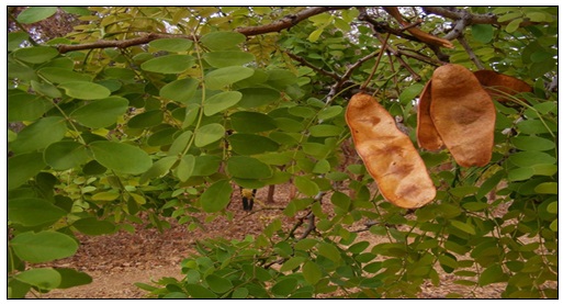
FIG. 1: THE LEAVES AND FRUITS OF ALBIZIA ANTUNESIANA 6
Medicinal uses of Albizia antunesiana: The exact medicinal uses and pharmacological activity of Albizia antunesiana are not scientifically known, however several studies have documented different uses. Several metabolic and non-metabolic disorders such as gonorrhea, ulcers, diabetes, sore eyes, cardiac problems, tuberculosis, sore throats, cuts and tonsilitis, are being cured by traditional healers using the roots and leaves of Albizia antunesiana 7. Chipiti et al.7 concluded that, the findings from their study supports, the use of particularly the roots of Albizia antunesiana that is being used as medicine by herbalists and traditional healers in the Mrewa district. Another scientific study by Chipiti et al.1 focusing on Albizia antunesiana use in diabetes concluded in their findings that, the ethanolic root extract of Albizia antunesiana is non-toxic in nature, and it possess effective α‐amylase and α‐glucosidase inhibitory effects hence this warrants further in vivo study using an animal model of diabetes. Chifamba et al.4 concluded in their study that, Albizia antunesiana possesses considerable anti-inflammatory and antibacterial activity, and it has a favourable safety profile.
In a study by Muvengwi et al.8, a total of 16 medicinal plant taxa were employed in treatment of Sexually Transmitted Infections (STIs) as shown in Fig. 2. Albizia antunesiana was one of the medicinal plants that was reported to be used in treating various conditions in their study 8. Fig. 2 below shows the various medicinal plant uses of Albiza genus (including Albizia antunesiana), that were recorded in Harare Zimbabwe. Albizia antunesiana had reported uses in the management of malaria and respiratory complaints. Despite all this extensive use of Albizia antunesiana, there are still limited reports on the pharmacological activity and scientific rationale for using it in traditional medicine 7. This warrants scientific research on Albizia antunesiana, because documentation of indigenous knowledge about medicinal plants used in various local communities could lead to the discovery of essential effective pharmaceutical drugs through ethnopharmacological screening 9.
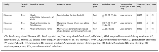
FIG. 2: LIST OF MEDICINAL PLANTS, THEIR USE CATEGORIES (TCD), USE REPORTS (TURS), AND CONSERVATION STATUS. REPRODUCED FROM 8
Traditional healers in Zimbabwe report using A. antunesiana roots and leaves to treat gonorrhea, ulcers, diabetes, sore eyes, cardiac problems, tuberculosis, sore throats, cuts, and tonsillitis 3. Chipiti et al.1 reported that ethanolic root extracts possess α‑amylase and α‑glucosidase inhibitory effects with non‑toxic properties. Muvengwi et al. documented the use of A. antunesiana in managing malaria and respiratory complaints. Despite these ethnomedicinal applications, limited pharmacological data exist for the leaf and bark materials, which are more sustainably harvestable than roots.
Plant secondary metabolites, including alkaloids, flavonoids, tannins, and terpenoids, serve as defense mechanisms against pathogens and have demonstrated antimicrobial and anti‑inflammatory properties in numerous species 6, 7. Phenolic compounds, particularly flavonoids, inhibit bacterial growth through membrane disruption and enzyme inhibition 8, while also reducing inflammation by blocking pro‑inflammatory cytokines and the NF‑κB pathway 9. Although traditional uses predominantly cite root preparations 3, leaves and bark were selected for this study based on three considerations: (1) sustainable harvesting without plant fatality; (2) preliminary data suggesting comparable phytochemical profiles across aerial and subterranean tissues in related Albizia species 10; and (3) local healer reports of occasional leaf and bark decoction use for wound care and topical inflammation (personal communication, Murewa district, 2024).
This study aimed to: (1) perform phytochemical screening of hydroethanolic leaf and bark extracts of A. antunesiana; (2) evaluate in vitro antibacterial activity against Staphylococcus aureus and Escherichia coli; (3) assess in-vitro anti‑inflammatory activity via protein denaturation inhibition; and (4) determine acute oral toxicity in a rodent model following OECD guidelines.
MATERIALS AND METHODS:
Materials, Equipment and Facilities: All chemicals, associated reagents, equipment and facilities for the in-vivo laboratory animal toxicity investigations and the bioactivity assays were obtained from the University of Zimbabwe, Faculty of Medicine and Health Sciences laboratories.
Albizia antunesiana Plant Material Collection and Preparation: Plant material was collected from the Lake Chivero area of Zimbabwe in the Mashonaland West province, which is about 37 kilometers southwest of the City of Harare (See Fig. 3 below).
The plant material was authenticated by the National Herbarium and Botanical Garden in Harare, Zimbabwe.
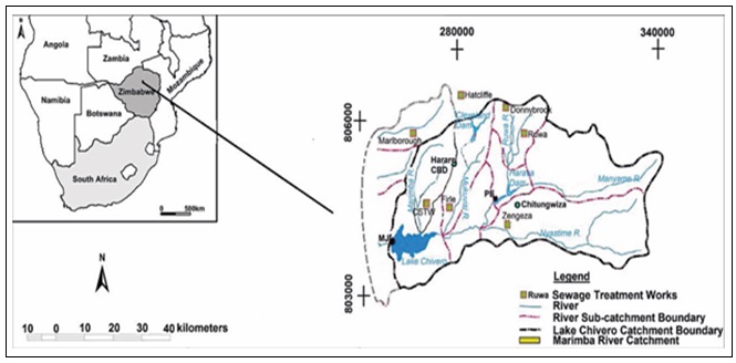
FIG. 3: LOCATION OF LAKE CHIVERO IN ZIMBABWE 47
To remove contaminants and debris, the leaves and barks were thoroughly washed using distilled water. Leaves and bark were collected separately, processed in parallel, and combined in equal proportions (1:1 w/w) for all extractions and assays described herein. Individual tissue analysis will be reported separately. After that, they were then shade dried at room temperature to constant weight and moisture of about 4% in a period of three weeks and then crash grinded to fine powder using mortar and pestle. The Phyto-extraction was done by adding 300g plant powder into 1000mL of 70% (v/v) hydro ethanolic mixture in a 2-litre sterile amber bottle and macerated for 3 days, with about 3 minutes of physical shaking twice a day. To obtain a filtrate from the solution, a muslin cloth was used which was further clarified by filtration using Whatman filter paper number 1. The filtrate was then evaporated 3 times under vacuum and low pressure using Rotavapor R-300, followed by freeze drying (lyophilization) under 140Pa pressure and -50 °C. The lyophilized extract was stored in an air-tight sample bottle.
Phytochemical Screening of Albizia antunesiana: In a 200mL flask, 5g of freeze-dried hydro-ethanolic extracts from Albizia antunesiana were mixed with 50mL of distilled water. This mixture was tested using various methods to check for the presence or absence of plant compounds that might have medicinal benefits. Several specific tests were performed on the liquid extract to identify these compounds. All procedures were carried out as per guidelines by Chifamba et al.48 with slight modifications. For comparative purposes, a separate distilled‑water extract was prepared by macerating 300 g of the combined leaf‑bark powder in 1000 mL of distilled water for 72 hours at room temperature, followed by filtration, evaporation, and lyophilization using identical parameters as the hydroethanolic extract. (Results for the distilled‑water extract will be reported separately.)
Detection for Alkaloids by the Iodine Test: The Iodine test was used to determine the presence of alkaloids. In this test, a few drops of iodine solution were slowly added along the sides of the test tube to 3mL of the lyophilized extract solution. The presence of alkaloids was then identified by the appearance of a blue colour, which disappears on boiling and reappears on cooling 49.
Detection of Tannins: The Braymer’s test was used to detect the presence of tannins. To 1mL lyophilized extract solution, 2mL of distilled water was added, followed by 4 drops of a 10% ferric chloride solution. The presence of tannins is confirmed by a blue-green colour 50.
Detection of Flavonoids by the Ammonia Test: Flavonoids were detected by means of the Ammonia test where 5mL dilute ammonia solution was added to 5mL of the lyophilized extract solution, followed by a few drops of concentrated sulphuric acid. Emergence of a yellow colour indicates the presence of flavonoids 51.
Detection of Glycosides: To 2mL of the extract, 3mL of chloroform was added followed by 3 drops of ammonia in a test-tube. Formation of a pink color indicates presence of the glycosides 52.
Detection of Phenolic Compounds: To 1mL of the extract, 2mL of distilled water was added followed by a few drops of ferric chloride. Formation of a blue-green color indicates presence of phenols 53.
Detection of Saponins by the Simplified Foam Test: The simplified foam test was used to determine the presence of saponins. In this assay 2mL of the extract was added to 20mL distilled water. The mixture was shaken in a graduated measuring cylinder for 15 minutes. The presence of saponins was confirmed by the formation of form with a head height of at least 1cm 54.
Detection of Phytosterols: The Salkowski test was used to determine the presence of phytosterols. To 2 mL of extract, 2 mL of chloroform and 2 mL of concentrated sulfuric acid were added along the test tube wall. The presence of phytosterols was indicated by a red coloration in the chloroform layer 11.
Detection of Steroids: The Liebermann‑Burchard test was used. To 2 mL of extract, 2 mL of acetic anhydride followed by 2 drops of concentrated sulfuric acid were added. A blue‑green color indicated the presence of steroids 12.
Detection of Coumarins: To 2 mL of extract, 3 mL of 10% sodium hydroxide was added. Formation of a yellow color indicated coumarins 13.
Detection of Terpenoids: The Salkowski test was used to detect the presence of terpenoids. To 2 mL of extract, 2 mL of chloroform and 2 mL of concentrated sulfuric acid were carefully added. A reddish‑brown interface indicated terpenoids 14.
Acute Oral Toxicity Evaluation of Albizia antunesiana:
Study Design and Ethical Framework: To determine the potential for immediate adverse effects following a single administration, the acute oral toxicity of lyophilized Albizia antunesiana extract was evaluated using the up‑and‑down procedure (OECD Technical Guideline 425) as described by Chifamba et al. 2025 55. Eight male and female Sprague‑Dawley rats (nulliparous, non‑pregnant, 8‑12 weeks old, weighing 180‑220 g) were used. Animals were randomly assigned using computer‑generated numbers. The study was approved by the Joint Parirenyatwa Research Ethics Committee (JREC/171/2024).
Acclimatization and Environmental Control: A cohort of eight Sprague-Dawley rats was monitored throughout the study. Before dosing began, the animals were given 10 days to habituate with the testing environment. They were housed under regulated conditions (specifically an average temperature of 25°C and 45% humidity) with access to standard rodent pellets and clean water. All welfare and care protocols were overseen by a qualified veterinary officer.
Dosing Protocol and Progression: Following a 20‑hour fast (water ad libitum), the extract was administered via oral gavage at 48‑hour intervals. A between‑subjects design was used: each animal received only one dose level.
The starting dose was 250 mg/kg. If the animal survived, a second animal received 500 mg/kg, then a third received 1000 mg/kg, then 2500 mg/kg, and finally 5000 mg/kg. A total of 5 treated animals (one per dose level) and 3 control animals (distilled water only) completed the study. Humane endpoints included ≥20% weight loss in 24 hours, hunched posture, piloerection, inability to access food/water, or moribund status, which would trigger immediate euthanasia via intraperitoneal pentobarbital (150 mg/kg). No animals met these endpoints.
Observation and Health Monitoring: Post-dosing surveillance lasted 14 days, with a veterinary specialist checking for morbidity and mortality twice daily. The first day involved intensive monitoring every hour for the first 12 hours, to observe any immediate clinical signs or behavioral changes. For the remaining part of the study, physical observations and weight measurements were recorded daily to track systemic health and to identify potential delayed toxic effects.
Antibacterial activities of Albizia antunesiana: The antibacterial potential of Albizia antunesiana extracts was evaluated through a qualitative screening process, utilizing the agar well diffusion method to measure zones of inhibition against Escherichia coli (MTCC443) and Staphylococcus aureus (MTCC 96). This approach allowed for a clear, direct observation of the inhibitory effects exerted by the extracts, providing concrete evidence of their antimicrobial properties.
Preparation of Nutrient Agar Culture Media: The preparation process began with weighing 11.2g of nutrient agar powder, which was then dissolved in 400mL of distilled water within a borosilicate glass beaker. Continuous stirring was maintained using a magnetic stirrer while heat was applied via a Bunsen burner, ensuring the powder reached full dissolution and the solution became clear. This mixture was transferred into a glass media bottle and sterilized in an autoclave at 121°C for 15 minutes. Following the sterilization cycle, the medium was moved to a workspace previously disinfected with 70% ethanol to cool. Once the temperature dropped to a range safe for handling without causing excessive condensation, 20mL portions of the sterile agar were aseptically poured into 20 sterile Petri dishes. These plates were then left to solidify on a level surface, providing a consistent medium for the subsequent bacterial inoculation.
Bacterial Inoculation: Bacterial strains of Staphylococcus aureus and Escherichia coli, which were previously cultured and supplied by the Department of Pharmacy and Pharmaceutical Sciences at the University of Zimbabwe, were inoculated onto the solidified nutrient agar. A sterilized inoculation loop was utilized to transfer the cultures using the standard streak plate technique. Each bacterial species was assigned four separate Petri dishes to ensure sufficient experimental replication and statistical reliability. These plates were carefully labeled on the base to avoid obscuring the observation of microbial growth and were subsequently placed in an incubator at 37°C for a 24-hour period.
Preparation of the Test Extract and Controls: Lyophilized extracts of Albizia antunesiana were carefully weighed and dissolved in distilled water to achieve the required concentrations for testing. For the positive control, a doxycycline capsule was dissolved in 2mL of distilled water, to make up a 100 mg concentration. All solutions were prepared immediately before the application phase to maintain the stability and chemical integrity of the active compounds.
Application of Test Solutions: Wells were bored into the freshly inoculated agar plates immediately after bacterial streaking (within 30 minutes), and test solutions were added without delay. Plates were then incubated at 37°C for 24 hours. Specific volumes of the prepared Albizia antunesiana extracts and doxycycline were introduced into these wells to reach final concentrations of 500, 250, and 150 µg/mL. This procedure was carried out for both E. coli and S. aureus plates to allow for a direct comparison of efficacy across different bacterial strains. Negative control wells were filled with distilled water to confirm that the solvent itself did not contribute to any observed inhibitory activity.
Incubation and Assessment: Once the test and control solutions were introduced, the plates were returned to the incubator and maintained at a constant temperature of 37°C for a further 24 hours. After this period, each plate was carefully examined for zones of inhibition around the wells, as these served as the primary metric for antibacterial efficacy. The diameters of these clear zones were measured precisely, and the resulting data was used to calculate the mean and standard deviation for every treatment group. These values were then compared against both the positive and negative controls to determine the relative potency of the Albizia antunesiana extracts.
Equation 1: % inhibition equation.
% inhibition = (Corrected ZOI of extract at concentration) / (Corrected ZOI of doxycycline at concentration) × 100
Determination of Minimum Inhibitory Concentration (MIC): The broth microdilution method was utilized to determine the Minimum Inhibitory Concentration (MIC) of the Albizia antunesiana lyophilized extract 3. Bacterial suspensions were standardized to 0.5 McFarland standard (≈1.5 × 10⁸ CFU/mL). Two‑fold serial dilutions of the extract were prepared in sterile nutrient broth in 96‑well U‑bottom plates, with 100 µL of bacterial suspension added to 100 µL of extract dilution. Final concentration ranged from 160,000 µg/mL down to 1.25 µg/mL. Doxycycline (160 µg/mL) served as positive control; distilled water with bacteria as negative control; extract with sterile broth as sterility control. After 24‑hour incubation at 37°C, 20 µL of 0.2% resazurin solution was added to each well. Blue‑to‑pink color change indicated bacterial growth. The MIC was recorded as the lowest concentration with no color change (blue) and no visible turbidity. Three independent experiments were performed, each with triplicate wells.
Anti-inflammatory Activity of Albizia antunesiana:
Assay Rationale and Methodology: The anti-inflammatory potential of Albizia antunesiana was evaluated by measuring its capacity to inhibit protein denaturation. This process serves as a biochemical indicator of how a substance manages structural damage during an inflammatory response. The study used a modified egg albumin assay following the methodology utilized by Chifamba et al.57, where the extract’s ability to protect proteins from heat-induced denaturation was used to quantify its effectiveness as a natural protective agent.
Preparation of Reaction Mixtures: The procedure involved combining 0.4 mL of fresh egg albumin with 1.0 mL of phosphate‑buffered saline (PBS, pH 7.4). To this mixture, 0.5 mL of lyophilized A. antunesiana extract dissolved in 0.4% DMSO was added. To this mixture, 5mL solutions of lyophilized A. antunesiana extract dissolved in 0.4% dimethyl sulfoxide (DMSO) were added. The experimental design employed a concentration gradient ranging from 100 to 500 µg/mL to determine how the intensity of the dose influenced protein protection.
Thermal Stress Protocol: Testing protein resilience required a two-stage heating process. Initially, the samples were incubated at a steady 37°C for 20 minutes. The temperature was then increased to 70°C for an additional 30 minutes to force the proteins to denature, simulating the thermal stress typically found in inflamed human tissues. After the heating cycles, the mixtures were cooled and filtered to prepare for final analysis.
Spectrophotometric Analysis and Controls: Absorbance of the resulting liquid was measured at 660 nm using a spectrophotometer, with a blank solution used as a reference. To ensure the accuracy of the findings, results were compared against negative controls containing albumin, DMSO, and phosphate-buffered saline (PBS), as well as a positive control using a standard anti-inflammatory medication, diclofenac sodium.
Quantification of Inhibition: The final capacity of the extract to guard the proteins was expressed as an Inhibition Percentage. This value was calculated by comparing the absorbance of the test samples against the control to determine the relative reduction in denaturation:
Equation 1: the percentage inhibition formula.
in𝑓𝑙𝑎𝑚𝑚𝑎𝑡𝑖𝑜𝑛𝑖𝑛ℎ𝑖𝑏𝑖𝑡𝑖𝑜𝑛𝑝𝑒𝑟𝑐𝑒𝑛𝑡𝑎𝑔𝑒𝑒𝑓𝑓𝑒𝑐𝑡 = (ABS control – ABS sample) / (ABS control) × 100
RESULTS AND DISCUSSION:
Phytochemical Analysis: The table below details the specific phytoconstituents identified in Albizia antunesiana.
TABLE 2: PHYTO CONSTITUENTS IN ALBIZIA ANTENUSIANA
| Test | Presence in hydro-ethanolic extract |
| Alkaloids | +++ |
| Phytosterols | ++ |
| Flavonoids | +++ |
| Saponins | +++ |
| Steroids | +++ |
| Coumarines | ++ |
| Phenolic compounds | +++ |
| Tannins | +++ |
| Glycosides | ++ |
| Terpenoids | +++ |
(-): Indicates the absence of the phytochemical, (+): Indicates the presence of the phytochemical, (++): Indicates moderate presence of the phytochemical, (+++): Indicates strong presence of the phytochemical
Phytochemical screening of the Albizia antunesiana extract confirmed a diverse profile of both primary and secondary metabolites. These results validate the plant’s complex chemical composition, identifying a rich repository of bioactive compounds that likely contribute to its therapeutic potential. These findings align with previous research by Balkrishna et al.58, which also identified a range of bioactive compounds in A. antunesiana such as alkaloids, terpenes, tannins, flavonoids, and coumarins. These secondary metabolites are highly relevant in the management of diseases, including bacterial infections such as gonorrhea. Our findings reveal that the availability these significant phytochemicals provide a strong scientific basis for the plant’s use in traditional medicine in the treatment of diseases such as bacterial infections like gonorrhea, and other medical conditions. Analysis of the hydro-ethanolic extract confirmed strong positive results across nearly all major phytochemical classes. The screening identified the presence of alkaloids, phytosterols, flavonoids, saponins, and steroids. Additionally, the extract showed a significant yield of coumarins, phenolic compounds, tannins, glycosides, and terpenoids, indicating a broad-spectrum profile of secondary metabolites.
This abundance of bioactive compounds suggests that the plant’s efficacy is likely derived from the synergistic interplay of multiple compound families 59. Among the detected secondary metabolites, phenolics were the most dominant group, with a notably high prevalence of flavonoids. For the plant itself, these secondary metabolites are primarily produced as a defensive response to possible biological stressors, such as microbial invasion 17. These therapeutic compounds also act as antioxidants, anti-inflammatory agents, and as anti-bacterial agents 21. Specifically, flavonoids, a key category within the phenolics group, are a class of polyphenolic compounds that include subgroups such as flavones, flavanols, and flavanones. These molecules are recognized for exhibiting potent antimicrobial activity 19.
It is important to note that the phytochemical screening employed semi quantitative colorimetric tests that indicate the presence of compound classes but do not identify specific active constituents, quantify absolute concentrations, or establish structure activity relationships. While the detection of alkaloids, flavonoids, and tannins is consistent with known antimicrobial and anti‑inflammatory mechanisms reported in the literature 8, 9, these preliminary findings require confirmation through chromatographic isolation (e.g., HPLC, GC‑MS), quantitative analysis, and target specific bioactivity guided fractionation before definitive therapeutic claims can be made.
Acute Oral Toxicity: In alignment with the OECD 425 technical guidelines, an acute toxicity assessment was conducted under the rigorous supervision of a qualified veterinary specialist to evaluate the safety profile of A. antunesiana. Our observations revealed that, oral administration of the extract at dosages reaching 5000 mg/kg of body weight resulted in no discernible clinical signs of distress, behavioral abnormalities, or mortality among the test subjects (as shown on Table 3).
TABLE 3: ACUTE ORAL TOXICITY STUDY OF A. ANTUNESIANA BEHAVIOURAL OBSERVATIONS
| Observed parameter | Dose of A. antunesiana in mg/kg body weight | |||||
| 250 | 500 | 1000 | 2500 | 5000 | Control | |
| Food intake | Normal | Normal | Normal | Normal | Normal | Normal |
| Water intake | Normal | Normal | Normal | Normal | Normal | Normal |
| Death | No deaths | No deaths | No deaths | No deaths | No deaths | No deaths |
| Breathing | Normal | Normal | Normal | Normal | Normal | Normal |
| Diarrhea | None | None | None | None | None | None |
| Urination | Normal | Normal | Normal | Normal | Normal | Normal |
| Skin color | Normal | Normal | Normal | Normal | Normal | Normal |
| Drowsiness | None | None | None | None | None | None |
Throughout the entirety of the monitoring period, the integrity of the study group remained intact, with no instances of animal withdrawal or unexpected attrition. These findings correlate closely with previous literature, such as the work of Chifamba et al.4, which investigated similar plant species. Under the conditions of this acute oral study in Sprague Dawley rats, the no-observed adverse effect level (NOAEL) was determined to be 5,000 mg/kg, as no treatment related clinical signs, behavioral abnormalities, or mortality were observed at any dose level. Extrapolation of this finding to humans or chronic dosing regimens requires additional investigation.
This absence of mortality or any clinical signs of toxicity following a single oral dose of 5 000 mg/kg, indicates that the median lethal dose (LD₅₀) of the plant extract exceeds this limit, thereby classifying it as practically non-toxic under the conditions of this study 60. The data also suggests that A. antunesiana possesses a favorable safety margin, indicating that the utilization of concentrated doses to achieve targeted pharmacological or bioactivity outcomes is unlikely to induce adverse toxicological effects. This high LD50 enables researchers to use Albiza antunesiana extract for therapeutic exploration without significant risk of acute systemic toxicity. Our biosafety and bioactivity studies therefore authenticate the use of A. antunesiana as a potential remedy in traditional medicine.
Rat Weights Observations: The assessment of body weight remains a fundamental yet highly sensitive proxy for systemic toxicity during the administration of novel substances to animal models. Because shifts in the expected growth curve frequently stem from physiological stress, nutritional deficits, or occult pathological responses, these metrics are vital for evaluating the safety profile of a compound. Within this experimental framework, the administration of lyophilized extracts resulted in no statistically relevant deviations from normal weight gain patterns (as shown on Fig. 4). The stability of the rats' physiological trajectory throughout the evaluation period indicates that the extracts did not interfere with the biological developmental processes or metabolic homeostasis at the analyzed dosages.
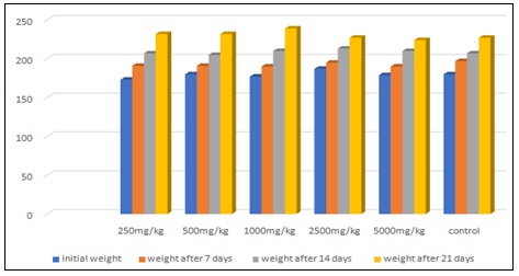
FIG. 4: OBSERVED SPRAGUE-DAWELY RATS WEIGHT VARIATION IN GRAMS DURING ACUTE ORAL TOXICITY STUDY OF A. ANTUNESIANA
Antibacterial Assay: The results of this study indicated that Albizia antunesiana lyophilized extracts possessed substantial antibacterial potential, effectively inhibiting the growth of both Staphylococcus aureus and Escherichia coli. S. aureus and E. coli were utilized as representatives of both Gram positive and Gram-negative models, to establish a baseline for the plant’s antimicrobial profile. Many traditional remedies used for sexually transmitted infections (STIs) function through broad spectrum antimicrobial activity rather than targeting a single pathogen. The ability of the extract to successfully penetrate the complex lipopolysaccharide outer membrane of E. coli suggests it contains bioactive compounds capable of overcoming the structural defenses common to many Gram-negative bacteria 61. The observed efficacy against S. aureus also confirmed that the plant harbors secondary metabolites that disrupt essential cellular processes in Gram-positive bacteria.
The antibacterial performance of A. antunesiana in this trial exceeded results reported for other species in the same genus, for example, prior studies on related Albizia species recorded moderate inhibition zones typically ranging between 10 mm and 17 mm when using crude aqueous extracts 62. The zone of inhibition of Albizia antunesiana extract in relation to its different concentrations, were recorded in this research. The zones observed reaching up to 22 mm for E. coli (as shown in Table 4) and 28 mm for S. aureus (as shown in Table 5), likely resulted from the lyophilization process. The determination of MIC values also supports the dose dependent nature of the antibacterial activity observed throughout the study 61. The ability of the extract to inhibit growth at concentrations of 40 mg/mL and 20.0 mg/mL (as shown in Table 6), suggests that the bioactive metabolites such as saponins and alkaloids, were successfully preserved during the lyophilization process. Lyophilization preserves unstable phytochemicals such as saponins and alkaloids more effectively than traditional air drying, which often leads to the thermal degradation of active ingredients 63.
TABLE 4: ANTIBACTERIAL ACTIVITY OF ALBIZIA ANTUNESIANA (WELL DIFFUSION METHOD USING E. COLI)
| Concentration
mg/ml |
Measured ZOI (mm) | Corrected ZOI (mm) | % Inhibition relative to doxycycline | |
| Positivecontrol doxycycline | 160 | 27 | 23 | - |
| 120 | 20 | 16 | ||
| 50 | 13 | 9 | ||
| Negative control distilled water | - | 0 | 0 | 0 |
| Albizia antunesiana | 160 | 22 | 18 | 81.8 |
| 120 | 15 | 11 | 73.3 | |
| 50 | 9 | 5 | 71.4 |
ZOI = Zone of Inhibition Well diameter = 4mm
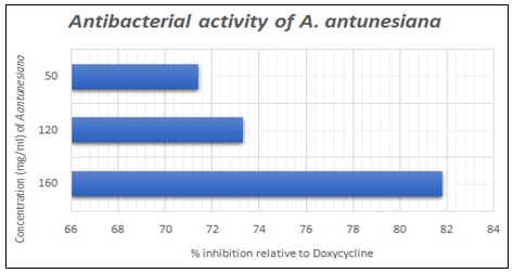
FIG. 5: GRAPHICAL PRESENTATION OF ANTIBACTERIAL ACTIVITY OF A. ANTUNESIANAAGAINST E. COLI
TABLE 5: ANTIBACTERIAL ACTIVITY OF ALBIZIA ANTUNESIANA (WELL DIFFUSION METHOD USING S. AUREUS)
| Concentration
mg/ml |
Measured ZOI (mm) | Corrected ZOI (mm) | % Inhibition relative to doxycycline | |
| Positive control doxycycline | 160 | 30 | 26 | - |
| 120 | 20 | 16 | ||
| 50 | 11 | 7 | ||
| Negative control distilled water | - | 0 | 0 | 0 |
| Albizia antunesiana | 160 | 28 | 24 | 92 |
| 120 | 18 | 14 | 87.5 | |
| 50 | 10 | 6 | 85.7 |
ZOI: Zone of Inhibition well diameter = 4mm
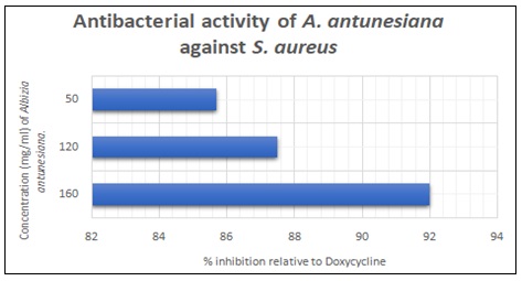
FIG. 6: GRAPHICAL PRESENTATION OF ANTIBACTERIAL ACTIVITY OF A. ANTUNESIANA AGAINSTS. AUREUS
Quantitative MIC Analysis: The broth microdilution assay provided a quantitative measurement of the antibacterial potency of Albizia antunesiana.
TABLE 6: QUANTITATIVE MIC ANALYSIS OF ALBIZIA ANTUNESIANA
| Test tube | Extract concentration (mg/ml) | Staphylococcus aureus | Escherichia coli |
| 1 | 160.0 | No visible growth | No visible growth |
| 2 | 80.0 | No visible growth | No visible growth |
| 3 | 40.0 | No visible growth (MIC) | No visible growth |
| 4 | 20.0 | Visible growth | No visible growth (MIC) |
| 5 | 10.0 | Visible growth | Visible growth |
| 6 | 5.0 | Visible growth | Visible growth |
| 7 | 2.5 | Visible growth | Visible growth |
| 8 | 1.25 | Visible growth | Visible growth |
| 9 | Positive Control | No visible growth | No visible growth |
| 10 | Broth (negative control) | Visible growth | Visible growth |
Anti-inflammatory Activity: The evaluation of the anti-inflammatory potential of Albizia antunesiana demonstrates a significant, dose-dependent protective effect. In this study, the plant extract was tested against heat-induced denaturation of egg albumin, a process used to simulate the structural damage and loss of biological function that proteins undergo during inflammatory responses.
The results indicate that as the concentration of the A. antunesiana extract increased from 100 µg/mL to 500 µg/mL, as shown in table 4, the percentage of inhibition rose from 36.67 ± 0.35% to 77.89 ± 0.53%. This progression is closely comparable to the performance of the standard anti-inflammatory drug, diclofenac sodium, which achieved a maximum inhibition of 80.23 ± 0.41% at 500 µg/mL shown in Fig. 7. This is likely due to the plant's rich profile of secondary metabolites, particularly phenolic compounds such as flavonoids, tannins, and gallotannins 4.
The extract demonstrated concentration dependent protein denaturation inhibition, achieving 77.89 ± 0.53% at 500 µg/mL compared to 80.23 ± 0.41% for diclofenac sodium. While these in-vitro findings suggest preliminary anti‑inflammatory potential, the egg albumin denaturation assay is a simple screening model that does not directly measure inflammatory mediator inhibition (e.g., COX‑2, TNF‑α, IL‑6). Confirmatory studies using carrageenan induced paw edema or cytokine specific assays are required before claiming therapeutic anti‑inflammatory efficacy.
TABLE 7: % INHIBITION OF BOTH THE STANDARD (DICLOFENAC) AND THE SAMPLE (A. ANTUNESIANA)
| Sample Number | Concentration (µg/ml) | % inhibition of standard | % inhibition of sample |
| 1 | 100 | 39.70±0.26 | 36.67±0.35 |
| 2 | 200 | 50.01±0.46 | 45.97±0.55 |
| 3 | 300 | 57.77±0.53 | 53.00±0.24 |
| 4 | 400 | 65.02±0.63 | 64.01±0.47 |
| 5 | 500 | 80.23±0.41 | 77.89±0.53 |
(*Average value of 3 replicates)
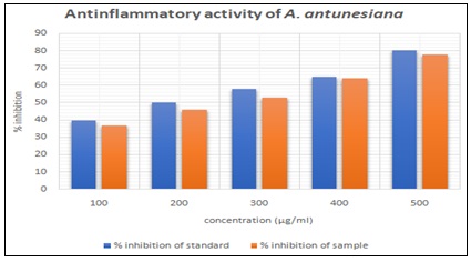
FIG. 7: PROTEIN DENATURATION ASSAY
CONCLUSION: This study of Albizia antunesiana confirms that this indigenous tree has a lot of therapeutic potential. The results of this research show that the lyophilized bark and leaf extracts possess the ability to neutralize bacterial pathogens like S. aureus and E. coli. In addition, it also reduces inflammation. A safety threshold of 5000 mg/kg confirmed the plant as practically non-toxic. In a nutshell, these findings bridge the gap between traditional healing practices and modern medicine, thereby paving way for the development of pharmaceutical products from this plant in the near future.
Disclaimer (Artificial Intelligence: The Authors hereby declare that NO generative AI technologies such as Large Language Models (ChatGPT, COPILOT, etc.) and text-to-image generators have been used during the writing or editing of this manuscript.
Ethical Approval: Prior to the investigations, animal use and research ethics approvals were obtained from the Joint Parirenyatwa Research Ethics Committee (JREC) which is the local research Institutional Review board for the University of Zimbabwe.
ACKNOWLEDGEMENTS: We wish to acknowledge the provision of laboratory facilities, equipment and study animals by the University of Zimbabwe, department of pharmacy and pharmaceutical sciences.
CONFLICTS OF INTERESTS: Authors have declared that no competing interests exist.
REFERENCES:
- Chipiti T, Ibrahim MA, Singh M and Islam MS: In-vitro α-amylase and α-glucosidase inhibitory effects and cytotoxic activity of Albizia antunesiana Pharmacogn Mag 2015; 11:231–6. doi:10.4103/0973-1296.166018.
- Ahmad F, Anwar F and Hira S: Review on medicinal importance of Fabaceae family. Pharmacologyonline 2016; 3: 151-7.
- Karuppannan K, Priyadharshini S and Sujatha V: Phytopharmacological properties of Albizia species: A review. Int J Pharm Pharm Sci 2013; 5: 70–3.
- Chifamba J, Chikore OT and Manyuchi G: Validation of biosafety and activity of Albizia antunesiana lyophilised leaf extracts. J Complement Altern Med Res 2025; 26(7): 132–42. doi:10.9734/jocamr/2025/v26i7680.
- Hills R: Albizia antunesiana. The IUCN Red List of Threatened Species. 2019:e.T60757728A60757732. doi:10.2305/IUCN.UK.2019-3.RLTS.T60757728A60757732.en. Cited 2025 Oct 17.
- Hyde MA, Wursten BT, Ballings P and Coates Palgrave M: Albizia antunesiana. Flora of Zimbabwe: species information and images. Available from: https://www.zimbabweflora.co.zw/speciesdata/image-display.php?species_id=125610&image_id=13. Cited 2026 Apr 1.
- Chipiti T, Ibrahim MA, Koorbanally NA and Islam MS: In-vitro antioxidant activities of leaf and root extracts of Albizia antunesiana Acta Pol Pharm 2013; 70(6): 1035–43.
- Muvengwi J and Mbiba M: Medicinal plants trade in Harare’s urban markets: diversity, conservation status, and economic significance. J Ethnobiol Ethnomed 2025; 21: 28. doi:10.1186/s13002-025-00778-0.
- Shopo B, Mapaya RJ and Maroyi A: Ethnobotanical study of medicinal plants traditionally used in Gokwe South District, Zimbabwe. S Afr J Bot 2022; 149: 29–48. doi:10.1016/j.sajb.2022.05.052.
- Bhatla SC and Lal MA: Secondary metabolites. In: Plant Physiology, Development and Metabolism. Singapore: Springer Nature Singapore 2023; 765–808. doi:10.1007/978-981-99-5736-1_33.
- Harborne JB: Phytochemical Methods: A Guide to Modern Techniques of Plant Analysis. 3rd ed. London: Chapman & Hall 1998.
- Ganjewala D and Srivastava AK: Phytochemicals in Ocimum sanctum: A review. J Med Plants Res 2009; 3(13): 1120-1126.
- Khan M, Bibi S and Zia M: Coumarin detection in medicinal plants. Pak J Bot 2015; 47(3): 1085-1090.
- Edeoga HO, Okwu DE and Mbaebie BO: Phytochemical constituents of some Nigerian medicinal plants. Afr J Biotechnol 2005; 4(7): 685-688.
- Bourgaud F, Gravot A, Milesi S and Gontier E: Production of plant secondary metabolites: a historical perspective. 2001. doi:10.1016/S0168-9452(01)00490-3.
- Fernie AR and Pichersky E: Focus issue on metabolism: metabolites, metabolites everywhere. Plant Physiol 2015. doi:10.1104/pp.15.01499.
- Radulovic NS, Blagojevic PD, Stojanovic-Radic ZZ and Stojanovic NM: Antimicrobial plant metabolites: structural diversity and mechanism of action. Curr Med Chem 2013; 20: 932–952. doi:10.2174/092986713805219136.
- Bai J, Wu Y, Bu Q, Zhong K and Gao H: Comparative study on antibacterial mechanism of shikimic acid and quinic acid against Staphylococcus aureus through transcriptomic and metabolomic approaches. LWT 2022; 153: 112441. doi:10.1016/j.lwt.2021.112441.
- Ecevit K, Barros AA, Silva JM and Reis RL: Preventing microbial infections with natural phenolic compounds. Future Pharmacol 2022; 2: 460–498. doi:10.3390/futurepharmacol2040030.
- Hodgson DA: Primary metabolism and its control in Streptomycetes: a most unusual group of bacteria 2000. Academic Press. doi:10.1016/s0065-2911(00)42003-5.
- Wink M: Introduction: biochemistry, physiology and ecological functions of secondary metabolites 2010. Wiley Blackwell. doi:10.1002/9781444320503.ch1.
- Khameneh B, Iranshahy M, Soheili V and Fazly Bazzaz BS: Review on plant antimicrobials: a mechanistic viewpoint 2019. doi:10.1186/s13756-019-0559-6.
- Cowan MM: Plant products as antimicrobial agents. Clin Microbiol Rev 1999; 12: 564–582.
- Kamal Z, Esa M, Sahar H, Ali A, Sher U and Jan AK: Exploring the impact of endophytes on secondary metabolite production in medicinal plants under abiotic stress. In: Symbiotic Association of Microorganisms with Medicinal and Herbal Plants. Boca Raton: CRC Press 2024; 71–82. doi:10.1201/b23155-4.
- Newman DJ and Cragg GM: Natural products as sources of new drugs over the nearly four decades from 01/1981 to 09/2019. J Nat Prod 2020. doi:10.1021/acs.jnatprod.9b01285.
- Fawole OA, Amoo SO, Ndhlala AR, Light ME, Finnie JF and Van Staden J: Anti-inflammatory, anticholinesterase, antioxidant and phytochemical properties of medicinal plants used for pain-related ailments in South Africa. J Ethnopharmacol 2010; 127: 235–241. doi:10.1016/j.jep.2009.11.015.
- Cavaillon JM: Once upon a time, inflammation. 2021. doi:10.1590/1678-9199-JVATITD-2020-0147.
- Beecher GR: Proanthocyanidins: biological activities associated with human health. Pharm Biol 2004; 42: 2–20. doi:10.3109/13880200490893474.
- Kiss AK and Piwowarski JP: Ellagitannins, gallotannins and their metabolites—the contribution to the anti-inflammatory effect of food products and medicinal plants. Curr Med Chem 2016; 25: 4946–4967. doi:10.2174/0929867323666160919111559.
- Pelzer LE, Guardia T, Juarez AO and Guerreiro E: Acute and chronic anti-inflammatory effects of plant flavonoids. Farmaco 1998; 53: 421–424. doi:10.1016/S0014-827X(98)00046-9.
- Ferraz CR, Carvalho TT, Manchope MF, Artero NA, Rasquel-Oliveira FS and Fattori V: Therapeutic potential of flavonoids in pain and inflammation: mechanisms of action, pre-clinical and clinical data, and pharmaceutical development. Molecules 2020; 25(3): 762. doi:10.3390/molecules25030762.
- Hadjipavlou-Litina D, Litinas K and Kontogiorgis C: The anti-inflammatory effect of coumarin and its derivatives. Antiinflamm Antiallergy Agents Med Chem 2008; 6: 293–306. doi:10.2174/187152307783219989.
- Siddique R, Mehmood MH, Iqbal A, Riaz M, Mehdi S and Shoukat B: Coumarin as a potential plant metabolite offering diverse range of anti-inflammatory and therapeutic effects. In: ZuD S, Aslam B, Faisal MN, Majeed W, editors. Ethnopharmacology and Ethnobotany of Medicinal Plants. Lahore: Fahum Sci 2024; 72–80. doi:10.61748/EEMP.2024/10.
- Li S, Liu X, Chen X and Bi L: Research Progress on Anti-Inflammatory Effects and Mechanisms of Alkaloids from Chinese Medical Herbs. Evid Based Complement Alternat Med 2020; 2020: 1303524. doi: 10.1155/2020/1303524
- Wijesekara T, Luo J and Xu B: Critical review on anti-inflammation effects of saponins and their molecular mechanisms 2024. doi:10.1002/ptr.8164.
- Sparg SG, Light ME and Van Staden J: Biological activities and distribution of plant saponins 2004. doi:10.1016/j.jep.2004.05.016.
- Poudel P, Petropoulos SA and Di Gioia F: Plant tocopherols and phytosterols and their bioactive properties. In: Natural Secondary Metabolites: From Nature, Through Science, to Industry. Springer 2023; 285–319. doi:10.1007/978-3-031-18587-8_8.
- Van Ee JH: Soy constituents: modes of action in low-density lipoprotein management 2009. doi:10.1111/j.1753-4887.2009.00192.x.
- Saw CLL, Guo Y, Yang AY, Paredes-Gonzalez X, Ramirez C and Yu S: The berry constituents quercetin, kaempferol, and pterostilbene synergistically attenuate reactive oxygen species: involvement of the Nrf2-ARE signaling pathway. Food Chem Toxicol 2014; 72: 303-311. doi: 10.1016/j.fct.2014.07.038.
- Yang X, Wang Y, Li L, Tang D, Yan Z and Li M: Berberine and its nanoformulations and extracts: potential strategies and future perspectives against multi-drug resistant bacterial infections. Frontiers in Microbiology 2025; 16.
- Sullivan DJ: Plasmodium Drug Targets Outside the Genetic Control of the Parasite. Curr Pharm Des 2013; 19(2): 282-289.
- Al-Khayri JM, Sahana GR, Nagella P, Joseph BV, Alessa FM and Al-Mssallem MQ: Flavonoids as potential anti-inflammatory molecules: a review. Molecules 2022; 27(9): 2901. doi: 10.3390/molecules27092901.
- Golenser J, Waknine JH, Krugliak M, Hunt NH and Grau GE: Current perspectives on the mechanism of action of artemisinins. Int J Parasitol 2006; 36(14): 1427-41.
- Sharifi-Rad J, Quispe C, Patra JK, Singh YD, Panda MK and Das G: Paclitaxel: Application in Modern Oncology and Nanotechnology-Based Cancer Therapy. Oxid Med Cell Longev 2021; 2021: 3687700.
- Ejiohuo O, Folami SO, Edi D and Jessica S: Polyphenol encapsulated nanofibers in wound healing and drug delivery. European Journal of Medicinal Chemistry Reports 2024; 12: 100184.
- Jiang M, Hong C, Zou W, Ye Z, Lu L and Liu Y: Recent advances in the anti-tumor activities of saponins through cholesterol regulation. Front Pharmacol 2025; 15: 1507102. doi: 10.3389/fphar.2024.1507102
- Alseekh S, Aharoni A, Brotman Y, Contrepois K, D'Auria J and Ewald J: Mass spectrometry-based metabolomics: a guide for annotation, quantification and best reporting practices. Nat Methods 2021; 18: 747-756. doi: 10.1038/s41592-021-01197-1.
- Li X, Zhang Y, Liu J, Wang H, Chen S and Zhao L: Nontargeted metabolomics combining with intestinal microbiota revealed the potential mechanisms of DHA/EPA-acetylated astaxanthin esters in regulating the abnormal lipid metabolism. Food Res Int 2025; 221: 117211. doi: 10.1016/j.foodres.2025.117211.
- Wishart DS: Metabolomics for investigating physiological and pathophysiological processes. Physiol Rev 2019; 99(4): 1819-1875. doi: 10.1152/physrev.00035.2018.
- Zhu X, Wang Y, Li M, Chen J, Liu S and Zhang W: Integrated metabolomics and lipidomics for in-depth understanding of dynamic metabolic changes in passion fruit with different ripeness. Food Chem 2026; 499: 147366. doi: 10.1016/j.foodchem.2025.147366.
- Dalu T, Tambara E, Chari LD, Moyo S and Nhiwatiwa T: A test of the Lake Habitat Survey method in Cleveland Reservoir and Lake Chivero (Manyame River Basin, Zimbabwe). Water SA 2016; 42(1): 102–11.
- Chifamba J, Zhou TK, Michael H and Chitambira P: Phytochemical Screening and Evaluation of Pharmaceutical Activity of Nymphaea nouchali. South Asian Research Journal of Natural Products 2025; 8(2): 357-369. doi:10.9734/sarjnp/2025/v8i2203.
- Safdar A, Iftikhar S, Karim M, Khan R, Hayat MQ and Afsar T: Rapid and cost-effective colorimetric detection of alkaloids in botanical extracts using a novel protocol with compressed tablets. Scientific Reports 2025; 15(1): 26332. doi:10.1038/s41598-025-11907-w.
- Falcão L and Araújo MEM: Tannins characterisation in new and historic vegetable tanned leathers fibres by spot tests. J Cult Herit 2011; 12(2): 149-156. doi:10.1016/j.culher.2010.10.005.
- Shah MD and Hossain MA: Total flavonoids content and biochemical screening of the leaves of tropical endemic medicinal plant Merremia borneensis. Arab J Chem 2014; 7(6): 1034-1038. doi: 10.1016/j.arabjc.2010.12.033.
- Aman U, Subhan F, Shahid M, Akbar S, Ahmad N and Ali G: Passiflora incarnata attenuation of neuropathic allodynia and vulvodynia apropos GABA-ergic and opioidergic antinociceptive and behavioural mechanisms. BMC Complement Altern Med 2016; 16: 77. doi: 10.1186/s12906-016-1048-6.
- Parimala M and Shoba FG: Phytochemical analysis and in-vitro antioxidant activity of hydroalcoholic seed extract of Nymphaea nouchali Burm. f. Asian Pac J Trop Biomed 2013; 3(11): 887-895. doi: 10.1016/S2221-1691(13)60174-4.
- Mohlakoana M and Moteetee A: Southern African soap plants and screening of selected phytochemicals and quantitative analysis of saponin content. Resources 2021; 10(10): 96. doi: 10.3390/resources10100096
- Raval K and Tirgar P: Acute oral toxicity evaluation of p-propoxybenzoic acid in Sprague-Dawley rats according to OECD guideline 425 – Up and down method. Indian J Physiol Pharmacol 2024; 68(4): 281-287. doi:10.25259/IJPP_21_2024
- Cordovana M and Ambretti S: Antibiotic susceptibility testing of anaerobic bacteria by broth microdilution method using the MICRONAUT-S Anaerobes MIC plates. Anaerobe 2020; 63: 102217. doi:10.1016/j.anaerobe.2020.102217.
- Chifamba J, Nhara TE and Zengeni S: Piliostigma thonningii biosafety and efficacy on common actinic damage endpoints. International Journal of Biochemistry Research & Review 2025; 34(3): 45-56. doi:10.9734/ijbcrr/2025/v34i41015.
- Balkrishna A, Sakshi, Chauhan M, Dabas A and Arya V: A comprehensive insight into the phytochemical, pharmacological potential, and traditional medicinal uses of Albizia lebbeck (L.) Benth. 2022 doi:10.1155/2022/5359669.
- Cushnie TPT and Lamb AJ: Recent advances in understanding the antibacterial properties of flavonoids. Int J Antimicrob Agents 2011. doi:10.1016/j.ijantimicag.2011.02.014.
- Hayes AW and Kruger CL: Hayes' Principles and Methods of Toxicology. 6th edition. Boca Raton, FL: CRC Press/Taylor & Francis 2014; 627-629.
- Marasini BP, Baral P, Aryal P, Ghimire KR, Neupane S and Dahal N: Evaluation of antibacterial activity of some traditionally used medicinal plants against human pathogenic bacteria. Biomed Res Int 2015; 2015: 265425. doi: 10.1155/2015/265425.
- Tchinda CF, Voukeng IK, Beng VP and Kuete V: Antibacterial activities of the methanol extracts of Albizia adianthifolia, Alchornea laxiflora, Laportea ovalifolia and three other Cameroonian plants against multi-drug resistant Gram-negative bacteria. Saudi J Biol Sci 2017; 24(4): 950-955. doi: 10.1016/j.sjbs.2016.01.033.
- Hossain N and Kumar P: Alchornea laxiflora, Laportea ovalifolia Yeong SW: A comparative study of phytochemical content in air dried and lyophilized Carica papaya leaves. International Journal of Green Pharmacy 2018; 10(4): 221.
How to cite this article:
Muparadzi N, Chifamba J and Zengeni S: Phytochemical screening, antibacterial and anti-inflammatory activities, and acute oral toxicity of Albizia antunesiana harms: a Zimbabwean medicinal plant traditionally used against infections and inflammation. Int J Pharm Sci & Res 2026; 17(8): 2382-95. doi: 10.13040/IJPSR.0975-8232.17(8).2382-95.
All © 2026 are reserved by International Journal of Pharmaceutical Sciences and Research. This Journal licensed under a Creative Commons Attribution-NonCommercial-ShareAlike 3.0 Unported License.
Article Information
12
2382-2395
1370 KB
645
English
IJPSR
Nicholas Muparadzi *, Joey Chifamba and Shingirayi Zengeni
Department of Pharmacy and Pharmaceutical Sciences, Faculty of Medicine and Health Sciences, University of Zimbabwe, P. O. Box MP167, Mt Pleasant, Harare, Zimbabwe.
nicholasmparadzi@gmail.com
10 April 2026
08 May 2026
19 June 2026
10.13040/IJPSR.0975-8232.17(8).2382-95
01 August 2026
 Download