




PREVALENCE OF VITAMIN D DEFICIENCY AND ITS ASSOCIATION WITH INSULIN RESISTANCE AMONG INDIAN WOMEN WITH POLYCYSTIC OVARY SYNDROME: EVIDENCE FROM A CROSS-SECTIONAL STUDY
HTML Full TextPREVALENCE OF VITAMIN D DEFICIENCY AND ITS ASSOCIATION WITH INSULIN RESISTANCE AMONG INDIAN WOMEN WITH POLYCYSTIC OVARY SYNDROME: EVIDENCE FROM A CROSS-SECTIONAL STUDY
Bhavin Vyas and Binal Patel *
Department of Pharmacology, Dr. Chunibhai Vallabhbhai Patel College of Pharmacy, Uka Tarsadia University, Tarsadi, Surat, Gujarat, India.
ABSTRACT: The current study aims to assess whether circulating levels of 25(OH) D are related to insulin resistance in PCOS individuals, or whether this association is confounded by other pathophysiological factors related to the syndrome. This cross-sectional study included 151 women who were diagnosed with PCOS based on the Rotterdam consensus, recruited from a tertiary healthcare center in South Gujarat, India. Data collection encompassed anthropometric parameter (BMI and WC) and biochemical indices (fasting blood glucose, fasting insulin, HOMA-IR, and serum 25(OH)D levels). Vitamin D levels were used to classify participants into three distinct categories: deficiency (serum 25(OH) D level <20 ng/mL), insufficiency (serum 25(OH)D level 20–32 ng/mL), and sufficiency (serum 25(OH)D level>32 ng/mL). From the total participants, 115 (76.15%) exhibited suboptimal serum 25(OH)D levels. Insulin resistance is characterized by HOMA-IR value (>2.5) and QUICKI value (<0.349), was observed in 69.5% of the cohort. While a substantial proportion74.3% of individuals with low level of vitamin D also demonstrated insulin resistance. Kruskal-Wallis test revealed a non-significant difference in IR across vitamin D categories. In supporting to that correlation coefficient was found very weak and non-significant among serum 25(OH)D concentrations and insulin resistance parameters, including fasting insulin and HOMA-IR, QUICKI. Despite a higher prevalence of insulin resistance amid individuals with deficient vitamin D, this investigation did not demonstrate a significant relationship between circulating 25(OH)D concentrations and insulin resistance in females affected by polycystic ovary syndrome. Larger controlled studies with adjustment for adiposity and other confounders are needed to confirm these findings.
Keywords: Polycystic ovary syndrome (PCOS), Vitamin D, Insulin resistance (IR), 25-hydroxyvitamin D [25(OH)D], Homeostatic Model Assessment for Insulin Resistance (HOMA-IR)
INTRODUCTION: Polycystic ovary syndrome is a complex endocrine disorder, impacting all age group but predominantly affects reproductive women age group 1.
This condition is marked by the presence of elevated concentrations of androgens in body called as hyperandrogenism (HA), irregularities in the ovulation process considered as ovulatory dysfunction (OD) and multiples cysts form in the ovaries known as polycystic ovaries (PCO) 2.
A significant proportion of women diagnosed with polycystic ovary syndrome show insulin resistance, compensatory hyperinsulinemia and either overweight or obese. PCOS patients have complicated pathophysiology of insulin resistance 3, 4. Patients characterised with higher BMI, waist circumference and obesity with elevated insulin resistance 5. PCOS patients have high prevalence of obesity which may show the association between obesity and insulin sensitivity 6. PCOS women may have high risk of infertility and negative pregnancy outcomes, which may develop severe health issues. Various complications like cardiovascular disease, diabetes mellitus and other metabolic disorders are higher in PCOS women who have insulin resistance 7. These circumstances significantly adversely affect the well-being of physical and mental health of women in their reproductive years, which in turn increases the social challenge and their burden 8.
Vitamin D is essential for basic metabolic processes, i.e., it helps in calcium and phosphorus absorption for healthy bones and teeth. Also, it supports immune function, mood regulation and inflammation reduction. Insufficient vitamin D can resist these vital functions. Detection of vitamin D receptor (VDR) in various components of the female reproductive system indicates that there is an involvement of vitamin D in regulatory function and synthesis of hormones in ovary. In female ovaries have granulosa cells (GC) which contain VDR in both their nuclei and cytoplasm, suggesting that it may have role for the physiological processes in ovarian follicles. 25(OH)D activates into 1,25(OH)2D metabolite and inducing metabolic effects by expressing VDR in reproductive tissues 9.
A growing global health concern, deficiency of vitamin D may lead to various metabolic imbalances, including effect on glucose metabolism, cardiovascular disorder, autoimmune illnesses and psychological disease 10. Findings indicate Hypovitaminosis D shifts cytokine balance to the proinflammatory levels 11. Evidence suggests Vitamin D may activate the insulin receptor’s expression, which improves insulin production and release, and inhibit proinflammatory cytokines 12. Vitamin D is involved in insulin synthesis and release and contribute for glucose metabolism by regulating intracellular Ca2+ physiology required for the same 13. In animal model research, it has been established that vitamin D reduces ROS increases antioxidant action which may preserve β cell function 14. Activation of VDR alters gene expression which controls β cell viability and insulin secretion 15. Likewise, vitamin D deficiency may cause metabolic and reproductive issues that are linked to PCOS. Low level of vitamin D may contribute to such dysfunctions through mechanisms that involve insulin resistance 16.
Various research has studied the relationship among low concentration vitamin D and women diagnosed with PCOS, also metabolic and clinical attributes of PCOS patients 17–19, but the exact nature of this link is still remaining unidentified in Indian population. Even though, there is an increasing number of intervention trials that are assessing the effect of vitamin D supplements in women diagnosed with PCOS, there are insufficient results which could assistance a connection among serum 25(OH)D concentrations and insulin resistance due to regional variations in lifestyle, sunlight exposure and genetic predisposition. Our research aimed to investigate the connection between a lack of vitamin D level and insulin resistance in individuals diagnosed with PCOS in an Indian clinical setting.
Patient and Method:
Patients: This cross-sectional analysis included 151 women diagnosed with PCOS from tertiary care Hospital, Navsari, Gujarat, India, between August 1, 2024, and January 31, 2025. Ethical clearance for the study was obtained from the Institutional Ethics Committee of Maliba Pharmacy College (Approval No: MPC/18/01/24-25), and informed consent was acquired from all participants prior to enrolment.
The assessment of PCOS was established based on the Rotterdam criteria (2003) 2. In accordance with Rotterdam criteria; two out of the three characteristics; ovulatory dysfunction, which is indicated by irregular menstruation; hyperandrogenism as indicated by clinical and biochemical parameter; and polycystic ovaries which are indicated by the existence of 12 follicle per ovary an elevated mean ovarian volume exceeding 10 ml; are necessary diagnosis.
Sample Size: As such separate a priori sample-size calculation was not performed specifically for the association between vitamin D status and insulin resistance, as this analysis was conducted using available data from the parent study. However, a post-hoc detectable-effect assessment indicated that a sample size of 151 participants would provide approximately 80% power at a two-sided α level of 0.05 to detect a correlation of about r = 0.23 or greater between vitamin D level and insulin-resistance indices.
Inclusion Criteria: Inclusion criteria were as follows: (1) Women aged 15-45 diagnosed with PCOS according to Rotterdam criteria (2003) (2) not receiving vitamin D supplements in any form from past three months (3) patients who are willing to provide informed consent.
Exclusion Criteria: Exclusion criteria were as follows: (1) already suffering from endocrine disorder like diabetes mellitus, thyroid disorders, or hyperprolactinemia (2) use of medications which altering glucose metabolism, insulin sensitivity, or hormone levels (3) at the time of enrolment either pregnant or lactation.
Anthropometric and Biochemical Assessments: Demographic data, including age, height in cm, weight in kg, and waist circumference in inches, were documented. Body mass index (BMI) was computed from these data. Blood samples were collected during the early follicular phase, between days 2 and 5 of menstrual bleeding, whenever spontaneous menstruation occurred. In women with oligomenorrhea, sampling was scheduled during days 2–5 following spontaneous menses where feasible. In women with amenorrhea, strict follicular-phase timing could not always be ensured, and sampling was performed at clinical evaluation. Fasting glucose and insulin were assessed using standard protocols. Insulin resistance indices were computed utilizing the homeostatic model assessment for insulin resistance (HOMA-IR) index and the quantitative insulin sensitivity check index (QUICKI) 20. In this study, IR was considered as HOMA-IR>2.5 and QUICKI<0.349, based on previous epidemiological studies 21, 22.
A commercially available rapid chromatographic immunoassay kit (AccuTest Vitamin D, Accurex Biomedical Pvt. Ltd., India) was used to test serum 25-hydroxyvitamin D[25(OH)D].Deficiency of vitamin D is characterized by a 25(OH)D concentration of below 20ng/ml, a level ranging between 20 to 32 ng/ml indicates an insufficiency of vitamin D, and sufficient vitamin D levels are detected with the concentration exceeds 32ng/ml; provided by assay manufacturer. These ranges were used as an assay-specific operational threshold to maintain consistency with the calibration and interpretation recommended for the diagnostic kit.
Statistical Analysis: Demographic values were presented as mean (standard deviation). Categorical variables were reported as frequencies and percentages. To identify the distinctions among the categories, first we applied the Kolmogorov-Smirnov test to check normality.
Comparisons among vitamin D categories were conducted Kruskal-Wallis (non-parametric) test. Spearman’s correlations were employed to ascertain relationship among variables. A data analysis was performed using SPSS version 27 software. Differences were considered significant when the p-value was less than 0.05.
RESULT:
Demographics and Clinical Characteristics: Overall, 151 PCOS patients were involved in this research. The mean BMI of the research cohort was 25.72 kg/m2 with standard deviation 3.91kg/m2. Likewise, mean fasting insulin and HOMA-IR value were found higher than their normal range Table 1.
TABLE 1: BASELINE FEATURES OF STUDY PARTICIPANTS
| Variable | Mean ± SD |
| Age (years) | 28.35 ± 6.01 |
| Weight (kg) | 63.49 ± 10.65 |
| Height (cm) | 157.02 ± 5.62 |
| BMI (kg/m2) | 25.72 ± 3.91 |
| Waist Circumference (in) | 35.37 ± 3.08 |
| Fasting Blood Sugar (mg/dL) | 91.60 ± 13.72 |
| Fasting Insulin (µU/mL) | 16.67 ± 10.86 |
| HOMA-IR | 3.71 ± 2.26 |
| QUICKI | 0.31 ± 0.02 |
Tests of Normality: Kolmogorov-Smirnov test was used to assess normality of data. Fasting blood sugar, fasting insulin, HOMA-IR and QUICKI values did not follow normal distribution (p<0.05). Hence, non-parametric methods like Kruskal-Wallis test is used to find out difference among categories.
Comparison across Vitamin D Categories: About 45.7% PCOS patients were having vitamin D deficient level, while 30.46% patients were having vitamin D insufficient level. Comparison of clinical and biochemical parameters across vitamin D categories (sufficient, insufficient, deficient) revealed no statistically significant differences in BMI, fasting glucose, fasting insulin, HOMA-IR, or QUICKI indices (p > 0.05, Kruskal–Wallis test). Although women with vitamin D deficiency tended to have slightly higher insulin levels and HOMA-IR values, these differences did not reach statistical significance Table 2.
TABLE 2: METABOLIC PARAMETERS BY VITAMIN D LEVEL
| Variable | Vitamin D deficient (n=69) | Vitamin D insufficient (n=46) | Vitamin D sufficient (n=36) | Kruskal-Wallis (H) | p value |
| BMI | 25.204 | 25.307 | 25.267 | 0.439 | 0.803 |
| Waist Circumferences | 34 | 35 | 36 | 3.693 | 0.158 |
| Fasting insulin | 14.32 | 14.95 | 14.61 | 0.049 | 0.976 |
| HOMA-IR | 3.12 | 3.64 | 3.10 | 0.119 | 0.942 |
| QUICKI | 0.32 | 0.31 | 0.32 | 0.99 | 0.952 |
Data are presented as median [IQR] because variables were non-normally distributed. Between-group comparisons were performed using the Kruskal–Wallis test. H = Kruskal–Wallis test statistic; IQR = interquartile range.
Box plot was generated to find out the distribution of HOMA-IR and to find out distinct among categories. Fig. 1 shows a box plot of HOMA-IR values among Vitamin D categories.
Women with insufficient group showed median close to the center of the box of HOMA-IR values compared to sufficient and deficient group. In contrast, the box plot for deficient group shows a longer upper whisker, indicating a right skewed distribution. Each category shows outliers, but insufficient group had extreme outlier.
While in Fig. 2 QUICKI value of deficient group displays symmetric distribution with balanced whiskers; while insufficient group showed a right-skewed distribution and sufficient group showed a left skewed distribution.
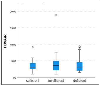
FIG. 1: BOX PLOT OF HOMA-IR VALUES ACROSS VITAMIN D CATEGORIES IN WOMEN WITH PCOS
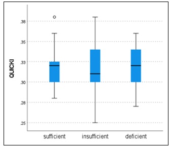
FIG. 2: BOX PLOT OF QUICKI VALUES ACROSS VITAMIN D CATEGORIES IN WOMEN WITH PCOS
Correlation Analysis: Spearman’s rank correlation presents weak correlation analysis of vitamin D with insulin resistance indicators, including BMI, waist circumference, fasting plasma insulin and HOMA-IR Table 3. Specifically, vitamin D showed a negative correlation with fasting blood sugar (r = –0.003, p > 0.05) and a positive correlation with fasting insulin (r = 0.002, p> 0.05), HOMA-IR (r = 0.004, p> 0.05) and QUICKI (r = 0.018, p > 0.05).
TABLE 3: RELATIONSHIP BETWEEN CIRCULATING 25(OH)D AND BIOMARKERS OF INSULIN RESISTANCE
| Variables | r | p value |
| BMI | 0.012 | 0.887 |
| Waist Circumference | -0.154 | 0.059 |
| Fasting Glucose | -0.003 | 0.967 |
| Fasting Insulin | 0.002 | 0.981 |
| HOMA-IR | 0.004 | 0.965 |
| QUICKI | 0.018 | 0.831 |
A statistically significant and strong positive correlation was identified between body mass index (BMI) and insulin resistance, indicated by HOMA-IR (r = 0.881, p < 0.001), implying increases in BMI are closely associated with elevated insulin resistance. Additionally, a moderate positive association was detected among BMI and fasting plasma glucose levels (r = 0.464, p < 0.001), implying a relationship between adiposity and glucose dysregulation. Furthermore, a strong positive relationship was found among BMI and fasting insulin concentrations (r = 0.864, p < 0.001), reinforcing the link between obesity and hyperinsulinemia. These reports suggest the important role of adiposity in modulating insulin sensitivity and glucose metabolism in women with PCOS Table 4.
TABLE 4: CORRELATION MATRIX OF BMI WITH MARKERS OF INSULIN RESISTANCE
| Variables | r | p value |
| Waist Circumference (inches) | 0.257 | 0.015 |
| Fasting Glucose (mg/dL) | 0.464 | <.001 |
| Fasting Insulin (µU/mL) | 0.864 | <.001 |
| HOMA-IR | 0.881 | <.001 |
TABLE 5: REGRESSION ANALYSIS
| Variable | B (Unstandardized) | SE | Beta (Standardized) | p value | 95% CI |
| Age | 0.018 | 0.031 | 0.047 | 0.575 | -0.045-0.080 |
| BMI | 0.175 | 0.059 | 0.302 | 0.003 | 0.058-0.291 |
| Waist Circumference | -0.179 | 0.073 | -0.244 | 0.015 | -0.3223- -0.036 |
| Vitamin D | 0.002 | 0.230 | 0.001 | 0.992 | -0.453-0.458 |
R2=0.075, Adjusted R2=0.050, F=2.971, p=0.021
Multiple linear regression analysis Table 5 demonstrated that BMI (β = 0.302, p=0.003) and waist circumference (β = -0.244, p = 0.015) were significant predictors of HOMA-IR. After adjusting for confounders, participants with vitamin D levels were not significantly (p = 0.992). Age was not significantly associated with HOMA-IR (p = 0.575). The overall model was significant (F = 2.971, p = 0.021) and explained 5% of the variance (adjusted R² = 0.050).
DISCUSSION: Nowadays, polycystic ovary syndrome (PCOS) is a widespread endocrine disorder which influence all age group women, may develop serious consequences like cardiovascular or metabolic disorder. A prevalent metabolic dysfunction observed in such conditions is insulin resistance. There is still uncertainty regarding molecular pathways that causes resistance of insulin in PCOS.
Recently considerable focus has been given on vitamin D status as a probable aspect in metabolic disorder. One of vitamin D deficiency pleiotropic effect may be consider as insulin resistance to its involvement in several cellular systems 5.
From our study it was determined that 30.46% of subjects had vitamin D insufficiency while 45.7% subjects had vitamin D deficiency. Numerous studies have demonstrated an association among vitamin D deficiency and women with PCOS 23. Yildizhan et al. reported 67% patient of PCOS were suffering from vitamin D 24. Our study revealed a significant vitamin D deficiency in PCOS patients, align to the previous research.
Our data also observed; in comparison to patients having insufficiency and sufficiency group respectively, patients having deficiency group had the highest HOMA-IR levels. Epidemiological findings report inverse correlation among 25(OH)D concentrations and insulin resistance and glucose intolerance 25. Despite of numerous researches among vitamin D deficiency and insulin resistance, mechanism is unclear due to undiscovered link at cellular level of these two parameters in PCOS. One of cohort study reported chronic inflammation with low severity; induces vitamin D deficiency and insulin resistance which might be absent in acute inflammation 26. At molecular level endocrine effects of vitamin D depend on tissue expression activated by enzymes and VDR; so directly systemic 25(OH)D may not capture local hormone or adipose signalling. Metabolic disorder could decrease vitamin D that do not reflect a causal vitamin D-IR pathway which raise question for interpreting of null hypothesis 27.
Although the correlation among vitamin D deficiency and insulin resistance has been well investigated, limited data are available from the Indian population, particularly from hospital-based cohorts. Our findings reported that there is no significant connection among vitamin D deficiency with insulin resistance in patients with polycystic ovary syndrome in this region. Several epidemiological studies have observed a positive association of glucose metabolism with vitamin D deficiency 28, 29. Hahn et al. found that lower levels of 25-OH vitamin D are linked with insulin resistance and obesity in patients with PCOS 30. Kotsa et al reported beneficial impact of cholecalciferol on glucose metabolism parameters in PCOS patients 31. But genetic variation in population alter vitamin D status and its metabolic consequences because of heterogeneity in cohorts. There are variants in the VDR and metabolising genes modify responses and may change the link between circulating 25(OH)D concentrations and insulin sensitivity 32. PCOS phenotypes may show vitamin D-IR relationships stronger but no correlation found in overall. So heterogenicity of phenotypes may reduce overall correlations 33.
Vitamin D-IR relationship is affected by co existing exposures and comorbidities which may hinder associations. Numerous observational studies report correlation is sensitive to adiposity, lifestyle and other covariates; therefore, null hypothesis for correlation between these two can be accepted if residual confounding factor is not properly controlled 34. According to studies, the majority of obese PCOS patients exhibit insulin resistance (IR). Furthermore, studies concluded that in obese patients, insulin resistance (IR) and low 25 (OH) D concentration are inversely correlated 12, 18. In compared to the control group for BMI, women who have been diagnosed with PCOS showed a tendency to have larger fat cells, as well as a higher waist to hip ratio 35. This indicates that women with PCOS not only have higher body fat distribution but also have its consequences like obesity and metabolic disorders. These changes are strongly related to lower insulin sensitivity. The major cause for insulin resistance in PCOS patient is declined level of serum adiponectin; a hormone that increase insulin sensitivity 36. In adipose tissue, reduction in adiponectin levels causes a rise in inflammatory cytokine which further contribute to insulin resistance 37.
Our result suggests vitamin D deficiency does not have direct link in insulin resistance development in this hospital based cross sectional sample using the present analytical approach. Prevalence of hypovitaminosis D, skin diseases, different sun exposure, for tified food intake and lifestyle changes in population may alter associations. These confounding factors may vary with vitamin D status and insulin tolerance which may fail to control bias result towards null hypothesis. Similarly, disease like metabolic syndrome or liver disease and drugs could change the insulin sensitivity or vitamin D concentrations by altering pharmacokinetic; can confound the correlation 32, 34.
The present findings should be viewed within the framework of the 2023 International Evidence-based Guideline for PCOS. The guideline emphasizes the increased cardiometabolic vulnerability of women with PCOS and supports assessment of cardiovascular risk factors and glycaemic status in this population. In this context, the frequent presence of insulin resistance in our study participants, particularly its relationship with BMI, underlines the clinical relevance of metabolic risk evaluation in PCOS. However, while insulin resistance is recognized as an important contributor to PCOS pathophysiology, the guideline cautions that available insulin assays have limited applicability in routine clinical care. Similarly, vitamin D status is not identified in the guideline as a diagnostic criterion or primary treatment target for PCOS 38. There were few limitations to our investigation. We solely examined PCOS patients; in lack of a control group in our research. This may have limited the depth of our analysis and the validation of our results because the control group could provide a comparative baseline to strengthen our results. Vitamin D thresholds vary among studies and clinical guidelines, and there is no universally accepted cut-off for sufficiency. In this study, a value of >32 ng/mL was selected because it was the reference threshold provided by the diagnostic kit used for serum 25(OH)D estimation. Therefore, comparisons with studies using alternative cut-offs, such as ≥20 ng/mL or ≥30 ng/mL, should be made carefully. Future studies may consider applying standardized or multiple threshold definitions to improve comparability across populations. Future studies should incorporate mechanistic analyses, such as proteomics or gene polymorphism evaluation, and Future research should include mechanistic studies, such as the assessment of proteomics or gene polymorphisms, along with randomized controlled trials that have larger participant groups.
CONCLUSION: In conclusion, no direct correlation was found among serum 25(OH)D concentrations and hyperinsulinemia in women with PCOS. Overall, vitamin D deficiency and insulin resistance were identified among the women with PCOS included in this study. However, due to the absence of a non-PCOS comparator group, no definitive conclusion can be drawn regarding whether these abnormalities are uniquely increased in PCOS. Further controlled studies are needed to confirm these observations.
Ethics Approval: The study was authorized by institutional ethics committee of Maliba Pharmacy College (Ref No. MPC/18/01/24-25)
CONFLICT OF INTEREST: The authors have no conflicts of interest regarding this investigation.
ACKNOWLEDGEMENTS: Nil
REFERENCES:
- Azziz R, Dumesic DA and Goodarzi MO: Polycystic ovary syndrome: an ancient disorder? Fertility and Sterility 2011; 95(5): 1544–8.
- Christ JP and Cedars MI: Current guidelines for diagnosing PCOS. Diagnostics 2023; 13(6): 1113.
- Hahn S, Fingerhut A, Khomtsiv U, Khomtsiv L, Tan S and Quadbeck B: The peroxisome proliferator activated receptor gamma Pro12Ala polymorphism is associated with a lower hirsutism score and increased insulin sensitivity in women with polycystic ovary syndrome. Clinical Endocrinology 2005; 62(5): 573–9.
- Lydic M and Juturu V: Dietary approaches and alternative therapies for polycystic ovary syndrome. Current Nutrition & Food Science 2008; 4(4): 265–81.
- Aronne LJ and Segal KR: Adiposity and fat distribution outcome measures: assessment and clinical implications. Obesity research 2002; 10(11): 14-21.
- Sam S: Obesity and polycystic ovary syndrome. Obesity Management 2007; 3(2): 69–73.
- Wekker V, Van Dammen L, Koning A, Heida KY, Painter RC and Limpens J: Long-term cardiometabolic disease risk in women with PCOS: a systematic review and meta-analysis. Human Reproduction Update 2020; 26(6): 942–60.
- Greenwood EA, Pasch LA, Cedars MI, Legro RS, Eisenberg E and Huddleston HG: Insulin resistance is associated with depression risk in polycystic ovary syndrome. Fertility and Sterility 2018; 110(1): 27–34.
- Thill M, Becker S, Fischer D, Cordes T, Hornemann A and Diedrich K: Expression of prostaglandin metabolising enzymes COX-2 and 15-PGDH and VDR in human granulosa cells. Anticancer Research 2009; 29(9): 3611–8.
- Holick MF: Vitamin D deficiency. The New England Journal of Medicine 357; 266–281.
- Netto EC, Silva AC, Pedroso C and Brites C: Hypovitaminosis D is associated with higher levels of inflammatory cytokines and with HAM/TSP in HTLV-infected patients. Viruses 2021; 13(11): 2223.
- Parikh G, Varadinova M, Suwandhi P, Araki T, Rosenwaks Z and Poretsky L: Vitamin D regulates steroidogenesis and insulin-like growth factor binding protein-1 (IGFBP-1) production in human ovarian cells. Hormone and Metabolic Research 2010; 42(10): 754–7.
- Argano C, Mirarchi L, Amodeo S, Orlando V, Torres A and Corrao S: The role of vitamin D and its molecular bases in insulin resistance, diabetes, metabolic syndrome, and cardiovascular disease: state of the art. International Journal of Molecular Sciences 2023; 24(20): 15485.
- Fathi FEZM, Sadek KM, Khafaga AF, Al Senosy AW, Ghoniem HA and Fayez S: Vitamin D regulates insulin and ameliorates apoptosis and oxidative stress in pancreatic tissues of rats with streptozotocin-induced diabetes. Environmental Science and Pollution Research 2022; 29(60): 90219–29.
- Bornstedt ME, Gjerlaugsen N, Olstad OK, Berg JP, Bredahl MK and Thorsby PM: Vitamin D metabolites influence expression of genes concerning cellular viability and function in insulin producing β-cells (INS1E). Gene 2020; 746: 144649.
- Teegarden D and Donkin SS: Vitamin D: emerging new roles in insulin sensitivity. Nutrition Research Reviews 2009; 22(1): 82–92.
- Gao B, Zhang C, Wang D, Li B, Shan Z and Teng W: Causal association between low vitamin D and polycystic ovary syndrome: a bidirectional mendelian randomization study. Journal of Ovarian Research 2024; 17(1): 95.
- Zerrouki D, Rami I, Assarrar I, Bouichrat N, Rouf S and Latrech H: Is there any association between vitamin D status and PCOS disease? Gynecological Endocrinology 2024; 40(1): 2381501.
- Singh P, Choudhary A, Saxena N and Bahuguna N: Correlation of Insulin Resistance and Anti-müllerian Hormone in Women with Polycystic Ovarian Syndrome. Journal of South Asian Federation of Obstetrics and Gynaecology 2024; 16(4): 397–401.
- Matthews DR, Hosker JP, Rudenski AS, Naylor BA, Treacher DF and Turner R: Homeostasis model assessment: insulin resistance and β-cell function from fasting plasma glucose and insulin concentrations in man. Diabetologia 1985; 28(7): 412–9.
- Nayeem J, Islam MT, Deeba F, Selim S, Ali L and Kabir Y: Insulin resistance and insulin secretory defect among Bangalee PCOS women: A case-control study. BMC Endocrine Disorders 2024; 24(1): 207.
- Biernacka-Bartnik A, Kocełak P, Owczarek AJ, Choręza PS, Markuszewski L and Madej P: The cut-off value for HOMA-IR discriminating the insulin resistance based on the SHBG level in women with polycystic ovary syndrome. Frontiers in Medicine 2023; 10: 1100547.
- Wehr E, Pilz S, Schweighofer N, Giuliani A, Kopera D and Pieber TR: Association of hypovitaminosis D with metabolic disturbances in polycystic ovary syndrome. European Journal of Endocrinology 2009; 161(4): 575–82.
- Yildizhan R, Kurdoglu M, Adali E, Kolusari A, Yildizhan B and Sahin HG: Serum 25-hydroxyvitamin D concentrations in obese and non-obese women with polycystic ovary syndrome. Archives of Gynecology and Obstetrics 2009; 280(4): 559–63.
- Ehrampoush E, Razzaz JM, Ghaemi A, Shahraki HR, Babaei AE and Osati S: The association of vitamin D levels and insulin resistance. Clinical Nutrition ESPEN 2021; 42: 325–32.
- Bennouar S, Bachir Cherif A, Aoudia Y and Abdi S: Additive interaction between insulin resistance, chronic low-grade inflammation and vitamin D deficiency on the risk of type 2 diabetes mellitus: a cohort study. Journal of the American Nutrition Association 2024; 43(7): 571–81.
- Nasser A, Papandreou D, Papadopoulou SK and Cheikh Ismail L: The role of vitamin D supplementation in type 1, type 2, and gestational diabetes: A comprehensive updated narrative review. Clinics and Practice 2025; 15(8): 148.
- Hassan NE, El-Orabi HA, Eid YM and Mohammed NR: Effect of 25-hydroxyvitamin D on metabolic parameters and insulin resistance in patients with polycystic ovarian syndrome. Middle East Fertility Society Journal 2012; 17(3): 176–80.
- Patra SK, Nasrat H, Goswami B and Jain A: Vitamin D as a predictor of insulin resistance in polycystic ovarian syndrome. Diabetes & Metabolic Syndrome: Clinical Research & Reviews 2012; 6(3): 146–9.
- Hahn S, Haselhorst U, Tan S, Quadbeck B, Schmidt M and Roesler S: Low serum 25-hydroxyvitamin D concentrations are associated with insulin resistance and obesity in women with polycystic ovary syndrome. Experimental and Clinical Endocrinology & Diabetes 2006; 114(10): 577–83.
- Kotsa K, Yavropoulou MP, Anastasiou O and Yovos JG: Role of vitamin D treatment in glucose metabolism in polycystic ovary syndrome. Fertility and Sterility 2009; 92(3): 1053–8.
- Yakout SM, Hussain SD, Alnaami AM and Al-Daghri NM: Association between triglyceride-glucose index and vitamin D levels in Saudi postmenopausal women: a cross-sectional study. International Journal of Clinical and Experimental Pathology 2025; 18(7): 375.
- Fu Y, Wang YH, Wang L and Huang MR: Correlation between follicular fluid of 25-hydroxyvitamin D level and endocrine function, ovarian function and insulin resistance in women with polycystic ovary syndrome. Journal of Physiology & Pharmacology 2025; 76(3).
- Abiri B, Valizadeh M, Ahmadi AR, Amini S, Nikoohemmat M and Abbaspour F: Association of vitamin D levels with anthropometric and adiposity indicators across all age groups: a systematic review of epidemiologic studies. Endocrine Connections 2024; 13(2).
- Helvaci N and Yildiz BO: Polycystic ovary syndrome as a metabolic disease. Nature Reviews Endocrinology 2025; 21(4): 230–44.
- Mannerås-Holm L, Leonhardt H, Kullberg J, Jennische E, Odén A and Holm G: Adipose tissue has aberrant morphology and function in PCOS: enlarged adipocytes and low serum adiponectin, but not circulating sex steroids, are strongly associated with insulin resistance. The Journal of Clinical Endocrinology & Metabolism 2011; 96(2): 304–11.
- Mladenović D, Vesković M, Šutulović N, Hrnčić D, Stanojlović O and Radić L: Adipose-derived extracellular vesicles–a novel cross-talk mechanism in insulin resistance, non-alcoholic fatty liver disease, and polycystic ovary syndrome. Endocrine 2024; 85(1):18–34.
- Legro RS, Lujan ME, Costello M and Dokras A: International, Evidence-Based Guideline for the Assessment and Management of Polycystic Ovary Syndrome 2023. In: ASRM 2023 Scientific Congress & Expo ASRM 2023.
How to cite this article:
Vyas B and Patel B: Prevalence of vitamin d deficiency and its association with insulin resistance among Indian women with polycystic ovary syndrome: evidence from a cross-sectional study. Int J Pharm Sci & Res 2026; 17(8): 2523-30. doi: 10.13040/IJPSR.0975-8232.17(8).2523-30.
All © 2026 are reserved by International Journal of Pharmaceutical Sciences and Research. This Journal licensed under a Creative Commons Attribution-NonCommercial-ShareAlike 3.0 Unported License.
Article Information
29
2523-2530
553 KB
301
English
IJPSR
Bhavin Vyas and Binal Patel *
Department of Pharmacology, Dr. Chunibhai Vallabhbhai Patel College of Pharmacy, Uka Tarsadia University, Tarsadi, Surat, Gujarat, India.
binalspatel206@gmail.com
02 April 2026
06 May 2026
08 May 2026
10.13040/IJPSR.0975-8232.17(8).2523-30
01 August 2026
 Download