




SPECTRUM OF HISTOPATHOLOGICAL FINDINGS IN CHOLECYSTECTOMY SPECIMENS WITH SPECIAL REFERENCE TO CARCINOMA OF GALLBLADDER – A 5-YEAR RETROSPECTIVE STUDY
HTML Full TextSPECTRUM OF HISTOPATHOLOGICAL FINDINGS IN CHOLECYSTECTOMY SPECIMENS WITH SPECIAL REFERENCE TO CARCINOMA OF GALLBLADDER – A 5-YEAR RETROSPECTIVE STUDY
K. K. Chelleng *, P. Jain and B. Kakati
Department of Pathology, Tezpur Medical College and Hospital, Tezpur, Assam, India.
ABSTRACT: Background: Cholecystectomy is one of the most frequently performed abdominal surgeries worldwide for symptomatic gallstones and inflammatory gallbladder lesions. Routine histopathological examination remains the gold standard for detection of inflammatory, premalignant and malignant lesions, while immunohistochemistry is reserved for selected diagnostically challenging malignant cases. Objective: To analyze the spectrum of histopathological lesions in cholecystectomy specimens and to perform detailed histopathological and immunohistochemical analysis of gallbladder carcinoma cases. Materials and Methods: This retrospective cross-sectional record-based study included all cholecystectomy specimens received over a 5-year period from 1st June 2019 to 31st May 2024. Histopathology records, gross descriptions, and microscopic slides were reviewed. Cases with incomplete records or autolyzed specimens were excluded. Missing clinicodemographic or gross data fields were recorded as unavailable and excluded from variable-specific analysis without excluding the case from overall histopathological spectrum analysis. Statistical analysis included Chi-square test and comparative clinicopathological correlation. Results: Among 2063 specimens, chronic cholecystitis was the most common lesion followed by cholesterolosis, acute on chronic cholecystitis and carcinoma gallbladder. Female predominance was observed in both overall lesions and carcinoma cases. Gallstones were significantly associated with carcinoma gallbladder. Premalignant lesions including dysplasia, intestinal metaplasia and ICPN (Intracholecystic papillary neoplasm) were also identified. A substantial proportion of carcinoma cases were incidental on microscopy without obvious gross growth. Conclusion: Routine gross and microscopic evaluation of all cholecystectomy specimens is essential for detecting incidental carcinoma and premalignant lesions. Immunohistochemistry serves as an adjunct in selected difficult malignant cases.
Keywords: Cholecystectomy, Chronic cholecystitis, Gallstones, Gallbladder carcinoma
INTRODUCTION: Cholecystectomy is one of the most commonly performed abdominal procedures globally, most often indicated for symptomatic gallstones or cholecystitis.
The spectrum of pathological lesions involving the gallbladder, which may or may not be associated with gallstones, encompass entities like chronic and acute cholecystitis, empyema, mucocele and ominous ones like gallbladder carcinoma 1.
Different studies have highlighted the strong association of cholelithiasis with gallbladder carcinoma (GBC). Chronic trauma and inflammation are known to induce epithelial dysplasia and explain the subsequent progression to carcinoma in-situ and invasive carcinoma 2.
Immunohistochemistry (IHC) is crucial for differentiating GBC from reactive or metastatic lesions 3, 4. Conducting a large-scale histopathological evaluation of cholecystectomy specimens allows for a comprehensive assessment of the prevalence and spectrum of gallbladder pathologies in a defined population. A large sample size improves the statistical reliability of observations and enables identification of relatively rare but clinically important lesions, including incidental carcinoma. The analysis of histopathological findings in relation to demographic parameters and gross features helps in understanding potential risk factors and disease patterns. The objective of the study is to analyze the spectrum of histopathological lesions in cholecystectomy specimens and to perform detailed histopathological and immunohistochemical analysis of gallbladder carcinoma cases in a tertiary care hospital.
MATERIALS AND METHODS:
Study Design and Period: This was a retrospective cross-sectional record-based study conducted in the Department of Pathology, Tezpur Medical College and Hospital, from 1st June 2019 to 31st May 2024. All consecutively received cholecystectomy specimens during the study period were retrieved from the histopathology register, laboratory information system, and archived slides/blocks.
Record Review Process: For each case, demographic details, gross findings, histopathological diagnosis, and carcinoma-related microscopic parameters were retrieved from pathology records and rechecked against archived reports. In carcinoma and premalignant lesions, original haematoxylin and eosin (H&E) stained slides were reviewed for confirmation.
Inclusion Criteria: All cholecystectomy specimens received for histopathological examination during the study period.
Exclusion Criteria: Specimens that were autolyzed and with incomplete clinical data were excluded from the study. For partially incomplete records, the case was retained for overall lesion spectrum analysis, while missing variables were excluded only from variable-specific statistical analysis.
Ethical Consideration: This study does not involve any in-vivo experiment on humans or animals. It is a retrospective record-based observational study for which ethical clearance was obtained from Institutional Human Ethical Committee, Tezpur Medical College and Hospital, Assam vide IEC Sl. No: 2024/083/TMC&H, dated 22/06/2024. As the study involved archival histopathology records and anonymized patient data, waiver of individual informed consent was granted by the ethics committee, and strict confidentiality was maintained.
Study Procedure: The specimens were processed routinely, fixed in 10% neutral buffered formalin followed by paraffin embedding, microtome sectioning at 4–5μm, and staining with haematoxylin and eosin (H&E) stain. Immunohistochemistry was performed as per standard protocol for cytokeratin 7(CK7), cytokeratin 19 (CK19) and cytokeratin 20 (CK20) in cases diagnosed as carcinoma of gallbladder on routine H&E stained slides.
Study Variables: The study variables included age, gender, gross morphology features like presence or absence of gallstones, wall thickness, mucosal changes and visible growth.
The microscopic findings included histopathological diagnosis, tumor differentiation, lymphovascular and perineural invasion and positivity/negativity of immunohistochemical markers in IHC slides.
Data Analysis: Data was collected from histopathological records and analysed using SPSS (Statistical Package for the Social Sciences) version 23.0 (Released 2015, IBM Corp, Armonk NY) software (IBM Corp 2015).
Association between gallstones and gallbladder carcinoma were evaluated using Chi-square test (significance at p <0.05). Visual representation included tables, charts, gross image and photomicrographs.
RESULTS: A total of 2063 cholecystectomy specimens were studied.
Gender-wise Case Distribution:
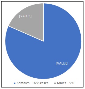
FIG. 1: PIE CHART DEPICTING GENDER-WISE DISTRIBUTION OF ALL CASES
Age-Specific Distribution of Gallbladder Lesions:
TABLE 1: TABLE DEPICTING AGE-SPECIFIC DISTRIBUTION OF GALLBLADDER LESIONS
| Age group (years) | Total cases (n=2063) | Percentage (%) | Carcinoma cases (n=21) |
| ≤20 | 35 | 1.7 | 0 |
| 21–30 | 285 | 13.8 | 1 |
| 31–40 | 620 | 30.1 | 4 |
| 41–50 | 710 | 34.4 | 7 |
| 51–60 | 295 | 14.3 | 5 |
| >60 | 118 | 5.7 | 4 |
Gross Pathological Findings in Gallbladder Lesions:
TABLE 2: TABLE DEPICTING GROSS PATHOLOGICAL FINDINGS IN GALLBLADDER LESIONS
| Gross parameter | Category | Cases (n=2063) | Percentage (%) |
| Gallstones | Present | 1520 | 73.7 |
| Absent | 543 | 26.3 | |
| Wall thickness | Normal | 580 | 28.1 |
| Thickened | 1483 | 71.9 | |
| Mucosal appearance | Normal | 738 | 35.8 |
| Congested/ulcerated | 1325 | 64.2 |
Histopathological Patterns:
TABLE 3: HISTOPATHOLOGICAL SPECTRUM OF GALLBLADDER LESIONS
| Diagnosis | Cases (n=2063) | Percentage (%) |
| Chronic cholecystitis | 1850 | 89.7 |
| Cholesterolosis | 97 | 4.7 |
| Acute on chronic cholecystitis | 39 | 1.9 |
| Carcinoma gallbladder | 21 | 1.0 |
| Acute cholecystitis | 12 | 0.6 |
| Adenomyomatosis | 10 | 0.5 |
| Dysplasia | 10 | 0.5 |
| Mucocele | 6 | 0.3 |
| Inflammatory polyp | 6 | 0.3 |
| Adenomatous polyp | 4 | 0.2 |
| Intestinal metaplasia | 4 | 0.2 |
| Intracholecystic Papillary Neoplasm [ICPN] | 4 | 0.2 |
Carcinoma Gallbladder Gender-Wise Distribution (n=21):
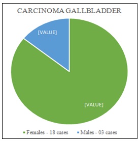
FIG. 2: PIE CHART DEPICTING CARCINOMA GALLBLADDER CASES WITH GENDER-WISE DISTRIBUTION
Gallstones in Carcinoma versus Non-Carcinoma Cases:
TABLE 4: TABLE DEPICTING GALLSTONES IN CARCINOMA VERSUS NON-CARCINOMA CASES
| Gallstones | Carcinoma | Non-carcinoma | Total |
| Present | 16 | 1504 | 1520 |
| Absent | 5 | 538 | 543 |
| Total | 21 | 2042 | 2063 |
Gross Findings in Carcinoma gallbladder:
TABLE 5: GROSS MORPHOLOGICAL PARAMETERS IN CARCINOMA GALLBLADDER CASES
| Gross parameter | Category | Cases (n=21) | Percentage (%) |
| Wall thickness | <8.25mm | 5 | 23.8 |
| >8.25mm | 16 | 76.2 | |
| Visible growth | Present | 8 | 38.1 |
| Absent | 13 | 61.9 | |
| Gallstones | Present | 16 | 76.2 |
| Absent | 5 | 23.8 |
Chi-Square Test - Association between Gallstones and Gallbladder Carcinoma:
TABLE 6: CHI-SQUARE TEST
| Parameter | Value |
| Chi-square (χ²) | 5.76 |
| Degrees of freedom (df) | 1 |
| p-value | 0.016 |
Histopathological Characteristics of Carcinoma Gallbladder:
TABLE 7: HISTOPATHOLOGICAL PARAMETERS OF CARCINOMA GALLBLADDER CASES
| Histopathological Parameter | Cases (n=21) | Percentage (%) |
| Tumor differentiation | ||
| Well differentiated adenocarcinoma | 8 | 38.1 |
| Moderately differentiated adenocarcinoma | 6 | 28.6 |
| Poorly differentiated adenocarcinoma | 7 | 33.3 |
| Lymphovascular invasion (LVI) | ||
| Present | 5 | 23.8 |
| Absent | 16 | 76.2 |
| Perineural invasion (PNI) | ||
| Present | 2 | 9.5 |
| Absent | 19 | 90.5 |
Subanalysis of Parameters of Carcinoma Cases:
TABLE 8: TABLE DEPICTING ADDITIONAL PARAMETERS IN GBC CASES
| Parameter associated with tumor | Cases (n=21) | Percentage (%) |
| Location | ||
| Fundus | 9 | 42.9 |
| Body | 7 | 33.3 |
| Neck | 5 | 23.8 |
| Pathological staging | ||
| pT1 | 4 | 19.0 |
| pT2 | 10 | 47.6 |
| pT3 | 7 | 33.4 |
| Margin positive | 3 | 14.3 |
| Associated dysplasia/metaplasia | 6 | 28.6 |
| Lymph node positivity | 2 | 9.5 |
Immunohistochemistry (IHC) Findings in Gallbladder Carcinoma Cases:
TABLE 9: IHC POSITIVITY RATES AND STAINING PATTERN
| IHC marker | Positive Cases (n=21) | Positivity Percentage (%) | Staining pattern |
| CK7 | 20 | 95.2 | Diffuse cytoplasmic |
| CK19 | 18 | 85.7 | Diffuse cytoplasmic |
| CK20 | 4 | 19 | Focal cytoplasmic |
Gross Morphology Image:
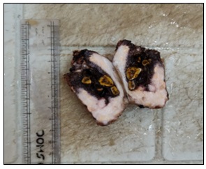
FIG. 3: GALLBLADDER SPECIMEN SHOWING GALLSTONES AND THICKENED WALL
Photomicrographs:
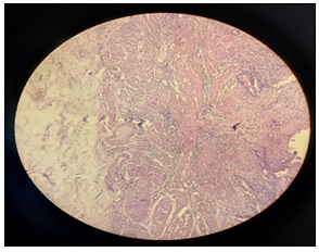
FIG. 4: CHRONIC CHOLECYSTITIS (H&E STAIN; MAGNIFICATION-10X; SCALE BAR: 1cm=200μm)
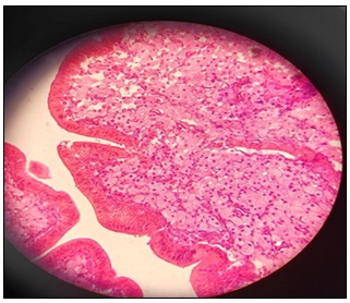
FIG. 5: CHOLESTEROLOSIS (H&E STAIN; MAGNIFICATION - 40X; SCALE BAR: 1cm=100μm)
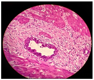
FIG. 6: GALLBLADDER CARCINOMA (H&E STAIN; MAGNIFICATION - 40X; SCALE BAR: 1cm=100μm)
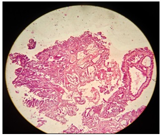
FIG. 7: ICPN (H&E STAIN; MAGNIFICATION-10X; SCALE BAR: 1cm=200μm)
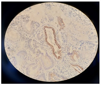
FIG. 8: IHC – CK7 POSITIVE (MAGNIFICATION-40X; SCALE BAR: 1cm=100μm)
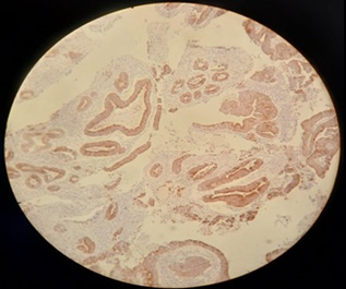
FIG. 9: IHC – CK19 POSITIVE (MAGNIFICATION-10X; SCALE BAR: 1cm=200μm)
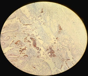
FIG. 10: IHC – CK20 POSITIVE (MAGNIFICATION-10X; SCALE BAR: 1cm=200μm)
DISCUSSION: A total of 2063 gallbladder specimens were evaluated histopathologically in the present study. Gallbladder diseases showed a marked female predominance, with females accounting for 81.6% and males 18.4%. Mondal et al. observed a higher prevalence of gallbladder disease among females undergoing cholecystectomy, with women constituting more than 80% of cases 5. Degloorkar et al. also reported a significant female predominance in gallbladder pathology in their retrospective study of cholecystectomy specimens 6. This female preponderance is widely attributed to hormonal influences, particularly estrogen, which increases cholesterol saturation in bile and predisposes to gallstone formation 7.
The age of presentation between the 4th and 5th decades was seen in majority of the cases (34.4%) in the present study with GBC cases (33.3%) occurring predominantly in the same age interval. This is in concordance with various other studies conducted world over 1, 3, 6, 7.
In the present study, chronic cholecystitis was the most common histopathological diagnosis, accounting for nearly 89.7% of cases, followed by cholesterolosis, acute on chronic cholecystitis, and GBC. Mondal et al. documented chronic cholecystitis in approximately 79.8% of cases 5. Gupta et al.10, Srinivasan and Sekar 11 and Kumar et.al12also reported chronic cholecystitis as the most frequent pathology in their histopathological study of gallbladder lesions. The predominance of chronic cholecystitis is largely attributed to the high prevalence of gallstones and chronic inflammation of the gallbladder mucosa. The incidence of gallbladder carcinoma in the present study was approximately 1%. This finding is comparable with previously published studies. Degloorkar et al.6 and Sangwan et al.8 reported gallbladder carcinoma in 0.46% and 1.9% respectively in their study of gallbladder specimens. Similarly, Mohan et al. observed carcinoma in a small percentage of cholecystectomy specimens, highlighting that incidental carcinoma remains an uncommon but clinically significant finding 9. Among GBC cases in the present study, females constituted the majority (85.7%) which is consistent with the known gender predisposition of gallbladder malignancy. Hundal and Shaffer reported that gallbladder carcinoma occurs two to six times more frequently in women compared to men14. Similarly, Misra et al. observed a strong female predominance in gallbladder carcinoma cases in their clinical study 19. Gallstones were identified in 76% of carcinoma cases in the present study, suggesting a strong association between cholelithiasis and gallbladder carcinoma. Lazcano-Ponce et al. reported that gallstones are present in approximately 70–90% of gallbladder carcinoma cases 13. Similarly, Dutta emphasized that chronic irritation and inflammation caused by gallstones play a major role in gallbladder carcinogenesis 17.
Chi-square analysis demonstrated a statistically significant association between gallstones and carcinoma gallbladder (χ² = 5.76, df = 1, p = 0.016). This statistical analysis concurs with similar observations made in other population based studies 13, 17. Additional exploratory analysis showed increasing age and gallbladder wall thickness were more frequently associated with GBC, though not subjected to multivariate testing.
In the present study, 76.2% of GBC cases demonstrated a wall thickness of >8.25mm suggesting significant association between wall thickness and GBC. Shagor et.al reported in their study that a wall thickness cut-off value of ≥8.25 mm showed 81.8% sensitivity and 72.7% specificity for predicting gallbladder carcinoma, indicating its usefulness as a predictive marker 20.
Premalignant lesions such as intestinal metaplasia and dysplasia were also observed in a considerable number (28.6%) of GBC cases in the present study highlighting their importance as harbingers of malignancy. Wistuba and Gazdar 18 and Randi et al.15 highlighted that chronic inflammation of the gallbladder mucosa can lead to metaplastic changes followed by dysplasia and eventual carcinoma. Another notable observation in the present study was that a considerable proportion of carcinoma cases (61.9%) did not show obvious gross tumour growth. This signifies the clinical importance of routine histopathological examination of all cholecystectomy specimens for detection of incidental GBC. Sangwan et al.18 and other studies 11 have reported that incidental gallbladder carcinoma may be detected only on microscopic examination even when gross examination appears normal. Regarding tumor differentiation, the adenocarcinomas in the present study demonstrated variable degrees of differentiation, including well-differentiated, moderately differentiated, and poorly differentiated tumours. Previous studies have also reported that adenocarcinoma is the most common histological type of gallbladder cancer, with varying grades reflecting differences in tumor aggressiveness and prognosis 16.
Subanalysis of additional parameters of GBC cases revealed fundal location (42.9%) of most tumours with majority of cases (47.6%) showing invasion of the subserosal connective tissue (pT2 staging). Several clinicopathological studies have similarly documented that the majority of gallbladder carcinomas arise in the fundus, with reported frequencies ranging widely but consistently demonstrating fundal predominance over other anatomical sites 21, 22. In addition, the distribution of tumor staging in the present study, with pT2 tumors forming the largest subgroup, is comparable to findings from surgical and histopathological series where T2 lesions constitute a major proportion of resectable gallbladder carcinomas 14, 22, 23.
Immunohistochemistry was performed only in histologically confirmed malignant cases where confirmation of primary biliary epithelial origin or exclusion of metastatic adenocarcinoma or reactive atypia was required. In the present study, out of a total of 21 cases, CK7 and CK19 showed diffuse cytoplasmic positivity in 95.2% and 85.7% cases respectively. Several studies have reported CK7 positivity in approximately 90–98% of gallbladder carcinomas, making it one of the most sensitive markers for tumours of pancreatobiliary origin 24-26. CK19, another marker of biliary epithelial differentiation, has also been shown to be positive in 85–98% of cases, supporting its diagnostic utility in biliary tract malignancies 24, 26.
In contrast, CK20 showed focal positivity in only 19% of cases in the present study. CK20 positivity has been reported in approximately 15–30% of cases, and when present, the staining is usually focal and less intense 24, 25. This immunohistochemical pattern is particularly useful in differentiating primary gallbladder carcinoma from metastatic colorectal carcinoma, which usually demonstrates a CK7-negative and CK20-positive staining pattern 25, 27, 28.
CONCLUSION: The present study demonstrates the importance of routine study of gross morphology and histopathological examination for the detection of premalignant lesions and incidental GBC. Immunohistochemistry plays a crucial role in differentiating between primary GBC and metastatic lesions and/or reactive atypia.
Collectively, these findings underscore the indispensable role of comprehensive pathological assessment in guiding appropriate clinical management and highlight the necessity of systematic evaluation of all cholecystectomy specimens, irrespective of the preoperative clinical impression. The study is limited by its retrospective design, single centre nature and lack of imaging correlation. However, the large sample size and methodical histopathological and immuno-histochemical evaluation reinforce the significance of the study findings.
ACKNOWLEDGMENT: We are thankful to our histopathology technicians and attendants for assisting us during the conduct of this research.
Funding Statement: No funding has been received from any organization, individual, or party for this research.
Authors’ Contribution: All the authors have significantly contributed in concept, study designing, data acquisition, data analysis, and writing of the manuscript.
Data Availability: Data are available with the authors and can be retrieved as and when required.
CONFLICT OF INTEREST: There are no conflicts of interest for this research work
REFERENCES:
- Almas T, Murad MF, Khan MK, Ullah M, Nadeem F, Ehtesham M and Zaidi SMJ: The spectrum of gallbladder histopathology at a tertiary hospital in a developing country: a retrospective study. Cureus 2020; 12(8): 9627.
- Dowling GP and Kelly JK: The histogenesis of adenocarcinomas of the gall bladder. Cancer. 1986; 58(8): 1702–1708. doi:10.1002/1097-0142(19861015)58:8<1702::AID-CNCR2820580821>3.0.CO;2-Z.
- Roa JC, Tapia O and Cakir A: Clinicopathologic analysis of gallbladder carcinoma: prognostic factors and evaluation of immunohistochemical markers. Hum Pathol 2011; 42(8): 1275–1283.
- Dursun N, Escalona OT and Roa I: Pathological features and molecular pathology of gallbladder carcinoma. World J Gastroenterol 2013; 19(42): 7497–7504.
- Mondal B, Maulik D, Biswas BK, Sarkar GN and Ghosh D: Histopathological spectrum of gallstone disease from cholecystectomy specimen in rural areas of West Bengal, India. Int J Community Med Public Health 2016; 3(11): 3229–3235.
- Degloorkar S, Parab S, Shah B, Prasad S and Gore S: A retrospective single-centre study on histopathological analysis of cholecystectomy specimens in a tertiary care centre in India. Int Surg J 2023; 10: 1–6.
- Noor S and Kumari N: Retrospective analysis of histopathological patterns in gallbladder diseases. Bioinformation 2025; 21(6): 1526–1529.
- Sangwan MK, Sangwan V, Garg MK, Singla D, Malik P and Duhan A: Incidental carcinoma of gallbladder in North India: is routine histopathology justified? Int Surg J 2015; 2: 465–470.
- Mohan A, Thakral R, Faiz A and Kumar R: Frequency and histological spectrum of gallbladder carcinoma in cholecystectomy specimens. IP J Diagn Pathol Oncol 2018; 3(3): 192–195.
- Gupta K, Faiz A, Thakral RK, Mohan A and Sharma VK: Spectrum of histopathological lesions in gallbladder in cholecystectomy specimens. Int J Clin Diagn Pathol 2019; 2(1): 146–151.
- Srinivasan G and Sekar ASI: Histopathological spectrum of gallbladder lesions in cholecystectomy specimens. Int J Res Med Sci 2019; 7: 1542–1546.
- Kumar H, Kini H and Tiwari A: Histological evaluation of cholecystectomy specimens. J Pathol Nepal 2015; 5: 720–724.
- Lazcano-Ponce EC, Miquel JF, Muñoz N, Herrero R, Ferrecio C and Wistuba II: Epidemiology and molecular pathology of gallbladder cancer. CA Cancer J Clin 2001; 51: 349–364.
- Hundal R and Shaffer EA: Gallbladder cancer: epidemiology and outcome. Clin Epidem 2014; 6: 99–109.
- Randi G, Franceschi S and La Vecchia C: Gallbladder cancer worldwide: geographical distribution and risk factors. Int J Cancer 2006; 118: 1591–1602.
- Shukla VK, Khandelwal C, Roy SK and Vaidya MP: Primary carcinoma of the gallbladder: a review of a 16-year experience. J Surg Oncol 1985; 28: 32–35.
- Dutta U: Gallbladder cancer: can newer insights improve the outcome? J Gastroenterol Hepatol 2012; 27: 642–653.
- Wistuba II and Gazdar AF: Gallbladder cancer: lessons from a rare tumor. Nat Rev Cancer 2004; 4: 695–706.
- Misra S, Chaturvedi A, Misra NC and Sharma ID: Carcinoma of gallbladder. Lancet Oncol 2003; 4: 167–176.
- Shagor S, Mitra TK and Rabbi H: Wall thickness is an important predictor of gallbladder malignancy in patients surgically treated for gallbladder disease. Int Surg J 2023; 10: 1–6.
- Kim WJ, Lim TW and Park PJ: Clinicopathological differences in T2 gallbladder cancer according to tumor location. Cancer Control 2020; 27(1): 1–9.
- Lee SE, Choi YS and Kim YH: Prognostic significance of tumor location in T2 gallbladder cancer. J Clin Med 2020; 9(5): 1–12.
- Shirai Y, Sakata J and Wakai T: Lymphatic spread and surgical strategy for T2 gallbladder carcinoma. Eur J Surg Oncol 2019; 45(4): 661–667.
- Chu PG and Weiss LM: Immunohistochemical characterization of biliary tract carcinomas including gallbladder carcinoma. Mod Pathol 2002; 15(2): 123–128.
- Lee H, Park SJ and Kim SW: Immunohistochemical analysis of cytokeratin expression patterns in gallbladder carcinoma. Cancers (Basel) 2021; 13(6): 1357.
- Jaszcz A, Kaczmarczyk M and Krawczyk M: Cytokeratin 7 and cytokeratin 20 expression in gallbladder carcinoma. Pol J Pathol 2011; 62(2): 70–75.
- Rosai J: Rosai and Ackerman's Surgical Pathology. 11th ed. Philadelphia: Elsevier 2018.
- WHO Classification of Tumours Editorial Board. Digestive System Tumours. 5th ed. Lyon: International Agency for Research on Cancer 2019
How to cite this article:
Chelleng KK, Jain P and Kakati B: Spectrum of histopathological findings in cholecystectomy specimens with special reference to carcinoma of gallbladder – a 5-year retrospective study. Int J Pharm Sci & Res 2026; 17(8): 2445-53. doi: 10.13040/IJPSR.0975-8232.17(8).2445-53.
All © 2026 are reserved by International Journal of Pharmaceutical Sciences and Research. This Journal licensed under a Creative Commons Attribution-NonCommercial-ShareAlike 3.0 Unported License.
Article Information
18
2445-2453
1967 KB
286
English
IJPSR
K. K. Chelleng *, P. Jain and B. Kakati
Department of Pathology, Tezpur Medical College and Hospital, Tezpur, Assam, India.
drkamal24@gmail.com
17 March 2026
17 April 2026
23 April 2026
10.13040/IJPSR.0975-8232.17(8).2445-53
01 August 2026
 Download