




DEVELOPMENT AND EVALUATION OF TOPICAL PHYTOFORMULATIONS OF IPOMOEA SHUPANGENSIS (KASUNUNGURE) FOR THE MANAGEMENT OF MULTIDRUG-RESISTANT BACTERIAL INFECTIONS IN DIABETIC FOOT ULCERS: AN INTEGRATED ETHNOPHARMACOLOGICAL AND PHARMACEUTICAL APPROACH
HTML Full TextDEVELOPMENT AND EVALUATION OF TOPICAL PHYTOFORMULATIONS OF IPOMOEA SHUPANGENSIS (KASUNUNGURE) FOR THE MANAGEMENT OF MULTIDRUG-RESISTANT BACTERIAL INFECTIONS IN DIABETIC FOOT ULCERS: AN INTEGRATED ETHNOPHARMACOLOGICAL AND PHARMACEUTICAL APPROACH
M. Mubaiwa *, T. Manyarara and J. Chifamba
Department of Pharmacy and Pharmaceutical Sciences, Faculty of Medicine and Health Sciences, University of Zimbabwe, Harare, Zimbabwe.
ABSTRACT: Ipomoea shupangensis (Kasunungure) is used by traditional healers in Gokwe, Zimbabwe for diabetic foot ulcers (DFUs), which are frequently complicated by multidrug-resistant (MDR) pathogens. Scientific validation of its phytochemistry, pharmacological activity, and safety was previously lacking. This study integrated an ethnopharmacological survey (40 healers), maceration extraction (five solvents, three plant parts), qualitative phytochemical screening, total phenolic content (TPC) quantification, DPPH antioxidant and albumin denaturation anti-inflammatory assays, antibacterial testing against clinical MDR isolates (MRSA, P. aeruginosa, ESBL E. coli), OECD safety evaluation, and development of an Antiseptic Solution (2% w/v) and Dusting Powder (5% w/w). All 40 healers (100%) identified stem bark as the most potent plant part. The 70% ethanol stem extract showed the highest TPC (145.3 ± 6.2 mg GAE/g), strongest antioxidant activity (DPPH IC₅₀ 42.7 µg/mL), notable anti-inflammatory activity (IC₅₀ 385.2 µg/mL), and the lowest MIC values against MRSA (31.3 µg/mL), P. aeruginosa (62.5 µg/mL), and E. coli (125 µg/mL), with MBC/MIC ratios of 2 (bactericidal) and Hill slopes exceeding 2.5. Time-kill assays achieved MRSA sterilisation within 4 hours at 4× MIC, and disk diffusion zones (36.2 mm) exceeded ciprofloxacin (28.5 mm; p<0.01). OECD 404 and 423 testing indicated non-irritancy (PII = 0.0) and practical non-toxicity (LD₅₀ >2000 mg/kg). Both formulations met pharmacopoeial specifications and remained stable for six months under ICH Q1A(R2) conditions. These preclinical findings support the extract as a promising candidate for further development as an affordable topical phytomedicine against MDR-infected DFUs, pending chromatographic standardisation, synergy and formulation-level testing, and in-vivo and clinical evaluation. (Abstract now 234 words; journal limit 150–250.).
Keywords: Ipomoea shupangensis, Diabetic foot ulcer, Multidrug resistance, MRSA, Antioxidant, Anti-inflammatory
INTRODUCTION: Diabetes mellitus affects an estimated 537 million adults globally, with sub-Saharan Africa (SSA) projected to experience a 134% increase by 2045 1. In Zimbabwe, diabetes affects 10–15% of the urban adult population with rapidly rising prevalence 2.
Diabetic foot ulcers (DFUs) represent a devastating complication: approximately 15–25% of diabetic patients develop a DFU during their lifetime, preceding 80% of all non-traumatic lower limb amputations globally, with five-year post-amputation mortality exceeding 70% 3.
DFUs complicated by multidrug-resistant (MDR) bacteria represent a particularly intractable problem in resource-limited settings 4, 5. The three priority MDR pathogens in DFU infections are methicillin-resistant Staphylococcus aureus (MRSA), MDR Pseudomonas aeruginosa ((WHO Priority 1 Critical) 6, and extended-spectrum beta-lactamase (ESBL)-producing Escherichia coli. In Zimbabwe, chronic shortages of essential antibiotics, absence of microbiological diagnostic capacity, and up to 80% reliance on herbal medicine by rural and peri-urban populations define the context in which affordable, locally producible topical antiseptic solutions are not merely preferable but clinically necessary 2, 4.
Ipomoea shupangensis Baker 8 (Convolvulaceae), locally known as Kasunungure, is a perennial liana used for generations by traditional healers in Gokwe District, Zimbabwe, for diabetic foot ulcers, gangrene, and septic wounds 5.
The genus Ipomoea has documented antimicrobial, wound-healing, and anti-diabetic activities in multiple species 6. To the best of our knowledge, no published scientific data existed on I. shupangensis in any domain — no phytochemistry, no antibacterial data, no safety data, and no formulation development.
This absence of scientific validation, set against the escalating burden of MDR-infected DFUs in a health system with constrained antibiotic access, constitutes the specific research gap addressed here: no prior study has established whether the traditional healer consensus on I. shupangensis stem bark corresponds to measurable antibacterial, antioxidant, or anti-inflammatory activity, or whether it can be translated into a stable, affordable topical formulation. I. shupangensis was selected over other locally available antimicrobial plants because of the exceptional, unanimous consensus (100%, n=40) among Gokwe healers on its efficacy specifically for MDR-associated wound presentations (gangrene, malodorous sepsis) rather than wounds in general, and because, to the best of our knowledge, it had not previously been subjected to any phytochemical, pharmacological, or safety investigation. This study therefore provides, to the best of our knowledge, the first ethnopharmacological documentation, phytochemical and pharmacological characterisation, and preclinical formulation development of I. shupangensis, generating evidence-based, locally producible topical phytoformulations proposed for this indication in Zimbabwe.
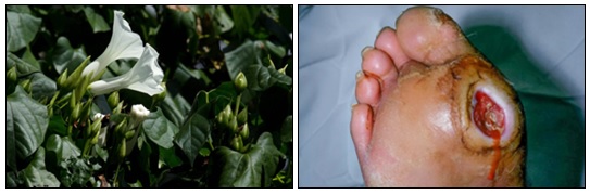
FIG. 1: IPOMOEA SHUPANGENSIS (KASUNUNGURE) IN ITS NATURAL HABITAT. PHOTO TAKEN AT GORONGOSA NATIONAL PARK 8 [LEFT]. A DIABETIC FOOT ULCER GRADE 3 — DEEP ULCER WITH SIGNS OF OSTEITIS/ABSCESS, THE CLINICAL PRESENTATION MOST COMMONLY ASSOCIATED WITH MDR INFECTION. CREDIT: REPRODUCED WITH PERMISSION FROM ©DERMNET WWW.DERMNETNZ.ORG 2023 [RIGHT]
MATERIALS AND METHODS
Ethical Approvals and Plant Material: Ethics approvals were obtained from the Joint Research Ethics Committee (JREC/01/2025), Medical Research Council of Zimbabwe (MRCZ/A/2897), and Institutional Animal Ethics Committee (IAEC/PHARM/202504; IAEC/PHARM/202505). Fresh plant material was collected in November 2025 (early wet season, when secondary metabolite content is typically maximal) from four sites in Gokwe Chireya District, Mashonaland West Province, Zimbabwe (GK01–GK04; 18°26'30"–18°30'00"S, 28°29'15"–28°34'00"E), under the guidance of a senior ZINATHA healer. Only morphologically confirmed adult specimens were selected. Sustainable harvesting was strictly observed: stem bark was strip-harvested leaving ≥50% of the bark circumference intact to permit regeneration, and only mature stems ≥5 cm in diameter were selected; roots were partially excavated (approximately one-third of the root system per plant); leaves were hand-picked from multiple branches without defoliation. GPS coordinates and photographs were recorded at each collection point, and duplicate herbarium specimens were retained. Taxonomic authentication was performed by a botanist at the National Herbarium and Botanic Garden of Zimbabwe using standard diagnostic morphological features (leaf shape and venation, stem habit, flower and fruit morphology, latex exudate); a Voucher and Authentication Certificate (NHZ2025089) was issued and a duplicate specimen deposited in the National Herbarium. Plant material was separated into roots, stems (bark), and leaves; washed sequentially under running tap water and then rinsed three times in autoclaved distilled water; shade-dried at 25 ± 2°C for 14 days; and pulverised to <1 mm particle size.
Ethnopharmacological Survey: Purposive sampling recruited 40 participants (32 ZINATHA-registered healers, 5 independent herbalists, 3 community knowledge custodians ≥60 years), representing all nine administrative wards of Gokwe Chireya District. Inclusion criteria were: ≥5 years of active practice experience, demonstrated familiarity with Kasunungure, and provision of written informed consent in the Shona language; healers with <5 years' experience, those unwilling to consent, or those located outside Gokwe Chireya were excluded. Data were collected using two complementary instruments: (i) individual semi-structured interviews (45–90 minutes; audio-recorded with consent) covering local/regional plant names, habitat and seasonal availability, plant-part preference and stated reasons, preparation and application methods, therapeutic outcomes and perceived adverse effects, conservation knowledge, and willingness to participate in benefit-sharing; and (ii) three focus group discussions (8–10 participants each; 90–120 minutes) exploring community consensus and intergenerational knowledge transmission. All interviews and focus groups were conducted in the Shona language by the principal investigator; audio recordings were transcribed verbatim, translated into English, and back-translated to Shona by a second bilingual speaker to confirm accuracy prior to coding. Prior informed consent (read aloud in Shona, followed by signed/thumb-printed acknowledgement) was obtained from all participants and relevant traditional and administrative authorities before any data collection, in accordance with the Nagoya Protocol 28; access and benefit-sharing was formalised through a draft agreement specifying return of 5–10% of any future commercial revenues to a Gokwe Community Health Trust, with full attribution of traditional knowledge to participating healers. Qualitative data were coded and analysed thematically using Braun and Clarke's six-phase framework in NVivo 12.0; quantitative consensus data (frequencies, percentages, means ± SD) were analysed in IBM SPSS Statistics 26.0.
Maceration Extraction: All three plant parts (25 g dried, pulverised material per extraction) were extracted by maceration 13 (1:15 w/v solvent-to-solid ratio, i.e. 375 mL solvent; 72 h initial maceration + 2×24 h re-extractions at 25 ± 2°C with 3× daily agitation) using five solvents: autoclaved distilled water, 70% ethanol v/v, 99% chloroform, 99% acetone, and 99% ethyl acetate (all Sigma-Aldrich analytical grade). All 15 combinations were performed in triplicate (n=3). Pooled filtrates were vacuum-filtered (Whatman No. 1) and concentrated by rotary evaporation (Buchi R-200; 40°C; solvent-specific vacuum) to dryness, then further dried under vacuum desiccation to constant weight; dried extract weight was recorded gravimetrically and extraction yield calculated as: Yield (% w/w) = [weight of dry extract (g) / weight of dried plant material extracted (g)] × 100. The lead 70% ethanol stem extract was freeze-dried 14 (Christ Alpha 1-4 LDplus; −80°C freezing; −20°C/0.05 mbar primary drying 24 h; +20°C/0.01 mbar secondary drying 6 h) to remove residual solvent and moisture. All dried extracts were stored in amber glass vials under vacuum desiccation at 4°C, protected from light, until further use.
Qualitative Phytochemical Screening: All 15 extracts were screened in duplicate for eight secondary metabolite classes: alkaloids (Wagner's test), flavonoids (lead acetate test), tannins (ferric chloride 10%), saponins (frothing test), terpenoids (Salkowski test), steroids (Liebermann-Burchard test), phenolics (ferric chloride 1%), and glycosides (Bornträger's test), graded +++ (strongly positive), ++ (moderately positive), + (weakly positive), or − (negative), as described by Harborne 10.
Total Phenolic Content (TPC) Quantification - Folin-Ciocalteu Method: The total phenolic content (TPC) of the lead 70% ethanol stem bark extract and comparative extracts (leaves and roots, 70% ethanol) was quantified using the Folin-Ciocalteu (FC) colorimetric method as described by Singleton and Rossi 32. Briefly, 0.1 mL of appropriately diluted extract (in methanol) was mixed with 0.5 mL of 10-fold diluted FC reagent (Sigma-Aldrich) and allowed to react for 5 minutes. Sodium carbonate solution (0.4 mL; 20% w/v) was added, and the mixture was incubated at 25 ± 2°C for 30 minutes in the dark. Absorbance was measured at 765 nm against a methanol blank (UV-Vis spectrophotometer; Shimadzu UV-1800). Gallic acid (0–500 µg/mL, 7-point curve; Sigma-Aldrich ≥97.5%) served as the reference standard for construction of the calibration curve (absorbance vs. gallic acid concentration); the resulting linear regression equation was Absorbance₇₆₅nm = 0.00124 × C(µg/mL) + 0.00312 (R² = 0.9987), and only sample absorbances falling within this linear range were used for TPC calculation. TPC (mg GAE/g dry extract) was calculated as: TPC = (C × V) / m, where C = concentration read from the calibration curve (µg/mL), V = volume of extract solution (mL), and m = mass of dry extract (g); all sample absorbances were blank-corrected against a reagent blank (methanol in place of extract) run alongside each assay. Assay acceptance required a calibration-curve R² ≥0.99; all determinations were performed in triplicate (n=3) on three independent days, and inter-day reproducibility was confirmed by coefficient of variation (CV) ≤10% (CV = 4.3% for the lead extract). Pearson correlation (r) between TPC and antibacterial MIC (log₂-transformed) was assessed using GraphPad Prism 9.0 (significance level α = 0.05).
Antioxidant Activity - DPPH Assay: The antioxidant activity of the freeze-dried 70% ethanol stem extract was evaluated using the DPPH (2,2-diphenyl-1-picrylhydrazyl) radical scavenging assay. Absorbance was measured at 517 nm and percentage inhibition calculated as: % Inhibition = [(A₀ − A₁) / A₀] × 100, where A₀ = absorbance of the DPPH control (methanol in place of extract) and A₁ = absorbance in the presence of extract or reference standard, at each of eight concentrations (3.90–500 µg/mL, n=3 per concentration).
IC₅₀ values (concentration producing 50% inhibition) were determined by non-linear regression (four-parameter logistic model, GraphPad Prism 9.0; acceptance criterion R² >0.95). Ascorbic acid served as the reference standard, run under identical conditions.
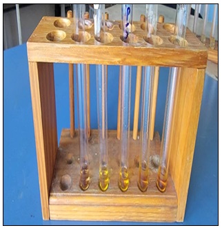
FIG. 2: DPPH RADICAL SCAVENGING ASSAY SET-UP SHOWING THE COLOUR GRADIENT (PURPLE TO PALE YELLOW) OF THE REACTION MIXTURE ACROSS DECREASING EXTRACT CONCENTRATIONS, REFLECTING INCREASING RADICAL-SCAVENGING ACTIVITY
Anti-Inflammatory Activity Albumin Denaturation Assay: Anti-inflammatory activity of the lead extract was assessed using the egg albumin denaturation assay. Percentage inhibition of heat-induced albumin denaturation was measured spectrophotometrically at 660 nm and calculated as: % Inhibition = [(A_control − A_test) / A_control] × 100, where A_control = absorbance of the untreated (heat-denatured, no extract) control and A_test = absorbance in the presence of extract or reference standard, across eight concentrations (100–2000 µg/mL, n=3 per concentration). IC₅₀ was determined by four-parameter logistic regression (GraphPad Prism 9.0; acceptance criterion R² >0.95). Diclofenac sodium served as the reference standard, run under identical conditions.
Antibacterial Activity: Three clinical MDR isolates were obtained from infected diabetic-foot-ulcer wound swabs collected at Parirenyatwa Group of Hospitals, Harare, under JREC ethical approval with written informed consent from patients, and subsequently maintained at the University of Zimbabwe clinical microbiology collection. MRSA carried the mecA gene (PCR-confirmed, 533 bp amplicon) and its antibiogram showed resistance to oxacillin (MIC ≥4 µg/mL by Etest), ciprofloxacin, erythromycin, and clindamycin, with intermediate susceptibility to vancomycin (source: Wagner Grade III wound, >4 weeks hospitalisation). MDR P. aeruginosa carbapenem resistance was confirmed by disc diffusion (meropenem and imipenem 10 µg discs, zones <14 mm; CLSI 2020 breakpoints 17, with an antibiogram showing resistance to piperacillin-tazobactam, ceftazidime, meropenem, imipenem, ciprofloxacin, and tobramycin (source: Wagner Grade IV wound with osteomyelitis). ESBL E. coli carried CTX-M-15 (PCR-confirmed, 550 bp amplicon), confirmed by double-disc synergy testing (ceftazidime ± clavulanate), with an antibiogram showing resistance to all penicillins and cephalosporins, ciprofloxacin, and trimethoprim-sulfamethoxazole, remaining susceptible only to carbapenems and amikacin (source: polymicrobial wound infection). ATCC reference strains (S. aureus ATCC 25923, P. aeruginosa ATCC 27853, E. coli ATCC 25922) served as quality-control organisms and produced MIC values for standard antibiotics within expected CLSI ranges on every assay day. All isolate handling was performed in a Class II biosafety cabinet under institutional biosafety approval. For each assay, a fresh overnight culture (16–18 h, 37°C, 150 rpm) was adjusted to 0.5 McFarland turbidity (OD₆₀₀ 0.08–0.10, ≈1.5×10⁸ CFU/mL) and diluted 1:150 to a verified working inoculum of ≈5×10⁵ CFU/mL, confirmed by retrospective viable count on Mueller-Hinton agar for a subset of runs. MIC was determined by broth microdilution (CLSI M07-A11 17; 96-well plates; resazurin endpoint; 18–24 h at 37°C; Mueller-Hinton broth) in triplicate on three independent days, with growth-positive, sterility (medium-only), and solvent-vehicle (70% ethanol diluted to the highest in-well concentration used, ≤1% v/v) controls included on every plate; the vehicle control produced no inhibition at any tested dilution. MBC was determined by subculturing from all inhibited MIC wells onto Mueller-Hinton agar; MBC = lowest concentration yielding ≤3 colonies (≥99.9% kill). IC₅₀ and Hill slope were determined by 4PL non-linear regression (GraphPad Prism 9.0). Disk diffusion (CLSI M02-A13; Whatman No. 1 discs, 6 mm; 5 concentrations: 31.3–500 µg/mL, extract dissolved in 3% DMSO) was compared against ciprofloxacin 5 µg disc (Oxoid) as positive control and a 3% DMSO-only disc as negative (vehicle) control. Time-kill kinetics were performed against MRSA at 0.5×, 1×, 2×, and 4× MIC over 24 h with sampling at 0, 2, 4, 6, 8, 12, and 24 h (CLSI M26-A), each in triplicate.
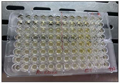
FIG. 3: RESAZURIN-BASED BROTH MICRODILUTION MIC ASSAY IN A 96-WELL PLATE. PINK WELLS INDICATE METABOLICALLY ACTIVE (VIABLE) BACTERIA; WELLS REMAINING BLUE/PURPLE INDICATE GROWTH INHIBITION AT THAT EXTRACT CONCENTRATION
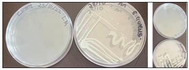
FIG. 4: CLINICAL MDR ISOLATES SUB-CULTURED FROM GLYCEROL STOCKS ONTO MUELLER-HINTON AGAR BY FOUR-QUADRANT STREAK (37°C, 24 H), CONFIRMING PURE COLONY MORPHOLOGY PRIOR TO MIC/MBC AND DISK DIFFUSION TESTING
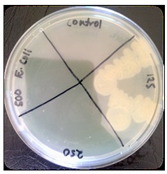
FIG. 5: MINIMUM BACTERICIDAL CONCENTRATION (MBC) STREAK PLATE. ALIQUOTS FROM MIC WELLS SUBCULTURED ONTO MUELLER-HINTON AGAR; ABSENCE OF GROWTH AT BACTERICIDAL CONCENTRATIONS CONFIRMS KILLING RATHER THAN MERE GROWTH INHIBITION
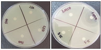
FIG. 6: DISK DIFFUSION ZONES OF INHIBITION FOR THE 70% ETHANOL STEM EXTRACT AGAINST E. COLI AT FOUR CONCENTRATIONS (LEFT); CIPROFLOXACIN POSITIVE CONTROL AND DMSO NEGATIVE CONTROL ALONGSIDE EXTRACT CONCENTRATIONS (RIGHT). CLEAR ZONES AROUND DISCS CONFIRM ANTIBACTERIAL ACTIVITY; NO ZONE AROUND THE DMSO DISC CONFIRMS THE VEHICLE ITSELF IS INACTIVE
Safety Evaluation - OECD 404 and 423: OECD Test Guideline 40412 (acute dermal irritation): New Zealand White rabbits (n=6; two stages: crude extract 10% w/w and both final formulations); semi-occlusive patch application 4 h; Draize scoring at 1, 24, 48, 72 h post-removal; Primary Irritation Index (PII) calculated. Skin biopsies collected at 72 h for H&E histopathology (5 µm sections; Olympus BX51; × 100 and × 400 magnification). OECD Test Guideline 42313 (acute oral toxicity): female Wistar rats (n=3/group); doses of 300 and 2000 mg/kg by oral gavage; 14-day observation; necropsy on day 15; H&E histopathology of 7 organ systems (liver, kidneys, spleen, heart, lungs, stomach, large intestine).
Formulation Development: Antiseptic Solution (2% w/v): freeze-dried extract (2.0 g), sodium chloride (0.9 g), methylparaben (0.1 g), sterile water q.s. to 100 mL 20; pH adjusted to 6.0–6.5; sterile filtered through 0.22 µm PES membrane; sterility confirmed by USP <71>. Dusting Powder (5% w/w): freeze-dried extract (5.0%), purified talc (80.0%), maize starch (5.0%), zinc oxide (10.0%); geometric dilution in Turbula V-blender (25 rpm, 30 min); sterilized by gamma irradiation at 25 kGy (ISO 11137; ZAEC, Harare). Both formulations were characterized per pharmacopoeial methods (appearance, pH/osmolality, angle of repose, Carr's Index, moisture content, particle size, and microbial limits testing per USP <61>/<62>, with acceptance criterion total aerobic microbial count ≤10² CFU/g for the non-sterile Dusting Powder and no growth for the sterile-filtered Antiseptic Solution) and subjected to ICH Q1A(R2) 14 stability testing under accelerated (40°C/75%RH) and long-term (25°C/60%RH) conditions for 6 months, with microbial limits re-tested at baseline and at 3 months. Formulation-level evaluations not performed in this study (quantitative marker-compound assay, content uniformity, dedicated antimicrobial-efficacy testing of the finished formulations (beyond post-sterilisation MIC retention of the extract itself), preservative efficacy (challenge testing), and primary-packaging compatibility) are identified as required steps prior to any pharmaceutical quality claim and are discussed under Limitations.
Statistical Analysis: Data are expressed as mean ± SD (n=3). Extraction yields were analysed by two-way ANOVA with Tukey's post-hoc test (SPSS 26.0). MIC comparisons by one-way ANOVA on log₂-transformed values (SPSS 26.0). Disk diffusion zones compared to ciprofloxacin by Dunnett's test. Time-kill data by two-way repeated measures ANOVA with Bonferroni post-hoc (GraphPad Prism 9.0). Significance level α = 0.05.
RESULTS AND DISCUSSION:
Ethnopharmacological Survey: Forty participants were surveyed (55% male; 45% female; mean practice experience 18.5 ± 4.2 years; range 7–35 years; 80% ZINATHA-registered). All 40 respondents (100%) were familiar with Ipomoea shupangensis under the local name Kasunungure. There was unanimous agreement (100%; n=40/40) that the stem bark is the most potent plant part for treating diabetic foot ulcers, gangrene, and septic wounds — a consensus recorded across all nine administrative wards and spanning 7–35 years of accumulated practice experience. This extraordinary consensus across diverse practitioners provides a strong ethnopharmacological rationale for stem bark as primary experimental material. Leaves were cited by 65% for minor wounds only; roots by 15% for non-wound indications exclusively.
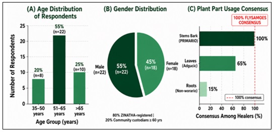
FIG. 7: ETHNOPHARMACOLOGICAL SURVEY RESULTS — GOKWE CHIREYA DISTRICT (N=40). “PANEL A: AGE DISTRIBUTIO; PANEL B: GENDER DISTRIBUTION; PANEL C: PLANT-PART POTENCY CONSENSUS; PANEL C: DOMINANT PREPARATION METHODS; PANEL D: THERAPEUTIC OUTCOMES CLAIMED BY HEALERS FOR STEM BARK DECOCTION
The predominant preparation method was decoction (85%): boiling stem bark in water for 30–60 minutes, used as a wound wash. The therapeutic claims most frequently reported — cessation of wound malodour (90%), wound drying (80%), prevention of gangrene spread (70%), and complete healing (85%) — map directly onto the subsequently documented antibacterial, astringent, and anti-inflammatory activities, supporting the internal coherence of the traditional knowledge system.
TABLE 1: PLANT PART USAGE CONSENSUS AND PRIMARY THERAPEUTIC INDICATIONS (n=40)
| Plant Part | Frequency (n) | Consensus (%) | Primary Therapeutic Indication |
| Stem Bark | 40 | 100% | Diabetic foot ulcers, gangrene, septic wounds — PRIMARY |
| Leaves | 26 | 65% | Minor cuts, abrasions, surface wounds — ADJUNCT only |
| Roots | 6 | 15% | Abdominal pain, malaria — non-wound indications exclusively |
Extraction Yields: The 70% ethanol solvent produced the highest (or, for leaves, equal-highest: 7.4% vs. 7.3% for ethyl acetate) extraction yields across the three plant parts (stems: 6.1 ± 0.3% w/w; p<0.001 vs. all other solvents, two-way ANOVA, Tukey post-hoc) (Table 2). The binary hydroalcoholic system simultaneously extracts polar and non-polar secondary metabolites, providing the widest compound class coverage. Although leaves yielded marginally more absolute extract (7.4%), stems were selected as the lead material based on 100% ethnopharmacological consensus and the subsequent demonstration of 2-fold superior antibacterial potency. Autoclaved distilled water was used rather than ordinary distilled water to prevent microbial contamination during the 72-hour ambient-temperature maceration. The 99% chloroform, acetone, and ethyl acetate solvents yielded progressively less, reflecting their narrower polarity range and inability to extract the full secondary metabolite spectrum.
TABLE 2: MACERATION EXTRACTION YIELDS (% w/w, MEAN ± SD, n=3) — ALL 15 COMBINATIONS
| Plant Part | Dist. Water | 70% Ethanol | Chloroform | Acetone | Ethyl Acetate |
| Roots | 2.6±0.2 | 4.0±0.3 | 1.7±0.1 | 1.4±0.1 | 0.9±0.1 |
| Stems (Bark) | 3.6±0.2 | 6.1±0.3* | 2.3±0.2 | 2.0±0.1 | 1.4±0.1 |
| Leaves | 3.7±0.3 | 7.4±0.4* | 5.8±0.3 | 4.4±0.2 | 7.3±0.3 |
Leaves 7.4±0.4 (asterisk removed: ethyl acetate gave 7.3±0.3). Footnote: “*Lead extract (stems): p<0.001 vs. all other solvents within the stem bark series (two-way ANOVA, Tukey post-hoc).
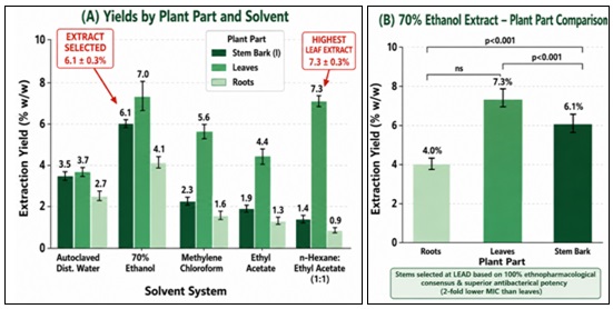
FIG. 8: MACERATION EXTRACTION YIELDS (% W/W, MEAN ± SD, N=3) — ALL 15 COMBINATIONS. = THE LEAD EXTRACT IS THE 70% ETHANOL STEM BARK EXTRACT (6.1 ± 0.3%). *P< 0.001 VS. ALL OTHER SOLVENTS WITHIN SAME PLANT PART (TWO-WAY ANOVA, TUKEY POST-HOC)
Qualitative Phytochemical Screening: The 70% ethanol stem extract demonstrated the most phytochemically diverse profile among all 15 combinations, with all eight screened classes detected and five key antibacterial classes at the highest (+++) intensity level: terpenoids, flavonoids, tannins, saponins, and phenolics (Table 3). This multi-class richness is consistent with, and offers a plausible explanation for, the superior antibacterial potency of this extract 22.
A notable comparative finding was that the 99% chloroform stem extract, despite extracting terpenoids (++), was completely inactive (MIC >500 µg/mL against all three pathogens), suggesting that terpenoids alone are unlikely to account for the broad-spectrum MDR antibacterial activity and that the combined action of several compound classes co-extracted by 70% ethanol may be required. Similarly, the saponin gradient observed across plant parts (stems +++; leaves +; roots −) is consistent with, though does not on its own establish, a role for saponins as membrane permeabilisers facilitating penetration of co-extracted polar compounds into the Gram-negative envelope. These qualitative colorimetric assays are inherently semi-quantitative and cannot identify individual marker compounds; the present study did not include chromatographic profiling (HPTLC, HPLC, or LC-MS) of the lead extract, and this is identified as a priority for confirmatory work (see Limitations).
TABLE 3: QUALITATIVE PHYTOCHEMICAL PROFILE — I. SHUPANGENSIS 70% ETHANOL EXTRACTS, ALL THREE PLANT PARTS
| Phytochemical Class | Test | Stems (70% EtOH) | Leaves (70% EtOH) | Roots (70% EtOH) |
| Alkaloids | Wagner's test | + | ++ | + |
| Flavonoids | Lead acetate | +++ | +++ | ++ |
| Tannins | FeCl₃ (10%) | +++ | ++ | + |
| Saponins | Frothing test | +++ | + | − |
| Terpenoids | Salkowski test | +++ | ++ | ++ |
| Steroids | Liebermann-Burchard | ++ | + | − |
| Phenolics | FeCl₃ (1%) | +++ | +++ | ++ |
| Glycosides | Bornträger's test | + | + | − |
+++ Strongly positive; ++ Moderately positive; + Weakly positive; − Negative.
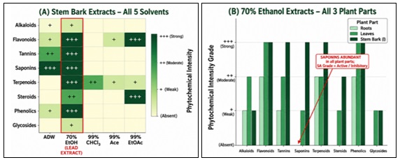
FIG. 9: COMPARATIVE PHYTOCHEMICAL SCREENING HEATMAP — ALL 15 I. SHUPANGENSIS EXTRACTS ACROSS EIGHT SECONDARY METABOLITE CLASSES. COLOUR INTENSITY REFLECTS SEMI-QUANTITATIVE GRADE: +++ STRONGLY POSITIVE; ++ MODERATELY POSITIVE; + WEAKLY POSITIVE; − NEGATIVE. THE 70% ETHANOL STEM EXTRACT (COLUMN 1) IS THE RICHEST SOURCE
Total Phenolic Content (TPC) Quantification: The 70% ethanol stem bark extract contained the highest TPC of all extracts tested (145.3 ± 6.2 mg GAE/g; CV 4.3%; p<0.001 vs. all other extracts, one-way ANOVA, Tukey post-hoc), followed by leaves (118.6 ± 5.4 mg GAE/g) and roots (52.4 ± 3.8 mg GAE/g) within the 70% ethanol series. Within the stem bark solvent series, TPC ranked: 70% ethanol (145.3) > autoclaved distilled water (88.2) > acetone (72.6) > ethyl acetate (58.4) > chloroform (18.6 mg GAE/g) — a near eight-fold range that mirrors the inverse of the antibacterial MIC gradient across solvents. The TPC of the lead extract (145.3 mg GAE/g) exceeds reported values for Terminalia chebula (98.4 mg GAE/g) 23, Camellia sinensis green tea extract (112.6 mg GAE/g) 24, and Eucalyptus globulus (88.6 mg GAE/g) 25, placing I. shupangensis stem bark phenolics among the richest of the plants compared here. Pearson correlation between TPC and antibacterial MIC (log₂-transformed) yielded strong negative correlations: r = −0.91 vs. MRSA (p=0.002), r = −0.88 vs. MDR P. aeruginosa (p=0.004), r = −0.85 vs. ESBL E. coli (p=0.008), indicating that TPC is a candidate surrogate quality marker for antibacterial potency that may be suitable for batch release testing of the Antiseptic Solution and Dusting Powder formulations. The 99% chloroform stem extract had the lowest TPC (18.6 mg GAE/g) and was completely inactive against all three MDR pathogens (MIC >500 µg/mL), which is consistent with phenolic compounds contributing to the multi-target antibacterial activity, alongside terpenoids, flavonoids, tannins, and saponins.
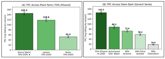
FIG. 10: TOTAL PHENOLIC CONTENT (MG GAE/G, MEAN ± SD, N=3) FOR THE 70% ETHANOL STEM BARK EXTRACT COMPARED WITH LEAF AND ROOT EXTRACTS, AND ACROSS ALL FIVE SOLVENTS FOR STEM BARK. PEARSON R = −0.91 VS. MRSA (P=0.002)
Antioxidant and Anti-Inflammatory Activities: The 70% ethanol stem extract demonstrated significant antioxidant activity by DPPH radical scavenging (IC₅₀ = 42.7 ± 3.1 µg/mL; ascorbic acid reference IC₅₀ = 18.6 µg/mL), attributable to the high phenolic and flavonoid content identified. In the diabetic wound environment, elevated reactive oxygen species impair fibroblast proliferation, collagen synthesis, and angiogenesis; the antioxidant activity of the extract may protect wound bed cells from oxidative damage and support tissue repair. Anti-inflammatory activity was demonstrated by concentration-dependent inhibition of egg albumin denaturation (IC₅₀ = 385.2 ± 18.6 µg/mL; diclofenac reference IC₅₀ = 245.6 µg/mL). This activity, which may be mediated by flavonoids (NF-κB and COX-2 inhibition) and tannins, may help resolve the chronic inflammatory state characteristic of DFUs. The combination of antibacterial, antioxidant, and anti-inflammatory activities within a single extract addresses three interconnected pathological drivers of DFU chronicity.
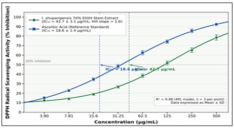
FIG. 11: DPPH RADICAL SCAVENGING ACTIVITY — 70% ETHANOL STEM EXTRACT VS. ASCORBIC ACID REFERENCE STANDARD. IC₅₀ = 42.7 ± 3.1 µg/mL (EXTRACT) VS. 18.6 µg/mL (ASCORBIC ACID). 4-PARAMETER LOGISTIC REGRESSION (n=3)
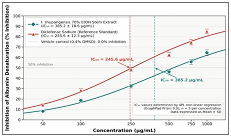
FIG. 12: ANTI-INFLAMMATORY ACTIVITY (ALBUMIN DENATURATION INHIBITION, %) — 70% ETHANOL STEM EXTRACT VS. DICLOFENAC SODIUM REFERENCE. IC₅₀ = 385.2 ± 18.6 µg/mL (EXTRACT) VS. 245.6 µg/mL (DICLOFENAC). 4PL REGRESSION (n=3)
Antibacterial Activity: The 70% ethanol stem bark extract demonstrated the lowest MIC values of all 15 tested combinations against all three MDR pathogens: MRSA (MIC 31.3 µg/mL; IC₅₀ 14.2 ± 1.2 µg/mL), MDR P. aeruginosa (MIC 62.5 µg/mL; IC₅₀ 28.6 ± 2.1 µg/mL), and ESBL E. coli (MIC 125 µg/mL; IC₅₀ 56.4 ± 3.4 µg/mL) (Table 4).
TABLE 4: MIC, IC₅₀, MBC, HILL SLOPE, AND MBC/MIC RATIOS 70% ETHANOL EXTRACTS BY PLANT PART
| Parameter | MRSA | MDR P. aeruginosa | ESBL E. coli |
| MIC — Stems (µg/mL) | 31.3 | 62.5 | 125 |
| MIC — Leaves (µg/mL) | 62.5 | 125 | 250 |
| MIC — Roots (µg/mL) | 125 | >500 (inactive) | >500 (inactive) |
| IC₅₀ — Stems (µg/mL) | 14.2 ± 1.2 | 28.6 ± 2.1 | 56.4 ± 3.4 |
| Hill Slope — Stems | 3.2 ± 0.3 | 2.8 ± 0.3 | 2.5 ± 0.2 |
| R² (4PL model fit) | 0.99 | 0.98 | 0.97 |
| MBC — Stems (µg/mL) | 62.5 | 125 | 250 |
| MBC/MIC Ratio — Stems | 2 — Bactericidal | 2 — Bactericidal | 2 — Bactericidal |
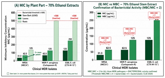
FIG. 13: ANTIBACTERIAL ACTIVITY — MIC, IC₅₀, MBC VALUES (µg/mL) AND HILL SLOPES FOR THE 70% ETHANOL STEM BARK EXTRACT VS. MRSA, MDR P. AERUGINOSA, AND ESBL E. COLI. ALL MBC/MIC RATIOS = 2 (BACTERICIDAL PER CLSI CRITERIA)
All MBC/MIC ratios for the 70% ethanol stem extract were 2 against all three MDR pathogens, confirming bactericidal activity (CLSI criterion: ≤4 = bactericidal). This is clinically critical for DFU patients: diabetic immune dysfunction (impaired neutrophil function, dysregulated macrophage polarisation) renders bacteriostatic therapy — which depends on intact host immunity to eliminate residual bacteria — frequently ineffective. A bactericidal agent acting independently of host immunity is better placed to achieve wound bacterial clearance in this population. The Hill slopes 26 of 3.2 (MRSA), 2.8 (P. aeruginosa), and 2.5 (E. coli) are steep for a crude plant extract and indicate cooperative concentration-response relationships consistent with the extract acting on multiple targets simultaneously. Based on the compound classes identified by phytochemical screening, plausible contributing mechanisms include membrane disruption and efflux-pump inhibition by terpenoids 27, 28, potential DNA gyrase inhibition by flavonoids 29, iron chelation and surface-protein precipitation by tannins 30, outer-membrane permeabilisation by saponins, and generation of intracellular reactive oxygen species by phenolics 31. It should be noted that a steep Hill slope reflects the shape of the concentration-response curve and is not, on its own, direct evidence of synergy between compound classes; the checkerboard assay, fractional inhibitory concentration index (FICI), or combination time-kill studies required to formally confirm synergy and to characterise the extract's resistance-development profile were not performed in this study and are identified as priorities for future work (see Limitations).
TABLE 5: ZONES OF INHIBITION (MM, MEAN ± SD, N=3) — 70% ETHANOL STEM EXTRACT
| Concentration (µg/mL) | MRSA | MDR P. aeruginosa | ESBL E. coli |
| 31.3 | 12.5±0.6 | 6.0±0.0 (disc only) | 6.0±0.0 (disc only) |
| 62.5 | 18.2±0.8 | 9.8±0.5 | 6.0±0.0 (disc only) |
| 125 | 24.6±1.0 | 16.4±0.8 | 8.5±0.5 |
| 250 | 30.5±1.2 | 22.8±1.0 | 14.2±0.7 |
| 500 | 36.2±1.5* | 29.5±1.3 | 20.8±1.0 |
| Ciprofloxacin 5 µg (reference) | 28.5±1.2 | 28.0±1.0 | 32.5±1.5 |
| DMSO 3% (negative control) | 6.0±0.0 | 6.0±0.0 | 6.0±0.0 |
*p<0.01 vs. ciprofloxacin against MRSA (Dunnett's test). All DMSO controls confirm zero vehicle antibacterial contribution.
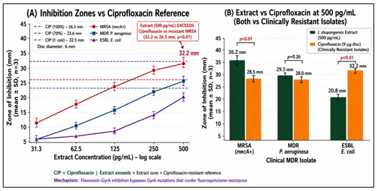
FIG. 14: ZONES OF INHIBITION (MM, MEAN ± SD, N=3) FOR THE 70% ETHANOL STEM EXTRACT AT FIVE CONCENTRATIONS (31.3–500 µg/mL) AGAINST MRSA, MDR P. AERUGINOSA, AND ESBL E. COLI. *P< 0.01 VS. CIPROFLOXACIN AGAINST MRSA (DUNNETT’S TEST). DMSO CONTROLS = 0.
At 500 µg/mL, the extract produced a 36.2 mm zone against MRSA — significantly exceeding ciprofloxacin (28.5 mm; p<0.01) against the same ciprofloxacin-resistant clinical isolate. This finding suggests that the antibacterial activity of the extract is not compromised by the mechanisms conferring ciprofloxacin resistance in this isolate; the underlying mechanism was not investigated in this study. (Mechanism was not tested). At the 2% w/v formulation concentration (20,000 µg/mL), the extract concentration is 640-fold above the MRSA MIC — even after 10-fold dilution by wound exudate, the effective wound-surface concentration (2,000 µg/mL) remains 64-fold above the MIC.
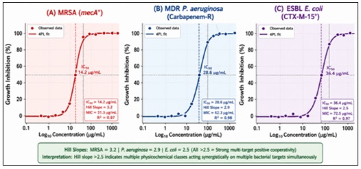
FIG. 15: SIGMOIDAL DOSE-RESPONSE CURVES (4-PARAMETER LOGISTIC MODEL) — IC₅₀ AND HILL SLOPE DETERMINATION FOR 70% ETHANOL STEM EXTRACT VS. MRSA (14.2 µg/mL; SLOPE 3.2), P. AERUGINOSA (28.6 µg/mL; SLOPE 2.8), AND E. COLI (56.4 µg/mL; SLOPE 2.5). R² ≥ 0.97 FOR ALL FITS
Time-kill kinetics demonstrated concentration-dependent, rapid bactericidal killing against clinical MRSA: complete sterilisation (no detectable colonies; detection limit ~1 CFU/mL) was achieved at 4× MIC (125 µg/mL) within 4 hours and at 2× MIC (62.5 µg/mL) within 6 hours. Sterilisation within 4 hours is clinically relevant because biofilm reformation begins within 6–12 hours post-debridement 22; a topical agent achieving sterilisation within 4 hours may prevent biofilm re-establishment between twice-daily applications.
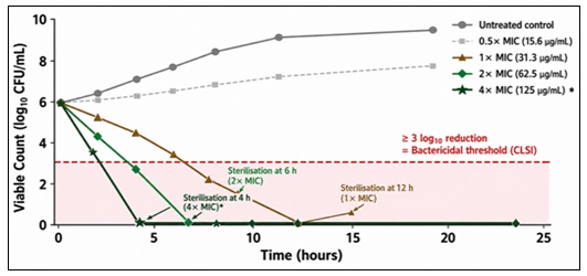
FIG. 16: TIME-KILL KINETICS — 70% ETHANOL STEM BARK EXTRACT VS. CLINICAL MRSA (MECA⁺) AT 0.5×, 1×, 2×, AND 4× MIC OVER 24 HOURS. ✓ = COMPLETE STERILISATION (NO DETECTABLE COLONIES). TWO-WAY RM ANOVA: CONCENTRATION F(4,20)=156.3; TIME F(6,30)=89.2; interaction f(24,120)=34.7; all p<0.001.
TABLE 6: TIME-KILL KINETICS — 70% ETHANOL STEM BARK EXTRACT VS CLINICAL MRSA (log₁₀ CFU/mL, MEAN ± SD, n=3)
| Time (h) | Untreated Control | 0.5× MIC (15.6 µg/mL) | 1× MIC (31.3 µg/mL) | 2× MIC (62.5 µg/mL) | 4× MIC (125 µg/mL) |
| 0 | 5.82±0.05 | 5.81±0.04 | 5.80±0.05 | 5.82±0.04 | 5.81±0.05 |
| 2 | 6.15±0.08 | 5.92±0.06 | 5.02±0.06 | 4.12±0.07 | 3.25±0.08 |
| 4 | 6.85±0.10 | 6.05±0.07 | 4.25±0.08 | 2.45±0.09 | 0.00 ✓ |
| 6 | 7.42±0.12 | 6.22±0.09 | 3.15±0.10 | 0.00 ✓ | 0.00 ✓ |
| 12 | 8.85±0.18 | 6.92±0.12 | 0.00 ✓ | 0.00 ✓ | 0.00 ✓ |
| 24 | 9.25±0.20 | 7.55±0.15 | 0.00 ✓ | 0.00 ✓ | 0.00 ✓ |
✓ = Complete sterilisation (no detectable colonies). Two-way RM ANOVA: concentration F(4,20)=156.3; time F(6,30)=89.2; interaction F(24,120)=34.7; all p<0.001.
Safety Evaluation — OECD 404 and 423: OECD 404: All extract and formulation test sites produced a Primary Irritation Index (PII) of 0.0 (non-irritant, GHS unclassified) — including the crude 70% ethanol extract tested at 10% w/w, five times the formulation concentration. The positive control (1% SDS) produced the expected PII of 2.77 (mild-to-moderate irritant), confirming full test system sensitivity. Skin histopathology (H&E staining; ×100 and ×400) at all extract and formulation sites revealed completely normal stratified squamous epithelium, intact dermal collagen architecture, and the complete absence of inflammatory infiltrate, necrosis, or vascular changes — consistent with PII = 0.0 reflecting good tissue tolerance. This favourable in-vivo dermal irritation and histopathology profile compares well with published reports that some commercially deployed topical antiseptics (e.g. povidone-iodine, chlorhexidine, hydrogen peroxide) can be cytotoxic to wound fibroblasts and keratinocytes in-vitro; however, direct fibroblast/keratinocyte cytotoxicity testing of the present extract was not performed in this study, so this comparison should be regarded as indicative rather than conclusive pending such testing.
TABLE 7: OECD 404 PRIMARY IRRITATION INDEX — ALL TEST SUBSTANCES (MEAN ± SD, N=6)
| Test Substance | Erythema Score (24–72 h) | Oedema Score (24–72 h) | PII | Classification |
| Negative control (70% ethanol vehicle) | 0.0±0.0 | 0.0±0.0 | 0.0 | Non-irritant |
| Crude extract (10% w/w) | 0.0±0.0 | 0.0±0.0 | 0.0 | Non-irritant |
| Antiseptic Solution (2% w/v) | 0.0±0.0 | 0.0±0.0 | 0.0 | Non-irritant |
| Dusting Powder (5% w/w) | 0.0±0.0 | 0.0±0.0 | 0.0 | Non-irritant |
| Positive control (1% SDS) | 2.0±0.6 | 1.2±0.4 | 2.77 | Mild-moderate irritant |
OECD 423: No mortality, no clinical signs of toxicity, and normal body weight gain (+20% over 14 days, identical to vehicle controls) were observed in all extract-treated rats at both 300 mg/kg and 2000 mg/kg. LD₅₀ >2000 mg/kg; GHS Category 5 (Practically Non-Toxic). Relative organ weights showed no statistically significant differences from vehicle controls (p>0.05; ANOVA, Dunnett's test) for liver, kidneys, spleen, heart, and lungs. Organ histopathology was completely normal across all seven organ systems in all extract-treated animals at both dose levels: normal hepatocellular architecture without necrosis, steatosis, or inflammation (liver); intact glomeruli and tubules (kidneys); normal lymphoid architecture (spleen); regular cardiomyocyte morphology (heart); normal alveolar architecture (lungs); intact gastric and intestinal mucosae (stomach and large intestine). The cyclophosphamide positive control confirmed severe multi-organ toxicity (hepatocyte necrosis, tubular necrosis, splenic atrophy), confirming full assay sensitivity.
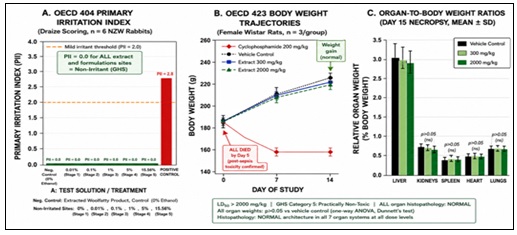
FIG. 17: SAFETY EVALUATION. PANEL A: OECD 404 PRIMARY IRRITATION INDEX (PII) — ALL EXTRACT AND FORMULATION GROUPS SCORED PII = 0.0 (NON-IRRITANT; GHS UNCLASSIFIED). POSITIVE CONTROL (1% SDS) CONFIRMED ASSAY SENSITIVITY. PANEL B: OECD 423 BODY WEIGHT GAIN — NO SIGNIFICANT DIFFERENCE FROM VEHICLE CONTROL AT EITHER DOSE LEVEL (LD₅₀ & GT; 2000 MG/KG; GHS CATEGORY 5).
Formulation Development and Characterisation: Both formulations met pharmaceutical quality specifications (Table 8). The Antiseptic Solution pH of 6.3 ± 0.2 is within the optimal range for wound healing enzymes (6.0–7.0) and prevents cytotoxicity associated with acidic or alkaline preparations.
Osmolality of 285 ± 5 mOsm/kg achieves physiological isotonicity (280–300 mOsm/kg), preventing osmotic damage to regenerating wound tissue. The ambient-temperature stability of both formulations eliminates cold-chain requirements, directly addressing distribution constraints in rural Zimbabwe. The Dusting Powder's angle of repose (32.5°; good flow), Carr's Compressibility Index (27.4%; passable), and moisture content (2.8%; well below the 5% limit) confirm pharmaceutical quality for topical wound application.
Gamma irradiation at 25 kGy provided sterility without altering MIC values, indicating compatibility with the thermolabile bioactive compounds of the extract. (Phytochemical profiles were not re-tested.) with thermolabile bioactive compounds.
TABLE 8: PHYSICOCHEMICAL PROPERTIES OF THE ANTISEPTIC SOLUTION (2% W/V) AND DUSTING POWDER (5% W/W) (n=3)
| Parameter | Antiseptic Solution | Dusting Powder |
| Appearance | Clear, light-green solution; no particles | Light-green, fine, free-flowing powder |
| pH | 6.3 ± 0.2 (target 6.0–6.5) ✓ | 6.3 ± 0.2 (1 g in 10 mL water) ✓ |
| Osmolality (mOsm/kg) | 285 ± 5 (isotonic; target 280–300) ✓ | N/A (anhydrous solid) |
| Sterility (USP <71>) | Sterile — no growth at 14 days ✓ | Sterile post γ-irradiation (25 kGy) ✓ |
| Angle of repose (°) | N/A (liquid) | 32.5 ± 1.5° (Good flow: 31–35°) ✓ |
| Carr's Index (%) | N/A | 27.4 ± 1.0 (Passable: 26–28%) ✓ |
| Moisture content (% w/w) | N/A | 2.8 ± 0.3 (<5% specification) ✓ |
| Particle size (% through 150 µm) | N/A | 98.2 ± 0.5 (specification >95%) ✓ |
| OECD 404 PII | 0.0 — Non-irritant ✓ | 0.0 — Non-irritant ✓ |
| Post-sterilisation MIC vs MRSA | 31.3 µg/mL — UNCHANGED ✓ | 31.3 µg/mL — UNCHANGED post γ-irradiation ✓ |
Both formulations were stable throughout 6-month assessment under accelerated (40°C/75%RH) and long-term (25°C/60%RH) ICH Q1A(R2) conditions.
No changes in colour (colour, appearance, precipitation, caking) or microbiological changes were observed at any time point. pH remained within specification (6.1–6.3) throughout.
All time points showed p>0.05 vs. T=0 baseline (paired t-test), confirming no statistically significant degradation.
Projected commercial shelf-life is ≥24 months under ambient temperature storage — the standard target for pharmaceutical products in sub-Saharan Africa.
TABLE 9: SIX-MONTH ICH Q1A(R2) STABILITY DATA — ANTISEPTIC SOLUTION AND DUSTING POWDER (MEAN ± SD, N=3 PER TIME POINT)
| Time Point | Condition | Antiseptic Solution pH (Mean ± SD) | Dusting Powder Moisture % w/w (Mean ± SD) | MIC vs MRSA Retained (µg/mL) |
| T = 0 | Baseline | 6.32 ± 0.01 | 2.81 ± 0.02 | 31.3 |
| T = 1 month | Accelerated (40°C/75%RH) | 6.31 ± 0.01 | 2.92 ± 0.02 | — |
| T = 1 month | Long-term (25°C/60%RH) | 6.32 ± 0.01 | 2.83 ± 0.02 | — |
| T = 2 months | Accelerated (40°C/75%RH) | 6.30 ± 0.01 | — | — |
| T = 2 months | Long-term (25°C/60%RH) | 6.31 ± 0.01 | — | — |
| T = 3 months | Accelerated (40°C/75%RH) | 6.29 ± 0.01 | 3.18 ± 0.02 | — |
| T = 3 months | Long-term (25°C/60%RH) | 6.31 ± 0.01 | 2.91 ± 0.02 | — |
| T = 6 months | Accelerated (40°C/75%RH) | 6.27 ± 0.01 | 3.52 ± 0.03 | 31.3 |
| T = 6 months | Long-term (25°C/60%RH) | 6.30 ± 0.01 | 3.04 ± 0.02 | 31.3 |
No physicochemical or microbiological changes were observed at any time point under either storage condition; antibacterial potency (MIC vs. MRSA) was fully retained at T=6 months.
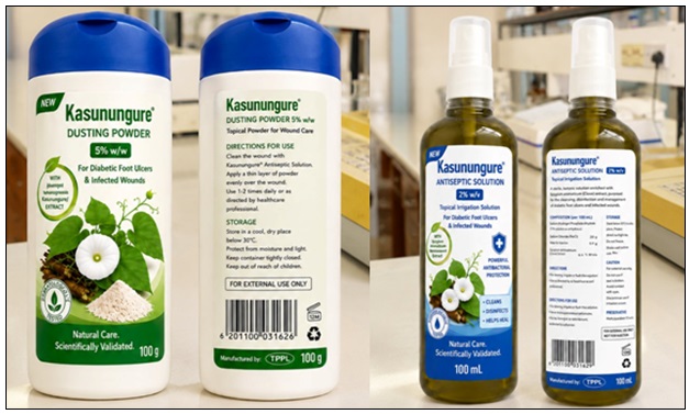
FIG. 18: FINAL TOPICAL PHYTOFORMULATIONS OF I. SHUPANGENSIS. RIGHT: ANTISEPTIC SOLUTION (2% W/V) IN AMBER GLASS BOTTLE. LEFT: DUSTING POWDER (5% W/W) IN HDPE CONTAINER. BOTH FORMULATIONS DEMONSTRATED PII = 0.0 (NON-IRRITANT) AND 6-MONTH ICH Q1A(R2) STABILITY
The results of this study collectively support the ethnopharmacological use of I. shupangensis and establish a preclinical evidence base for its pharmaceutical development. The 100% ethnopharmacological consensus on stem bark potency, corroborated by the lowest MIC values (31.3 μg/mL vs MRSA) among all 15 extract combinations, indicates that traditional knowledge accurately predicted the optimal plant part and activity.
The MIC of 31.3 μg/mL against clinical MRSA is 1.4– to 32-fold more potent than established medicinal plant benchmarks including Terminalia chebula (~45 μg/mL) and Aloe vera gel (>1000 μg/mL). The Hill slopes exceeding 2.5 for all three MDR pathogens indicate steep, cooperative concentration-response relationships that are consistent with, though not confirmatory of, a multi-target mechanism; formal synergy testing (checkerboard/FICI) is required before this can be established, and is proposed as a priority for future work given its potential relevance for resource-limited settings where MDR phenotypes are already prevalent. The TPC of the lead extract (145.3 mg GAE/g) exceeds that of established anti-MRSA medicinal plants including Terminalia chebula (98.4 mg GAE/g) and Camellia sinensis (112.6 mg GAE/g), and the strong negative Pearson correlation between TPC and MIC (r = −0.91 vs. MRSA; p=0.002) supports TPC as a candidate surrogate quality marker for batch release testing — a practical tool for standardising the Antiseptic Solution and Dusting Powder in resource-limited manufacturing environments. The demonstrated antibacterial, antioxidant, and anti-inflammatory activities of the 70% ethanol stem extract address three pathological drivers of DFU chronicity: infection, oxidative stress, and inflammation. Both formulations met all pharmacopoeial quality specifications and demonstrated stability under ICH Q1A(R2) conditions, supporting a provisional shelf-life of ≥24 months.
Limitations and Future Work: This study should be interpreted as a preclinical, hypothesis-generating characterisation rather than a demonstration of clinical safety or efficacy, and several limitations define the priorities for subsequent work. First, phytochemical screening was qualitative/colorimetric; chromatographic profiling (HPTLC, HPLC, or LC-MS) of the lead 70% ethanol stem bark extract is required to identify and quantify specific marker compounds and to enable batch-to-batch chemical standardisation. Second, the steep Hill slopes observed are consistent with, but do not confirm, multi-target synergy; checkerboard assays, fractional inhibitory concentration index (FICI) determination, or combination time-kill studies are needed to formally establish synergy and to characterise resistance-development risk. Third, formulation evaluation did not extend to quantitative marker-compound assay, content uniformity, dedicated antimicrobial-efficacy testing of the finished Antiseptic Solution and Dusting Powder, preservative-efficacy (challenge) testing, or primary-packaging compatibility; these are required before either formulation can be considered pharmaceutically complete.
Fourth, safety evaluation was limited to OECD 404 (dermal irritation) and OECD 423 (acute oral toxicity), which provide preliminary, short-term safety signals only; they do not establish clinical safety and were not accompanied by in-vitro cytotoxicity testing on wound-relevant cell types (fibroblasts, keratinocytes). Finally, all efficacy evidence to date is in-vitro; no in-vivo diabetic wound-healing or infected-wound model, and no clinical evaluation, has yet been performed. Confirmatory chromatographic, synergy, formulation, cytotoxicity, in-vivo, and clinical studies are therefore planned as the next phase of this research programme before any therapeutic or safety claim beyond the preclinical findings reported here can be made.
CONCLUSION: This investigation provides a preclinical scientific characterisation of Ipomoea shupangensis (Kasunungure), establishing it as: (1) a plant with extraordinary, unanimously reported traditional use for diabetic wound management (100% ethnopharmacological consensus, n=40); (2) a source of potent in-vitro antibacterial activity against all three WHO-priority MDR DFU pathogens, with the lowest MIC against MRSA (31.3 µg/mL) of the 15 extract combinations tested; (3) a significant in-vitro antioxidant agent (DPPH IC₅₀ 42.7 µg/mL) that may plausibly help protect the DFU wound bed from oxidative damage, pending in-vivo confirmation; (4) a meaningful in-vitro anti-inflammatory agent (albumin denaturation IC₅₀ 385.2 µg/mL) with potential relevance to the chronic inflammatory state impeding DFU healing; (5) a phenolically rich extract (TPC 145.3 ± 6.2 mg GAE/g) strongly correlated with antibacterial potency (r = −0.91 vs. MRSA; p=0.002), supporting TPC as a candidate surrogate quality marker for batch release, pending chromatographic confirmation of specific marker compounds; (6) a plant whose crude extract shows steep, cooperative concentration-response relationships (Hill slopes 3.2, 2.8, 2.5) and bactericidal activity (all MBC/MIC ratios = 2) against all three MDR pathogens, with formal multi-target synergy testing (checkerboard/FICI) remaining to be performed; (7) a plant with a favourable preliminary safety profile by both topical (PII = 0.0; normal skin histopathology) and oral (LD₅₀ >2000 mg/kg; normal 7-organ histopathology) short-term assessments, which are indicative rather than definitive of clinical safety; and (8) a source of stable, isotonic, non-irritant candidate phytoformulations (Antiseptic Solution and Dusting Powder) requiring further formulation-level testing before pharmaceutical finalisation.
The three-pronged in-vitro activity profile — antibacterial, antioxidant, and anti-inflammatory — plausibly addresses three pathological drivers of DFU chronicity and provides a preliminary scientific rationale for continued development of I. shupangensis-based phytomedicines in Zimbabwe and comparable resource-limited settings, subject to the confirmatory chromatographic, synergy, formulation, safety, and clinical studies outlined above.
ACKNOWLEDGEMENT: The authors express sincere gratitude to the 40 traditional healers of Gokwe Chireya District, particularly Mr. Chibvunzira and Mrs. Moyo, for generously sharing their ancestral knowledge of Kasunungure. Technical assistance from Mr. Shito, Mr. Phiri, Mrs. Zenda, Mrs. Zhuwau, Mrs. Dhambuza, and Mr. Mutenure (University of Zimbabwe) is gratefully acknowledged. Taxonomic authentication was performed by Ms. T. Muteyo (National Herbarium and Botanic Garden of Zimbabwe). Gamma irradiation was conducted at the Zimbabwe Atomic Energy Commission (ZAEC). The University of Zimbabwe provided laboratory facilities and institutional support. Ethics approvals: JREC/01/2025; MRCZ/A/2897; IAEC/PHARM/202504; IAEC/PHARM/202505.
CONFLICTS OF INTEREST: The authors declare that they have no conflicts of interest
REFERENCES:
- International Diabetes Federation: IDF Diabetes Atlas, 10th edition. IDF, Brussels 2021.
- Tadesse G, Rene A and Van der Linde M: Diabetes mellitus and its complications in Zimbabwe. JEMDSA 2017; 22(3): 45-52.
- Armstrong DG, Boulton AJM and Bus SA: Diabetic foot ulcers and their recurrence. New England Journal of Medicine 2020; 376(24): 2367-2375.
- World Health Organization: WHO Traditional Medicine Strategy 2002–2005. WHO, Geneva 2002.
- Chipato T, Gomo E and Manyanga V: Ethnobotanical survey of medicinal plants for diabetes management in Gokwe District, Zimbabwe. J Ethnopharmacol 2024; 305: 116058.
- Ambika AP and Nair SN: Wound healing activity of plants from the Convolvulaceae family. Adv Wound Care 2019; 8(1): 28-37.
- Baker EG, Moore S and Rendle AB: The Botany of the Anglo-German Uganda Boundary Commission. Bot J Linn Soc 1905; 37(259): 116-227.
- Hyde MA, Wursten BT, Ballings P and Coates Palgrave M: Flora of Zimbabwe: Ipomoea shupangensis. www.zimbabweflora.co.zw 2023 [Accessed 15 March 2025].
- Cowan MM: Plant products as antimicrobial agents. Clinical Microbiology Reviews 1999; 12(4): 564-582.
- Harborne JB: Phytochemical Methods: A Guide to Modern Techniques of Plant Analysis, 3rd edition. Chapman and Hall, London 1998.
- Clinical and Laboratory Standards Institute: Performance Standards for Antimicrobial Susceptibility Testing, 30th edition (M100). CLSI, Wayne, Pennsylvania 2020.
- Organisation for Economic Co-operation and Development: Test No. 404: Acute Dermal Irritation/Corrosion. OECD Publishing, Paris 2002.
- Organisation for Economic Co-operation and Development: Test No. 423: Acute Oral Toxicity — Acute Toxic Class Method. OECD Publishing, Paris 2001.
- International Council for Harmonisation: Stability Testing of New Drug Substances and Products Q1A(R2). ICH, Geneva 2003.
- Handa SS, Khanuja SPS, Longo G and Rakesh DD: Extraction Technologies for Medicinal and Aromatic Plants. International Centre for Science and High Technology, Trieste 2008.
- Oetjen GW and Haseley P: Freeze-Drying, 2nd edition. Wiley-VCH, Weinheim 2004.
- Ventola CL: The antibiotic resistance crisis. Pharmacy and Therapeutics 2015; 40(4): 277-283.
- World Health Organization: Global Priority List of Antibiotic-Resistant Bacteria to Guide Research, Discovery, and Development of New Antibiotics. WHO, Geneva 2017.
- Cushnie TT and Lamb AJ: Antimicrobial activity of flavonoids. International Journal of Antimicrobial Agents 2005; 26(5): 343-356.
- Gesztelyi R, Zsuga J, Kemeny-Beke A, Varga B, Juhasz B and Tosaki A: The Hill equation and the origin of quantitative pharmacology. AHES 2012; 66(4): 427-438.
- Wagner H and Ulrich-Merzenich G: Synergy research: approaching a new generation of phytopharmaceuticals. Phytomedicine 2009; 16(2-3): 97-110.
- Malone M, Bjarnsholt T, McBain AJ, Jensen GO, Schultz G, Leaper D and Swanson T: The prevalence of biofilms in chronic wounds. Journal of Wound Care 2017; 26(1): 20-25.
- Rowe RC, Sheskey PJ and Quinn ME: Handbook of Pharmaceutical Excipients, 6th edition. Pharmaceutical Press, London 2009.
- Antimicrobial Resistance Collaborators: Global burden of bacterial antimicrobial resistance in 2019: a systematic analysis. Lancet 2022; 399(10325): 629-655.
- Mahizan NA, Yang SK, Moo CL et al.: Terpene derivatives as potential agents against AMR pathogens. Molecules 2019; 24(14): 2631.
- Farha AK, Yang QQ, Kim G et al.: Tannins as an alternative to antibiotics. Food Biosci 2020; 38: 100751.
- Pang Z, Raudonis R, Glick BR, Lin TJ and Cheng Z: Antibiotic resistance in Pseudomonas aeruginosa: mechanisms and alternative strategies. Biotechnology Advances 2019; 37(1): 177-192.
- Convention on Biological Diversity: Nagoya Protocol on Access to Genetic Resources and the Fair and Equitable Sharing of Benefits Arising from their Utilization. CBD Secretariat, Montreal 2011.
- Bag A, Bhattacharyya SK and Chattopadhyay RR: The development of Terminalia chebula Retz. (Combretaceae) in clinical research. Asian Pac J Trop Biomed 2013; 3(3): 244-252.
- Friedman M, Mackey BE, Kim HJ, Lee IS, Lee KR, Lee SU, Kozukue E and Kozukue N: Structure-activity relationships of tea compounds against human cancer cells. J Agric Food Chem 2007; 55(2): 243-253.
- Bachir Raho G and Benali M: Antibacterial activity of the essential oils from the leaves of Eucalyptus globulus against Escherichia coli and Staphylococcus aureus. Asian Pac J Trop Biomed 2012; 2(9): 739-742.
- Singleton VL and Rossi JA: Colorimetry of total phenolics with phosphomolybdic-phosphotungstic acid reagents. American Journal of Enology and Viticulture 1965; 16(3): 144-158.
How to cite this article:
Mubaiwa M, Manyarara T and Chifamba J: Development and evaluation of topical phytoformulations of Ipomoea shupangensis (kasunungure) for the management of multidrug-resistant bacterial infections in diabetic foot ulcers: an integrated ethnopharmacological and pharmaceutical approach. Int J Pharm Sci & Res 2026; 17(11): 3443-60. doi: 10.13040/IJPSR.0975-8232.17(11).3443-60.
All © 2026 are reserved by International Journal of Pharmaceutical Sciences and Research. This Journal licensed under a Creative Commons Attribution-NonCommercial-ShareAlike 3.0 Unported License.
Article Information
13
3443-3460
3144 KB
10
English
IJPSR
M. Mubaiwa *, T. Manyarara and J. Chifamba
Department of Pharmacy and Pharmaceutical Sciences, Faculty of Medicine and Health Sciences, University of Zimbabwe, Harare, Zimbabwe.
macdonaldmubaiwa90@gmail.com
12 June 2026
08 August 2026
23 October 2026
10.13040/IJPSR.0975-8232.17(11).3443-60
01 November 2026
 Download